
Abstract
We determined the prevalence of diseases and pathogens associated with mortality in beef cattle in the State of Rio Grande do Sul, Brazil, based on pathology laboratory submissions. Postmortem examinations were conducted on 1,277 beef cattle that died between 2008 and 2018. Information regarding age, time of the year, breed, and regional location were analyzed statistically. Most cattle were from the surrounding region of Porto Alegre, and 78.7% of the analyzed cases had diagnostic value. The diagnostic category with most cases was infectious and/or parasitic diseases (60%), followed by toxic and toxicoinfectious (25%). Most cases occurred in the fall. Major disease conditions identified included hemoprotozoal infection (18.2%), rabies (8.2%), and plant intoxications by Senecio spp. (8.5%) and Pteridium arachnoideum (4.6%). Hemoprotozoal infection occurred at a higher frequency in young cattle, mainly in animals up to 1 y old. Intoxication by Senecio spp. was more frequent in cattle 2–3 y old.
Keywords
Mortality among beef cattle is a serious problem that leads to financial losses, especially in Brazil, one of the largest producers of beef in the world. In 2018, the beef industry represented one of the most significant segments of Brazilian agribusiness and, consequently, of the national economy, with the production of 11 million tonnes of meat. 1 The State of Rio Grande do Sul is one of the largest producers of meat nationwide. The efficiency of production is related to herd health; disease can lead to decreased production, increased treatment costs, and mortality of cattle.33,35
In Brazil, it is estimated that annually at least 5% of the herd die as a result of various causes, 29 especially from infectious diseases such as hemoprotozoal infection and rabies.2,24,30,35 In addition, it is estimated that plant intoxications are responsible for 7.4–15.8% of cattle deaths 20 ; given the predominance of extensive grazing systems, animals are frequently kept in native pasture, facilitating access to toxic plants.20,29 Southern Brazil is considered subtropical with well-defined seasons, including winter, which contributes to a severe decrease of native pastures. This decrease can lead to animal starvation, one of the main factors leading to plant intoxication. 29 In contrast, in countries such as the United States and Canada, causes of mortality are related to the bovine respiratory disease complex, which includes viral agents such as infectious bovine rhinotracheitis virus (Bovine alphaherpesvirus 1, BoHV1), bovine viral diarrhea virus (BVDV), bovine parainfluenza virus 3 (BPIV3; Bovine respirovirus 3), and bovine respiratory syncytial virus (BRSV; Bovine orthopneumovirus), as well as bacteria such as Mannheimia haemolytica, Pasteurella multocida, Histophilus somni, and Mycoplasma bovis.12,19,22,37
Although studies have been conducted to determine the main causes of death in beef and dairy cattle20,24 in southern Brazil, we found no studies related specifically to beef cattle. We aimed to determine the main causes of death in beef cattle in the state of Rio Grande do Sul, Brazil.
Materials and methods
We reviewed the postmortem records of all beef cattle submitted to the Department of Veterinary Pathology of the Federal University of Rio Grande do Sul (SPV-UFRGS) from January 2008 to December 2018 to determine the primary causes of death. Information related to breed, place of origin of the cattle, and cause of death was obtained through the evaluation of postmortem reports. The causes of death were categorized into 6 main groups: infectious and/or parasitic diseases, neoplastic diseases, toxic diseases, nutritional and metabolic diseases, physical agents, and others. The cases in which examinations did not establish a definitive diagnosis were termed inconclusive.
Postmortem procedures included gross and microscopic examination of major organs in each case. Tissues were fixed in 10% neutral-buffered formalin, processed routinely, and stained with hematoxylin and eosin for microscopic evaluation. As well, organ samples were collected and refrigerated or frozen for further examinations. In most cases, the macroscopic and histologic diagnosis defined which complementary examinations were requested. Complementary testing included virology, bacteriology, immunohistochemical, molecular (PCR), parasitology, and other (e.g., mineral analysis and toxicology) tests.
Data regarding the 4 most prevalent disease groups were used for statistical analysis. A Poisson regression model with robust variance was used to compare death numbers between seasons at the time of death (summer, fall, winter, spring), and the interaction between the causes of deaths within each age group (0–11, 12–23, 24–35, 36–59, 60–83, 84–144 mo). The interaction between these factors was considered, with only significant effects remaining in the model. Multiple comparisons were performed using the Bonferroni test with the slicing technique. For all analyses, a significance level of p ≤ 0.05, was adopted, except for testing the effect of the interaction of factors, in which a significant level of p ≤ 10% was used to favor details of the findings of the interaction term. The analysis was performed using R v.3.6.3 (https://www.r-project.org/), with basic features emmeans (v.1.4.5), car (v.3.0-7), and sandwich (v.2.5-1).11,41
Results
From January 2008 to December 2018, tissues from 1,277 beef cattle were examined: 742 postmortem examinations were performed by veterinary pathologists from SPV-UFRGS, and 535 postmortem examinations were performed by field veterinarians. From the 1,277 cases, 1,005 (78.7%) had a definitive diagnosis, and 272 (21.3%) were classified as inconclusive. Of the 272 inconclusive cases, 181 had been collected by field veterinarians (181 of 535 cases = 34% inconclusive), and 91 had been collected by a pathology team from SPV-UFRGS (91 of 742 cases = 12% inconclusive).
Regarding the main bovine breeds, 724 of 1,277 (56.7%) were Bos taurus taurus, most of which were Angus (565 of 724). Mixed breed (Angus × Hereford) comprised 498 of 1,277 (39%) cases, and Bos taurus indicus 55 of 1,277 (4%) cases. Most of the samples originated from the metropolitan region of Porto Alegre.
The final primary diagnoses were classified into categories, as well as the main diseases diagnosed, in the categories of infectious and/or parasitic diseases, toxic, and diseases of nutritional/metabolic origin (Fig. 1). The “pneumonia” diagnosis included bacterial pneumonia, aspiration pneumonia, and pleuropneumonia, mostly by P. multocida and Mannheimia haemolytica, viral interstitial pneumonia (without etiology identification), parasitic pneumonia (Dictyocaulus viviparus), and fungal pneumonia. The enteritis diagnosis refers to bacterial enteritis, caused mainly by Escherichia coli or Salmonella spp., parasitic enteritis caused by Eimeria sp., and suspected viral enteritis, mainly coronavirus and rotavirus. PCR and reverse-transcription PCR (RT-PCR) were performed for the identification of coronavirus and rotavirus (enteric disease). PCR was performed for BoHV1, BRSV, and BPIV3 (respiratory disease). These analyses were performed in all suspect samples (4 of 49 respiratory disease cases; 13 of 46 enteric disease cases). However, these agents were not identified in any of these cases. Bacteriologic analysis was used to confirm bacterial pneumonia (38 of 49 cases), as well as bacterial enteritis (21 of 46 cases). Aerobic and anaerobic bacteriology were often performed on the samples, and in some cases, such as salmonellosis, immunohistochemistry (IHC) was performed to confirm the detection of the agent.

Presumptive causes of death in beef cattle in Rio Grande do Sul, southern Brazil.
All confirmed rabies cases (n = 83) were submitted and confirmed by direct immunofluorescence and IHC. In the category of “physical agents,” gaseous bloat by an esophageal obstruction (foreign body) and traumatic reticulopericarditis were the main conditions. Disorders of the gastrointestinal tract (acidosis, ketosis, bloat) were diagnosed based on epidemiology (e.g., a history of grain overload), gross examination, and histologic features. Lymphoma was the main cause of death in the “neoplastic” category. In the category of “other diseases,” the main conditions were congenital malformations of undefined origin, including cardiac defects, such as patent foramen ovale, and arthrogryposis.
Diagnoses of parasitic disease were based on the gross morphology of the parasites (Haemonchus sp. and Dictyocaulus sp.), as well as by fecal flotation examination (Eimeria spp.). With regard to viral diseases, in general, only BVDV diagnoses (n = 21) were confirmed by virology analysis (RT-PCR). However, in other conditions in which a viral agent was suspected, the diagnoses could not be confirmed given our limited access to virology testing.
The diagnoses of hemoprotozoal infection were based on the association of clinical signs, macroscopic and microscopic findings, and in cases in which the animal had not been treated, a sample of blood was withdrawn to directly observe the hematozoan agents that were morphologically compatible with Anaplasma marginale or Babesia bovis or B. bigemina. To establish the diagnosis of plant intoxication, we used the association of epidemiology, such as access to the plant and visualization of the plants with signs of animal consumption, with the gross examination and histology features. Copper deficiency was confirmed by quantification of copper in the liver of animals by flame atomic absorption (Unicam 989; Thermo Electron). Strychnine and coumarin poisoning were confirmed with toxicologic analysis (chemical analysis by thin layer chromatography and gas chromatography, respectively).
Among 1,005 final primary diagnoses, the 4 most common causes of death in cattle were hemoprotozoal infection (A. marginale, Babesia bigemina, B. bovis; 183 cases; 18.2%), Senecio sp. intoxication (86 cases; 8.5%), rabies (83 cases; 8.2%), and acute and chronic tropical bracken fern intoxication (Pteridium arachnoideum; 47 cases; 4.6%). Of the 4 main diseases diagnosed, most cases occurred in the fall, and there were no differences in the number of deaths among winter, spring, and summer (Fig. 2). Cases in which the age was not stated are not included in these data. The mean age observed was 30 mo, and the median was 24 mo. The Poisson regression model with robust variance analysis demonstrated an interaction between the cause of deaths within each age group. Hemoprotozoal infection was the most common cause of death in cattle up to 12 mo old; rabies was more common in cattle 12–24 mo old. Senecio sp. intoxication usually affected cattle 24–36 mo old. Among cattle ≥36 mo old, there was no significant difference in age-related deaths among the different causes (Fig. 3).
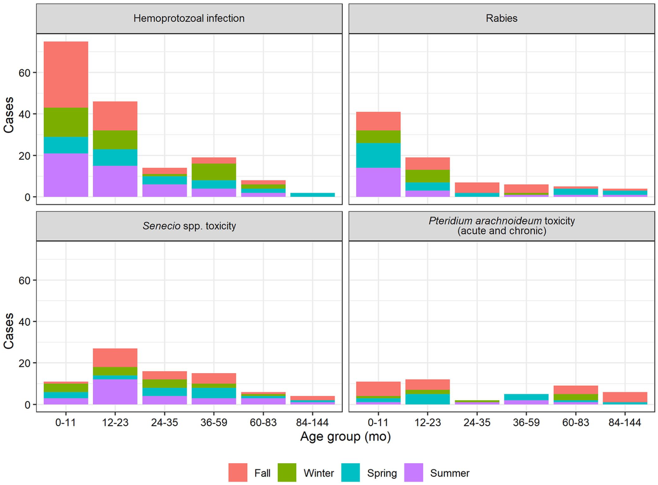
Association of the diagnoses of the 4 main causes of death in beef cattle, with age and season in which the cases occurred.
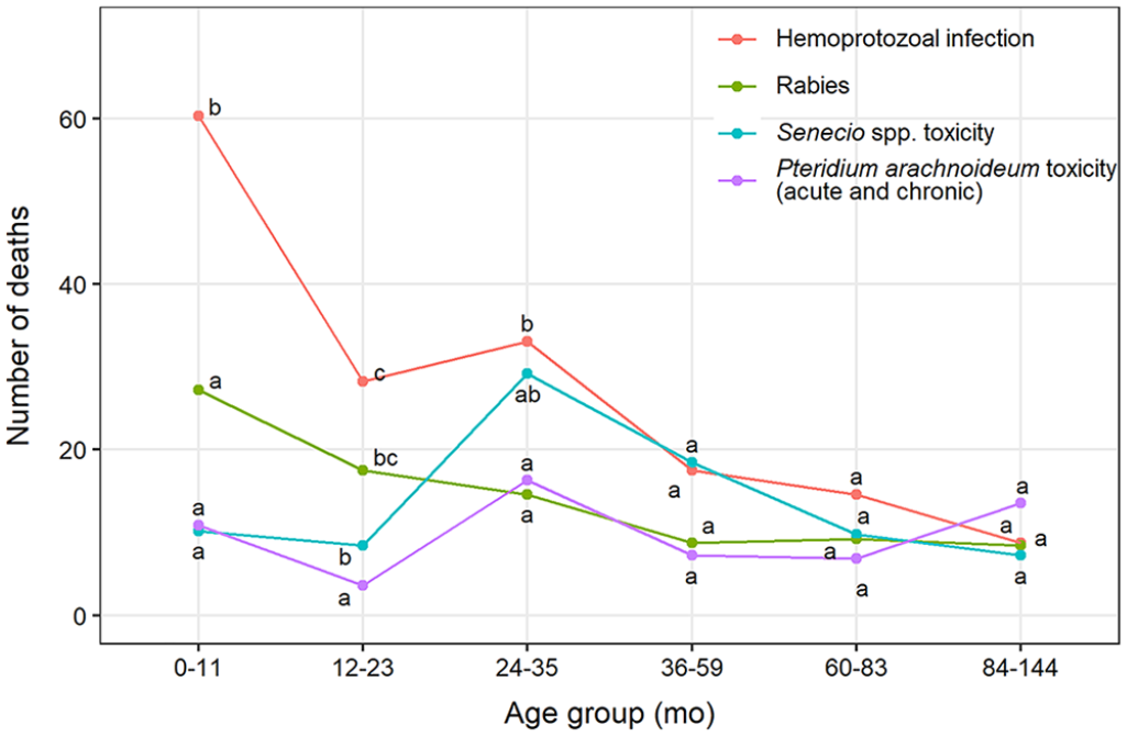
Frequencies of the 4 main causes of death in beef cattle according to age group (0–11, 12–23, 24–35, 36–59, 60–83, 84–144 mo). Different letters (a, b, c, d) within each age group indicate a statistical difference of ≥5%.
Discussion
A final diagnosis of the primary cause of death in beef cattle was possible in 78.7% of cases. This percentage is high compared to 49.3% and 46.2% observed in the central-west region of Brazil.29,33,35 In one study of mortality in beef cattle in Canada, final diagnoses were reached in 70% of cases. 40 In many of our cases, incomplete samples and clinical histories sent to the laboratory, or severe autolysis, interfered with the diagnosis. The conditions that we diagnosed were observed mainly on large properties with an extensive grazing system, where animals are not observed as frequently as in feedlots. 35
The most frequent cause of death was infectious and parasitic disease, with hemoprotozoal infection the most common diagnosis. Hemoprotozoal infection (“tick fever”) in Brazil denotes the presence of A. marginale, B. bovis, and/or B. bigemina, and represents an important cause of disease, death, and major economic loss.2,14,16,24 The disease is restricted to tropical and subtropical regions, where the tick Rhipicephalus microplus is endemic.2,13,27 In southern Brazil, as a consequence of climatic conditions, the presence of the tick is subject to enzootic instability. Depending on whether the case occurs at a time when the tick is present, the level of antibodies and morbidity and mortality from hemoprotozoal infection vary, as shown in previous studies.2,13 The disease occurred most frequently in the fall, similar to reports in a previous study. 2 In other Brazilian regions, this disease is not as frequent, given that temperature variation is minimal. In countries such as the United States, most regions are considered free of the vector; however, isolated outbreaks can occur. 5
The age group of cattle most affected by hemoprotozoal infection was <3 y old, and hemoprotozoal infection was especially significant in cases up to 1 y old. Similar data were described in 2019 in the central-west region of Brazil, in which the most affected age group of cattle was <1 y old. 30 Bos taurus indicus cattle are resistant to tick infestation and babesiosis,2,4,5,30 but they can develop severe illness and die as a result of Babesia spp. infection. 30 We observed hemoprotozoal infection in all breeds and crosses.
The second most common disease was rabies; this disease is endemic in the state of Rio Grande do Sul, as well as in other regions of Brazil.6,7,20,21,24 In South and Central America, rabies in herbivores occurs as outbreaks and is usually transmitted by the hematophagous bat Desmodus rotundus, 8 the most important natural reservoir of rabies virus in tropical and subtropical areas, from northern Mexico to northern Argentina and Chile, and some European countries.8,15,17,34 A higher incidence of rabies has been described in cattle in the southern region of Brazil in late summer, 35 which we also observed. However, in a study of dairy cows in southern Brazil, most cases of rabies occurred in the fall. 24 The age of animals affected by rabies can vary from 2 mo to 17 y. 34 We found that most animals affected by rabies were up to 2 y old, and especially <12 mo old. We suspect that rabies is an underdiagnosed disease given the zoonotic risk, which results in non-collected or incomplete materials essential to the diagnosis.
Pneumonia was responsible for 5% of cases, and similar results have been observed in other Brazilian regions, 35 suggesting an occurrence of lung disease lower than that in other countries, mainly because of grazing systems, and in the case of Brazil, a traditional extensive grazing system. Studies performed in many Brazilian regions have demonstrated the low prevalence of common respiratory agents associated with clinical disease, such as Mycoplasma sp., BoHV1, BRSV, and BPIV3. 25 Although some of our cases of pneumonia were suspected to have a viral cause, and virology for the above-mentioned viruses was performed, no agents were identified. Unfortunately, we do not have molecular tests for Mycoplasma spp. in our laboratory or region, so the prevalence of this agent in our study is unknown. In studies performed in several states of the United States with beef cattle feedlots, respiratory diseases were responsible for 44–67% of the total deaths in cattle9,19,39,40; in Ontario, Canada, the rate of lung disease deaths was ~70% in beef feedlots. 12 In feedlots, infectious agents, stress factors, adverse weather conditions, post-weaning, transport, and commingling cattle are predisposing factors that contribute to the mortality rate. 32
Ruminal acidosis is observed frequently in feedlot cattle following the intensification of production in association with an increase in grain feeding, which can disturb the digestive system of cattle. 18 In our study, this condition was not frequent, given that animals are kept in an extensive grazing system, with little or no grain in the diet.
In Brazil, extensive or semi-extensive grazing systems allow access to toxic plants, leading to intoxication outbreaks in herds.20,28 Senecio spp. (pyrrolizidine alkaloids) is the main toxic plant that causes economic losses in Rio Grande do Sul, with percentages up to 11.4% of cattle deaths, 26 and in our study up to 8.6% of deaths. We notice that toxic plant deaths were most common in cattle up to 12 mo old, except for Senecio sp. intoxication, which reached a mortality peak in cattle 24–36 mo old, in accordance with that previously described in older cows 26 ; this intoxication is uncommon in calves. 26 This age effect is mainly related to the quantity of the plant that cattle ingest, the growth stage at which the plant is ingested, and the duration of ingestion, given that deaths can occur months or years after the last intake of the plant. 26 It is believed that the ingestion of Senecio spp. is related to seasons in which there is a scarcity of feed, such as winter. 26 We could not determine the time of consumption of the plant, but deaths occurred more frequently in the fall.
The second most important toxic plant was the tropical bracken fern, P. arachnoideum. This intoxication is related to ptaquiloside, a norsesquiterpene glucoside, the active principle, and exhibits a peak in cases at 24–36 mo old, and then again at 84 mo old, which coincides with acute and chronic intoxication, respectively.3,10 Our data are compatible with those in previous studies.3,10,36 We observed that the highest number of bracken fern deaths occurred in the fall.
We also found cases of clostridial disease, including bacillary hemoglobinuria (Clostridium haemolyticum), botulism (Clostridium botulinum), and tetanus (Clostridium tetani). The diagnoses of botulism and tetanus were reached through the association of clinical signs, epidemiology, and lack of gross and histologic lesions. Eradication of clostridial microorganisms is almost impossible; however, control measures can be applied to prevent disease, such as vaccination programs. 38
Copper deficiency was the most frequent cause of death in the nutritional and metabolic disease category. Copper deficiency is an endemic disease, reported in several regions around the world, leading to economic losses as a result of weight loss, diarrhea, and inefficient immune response. 23 This deficiency can be caused by 2 main factors: high concentration of molybdenum in fodder, 23 or copper deficiency in the soil, which occurs mainly in flooded regions. 31 The coastal metropolitan region of southern Brazil, and the Salado River watershed in the province of Buenos Aires, Argentina, are known copper-deficient regions. 31 Most of our copper deficiency cases occurred in the coastal region of the state, where the animals were grazed extensively, with insufficient mineral supplementation.
Footnotes
Acknowledgements
We thank the Conselho Nacional de Desenvolvimento Científico e Tecnológico (CNPq) and Coordenação de Aperfeiçoamento de Pessoal de Nível Superior (CAPES) for the support of our study.
Declaration of conflicting interests
The authors declared no potential conflicts of interest with respect to the research, authorship, and/or publication of this article.
Funding
The authors received no financial support for the research, authorship, and/or publication of this article.