
Abstract
Protothecosis is an uncommon disease caused by algae of the genus Prototheca. In dogs, the infection is usually first localized to the colon but has the propensity to later disseminate hematogenously to many other organs, with marked tropism for the eyes and central nervous system. Diagnosis is established by culture and/or evidence of Prototheca organisms in cytologic or histologic preparations. Species characterization, however, requires molecular investigations. Our laboratory set up a real-time PCR targeting portion D1/D2 of the 28S rRNA for identification of Prototheca species from both positive cultures (of rectal swabs and urine) and formalin-fixed, paraffin-embedded tissue. Prototheca bovis, P. ciferrii, and P. wickerhamii were characterized in 11 dogs with systemic or cutaneous protothecosis. Prototheca identifications were phylogenetically consistent with the new taxonomy proposed for this genus based on the mitochondrial cytochrome b gene. As a pilot study, we screened feces and rectal scrapes from 200 asymptomatic dogs, using 2 cohorts of stray and owned animals, to determine the prevalence of intestinal carriage of Prototheca spp. The Prototheca-negative results from both cohorts of healthy dogs suggest that predisposing factors related to the host probably contribute more to the acquisition of clinical disease than exposure to contaminated environments.
Protothecosis is a disease caused by the non-photosynthetic microalga of the genus Prototheca. This alga is known to be ubiquitous in the environment, 13 and the infection occurs worldwide as localized or disseminated disease in humans and other animals.7,19 The disease is viewed with increased interest in the veterinary field because of bovine infections associated with mastitis causing economic losses incurred via reduced milk production and culling of infected cows, and sporadic life-threatening infections in dogs.8,17 To date, only 4 of the 14 recognized Prototheca species 7 are involved in canine cases: P. bovis, P. ciferrii, P. wickerhamii, and P. zopfii.10,16,17,20
Protothecosis in dogs most commonly occurs as a systemic disease following primary infection of the colon.10,16,17,20 Less commonly, dogs can develop focal or multifocal localized cutaneous disease following penetrating trauma.2,11,20 Canine patients with disseminated disease generally show signs referable to the gastrointestinal tract, particularly the colon, as well as involvement of ocular and/or neural tissues. The critical clinical sign is large bowel diarrhea, with excess mucus, blood, and tenesmus, all referable to colitis that is refractory to typical therapeutic measures.10,16,17,20 In many canine patients, 17 colitis was present for several months before the development of signs referable to dissemination. The tendency of Boxers, French Bulldogs, and their hybrids11,17 to develop protothecosis may be linked circumstantially to the occurrence of granulomatous colitis, such as that attributable to adherent-invasive Escherichia coli infections, 15 which then predisposes to Prototheca invasion of the colonic wall.
According to the clinical signs, rectal scrapings or colonic biopsy (when colitis is present), aspirates of lymph nodes and skin lesions (for cutaneous infections), 17 and urine (collected via cystocentesis) all represent specimens suitable for culture or cytologic or histologic examination to establish a diagnosis of protothecosis. 17 Prototheca colonies from positive cultures can be characterized at the species level by using molecular tools or matrix-assisted laser desorption/ionization time-of-flight mass spectrometry. 1 When specimens for culture are not available, which is commonly the case in canine medicine, characterization of Prototheca organisms detected in cytology or histology preparations can be reached by using molecular investigations. 14
We developed a SYBR green real-time PCR (rtPCR) targeting the D1/D2 portion of 28S rRNA that was able to detect and differentiate Prototheca DNA from both cultures and formalin-fixed, paraffin-embedded (FFPE) tissue specimens. Given that the environmental source of infection for canine protothecosis is generally not determined, we screened feces and rectal scrapings from 200 normal asymptomatic dogs to determine if healthy dogs might be colonized transiently after environmental exposure without developing infection.
From 11 dogs suspected of having systemic protothecosis, rectal scrapings (n = 7), colonic brush preparation (n = 1), urine (n = 3), and fresh tissue biopsy specimens (n = 2; 1 tibia and 1 lymph node) were submitted to the Mycology Unit of the Parasitology Laboratory (IZSVe; Legnaro, Padua, Italy) for cytologic and histologic evaluation and microbial culture (Table1). Histologic sections with organisms consistent with Prototheca species (Grocott–Gomori methenamine silver stain) were also available for 3 dogs (cases 7–9; Table 1). For the remaining 2 dogs, the detection of Prototheca was based on histologic examination alone, and FFPE tissue blocks were the only specimens available for further characterization (cases 10, 11; Table 1). Given that the samples used were convenience samples obtained during standard clinical investigations, specific client consent and animal ethics approval were not required as per institutional policy.
Details of the 11 canine clinical cases of protothecosis included in our study.
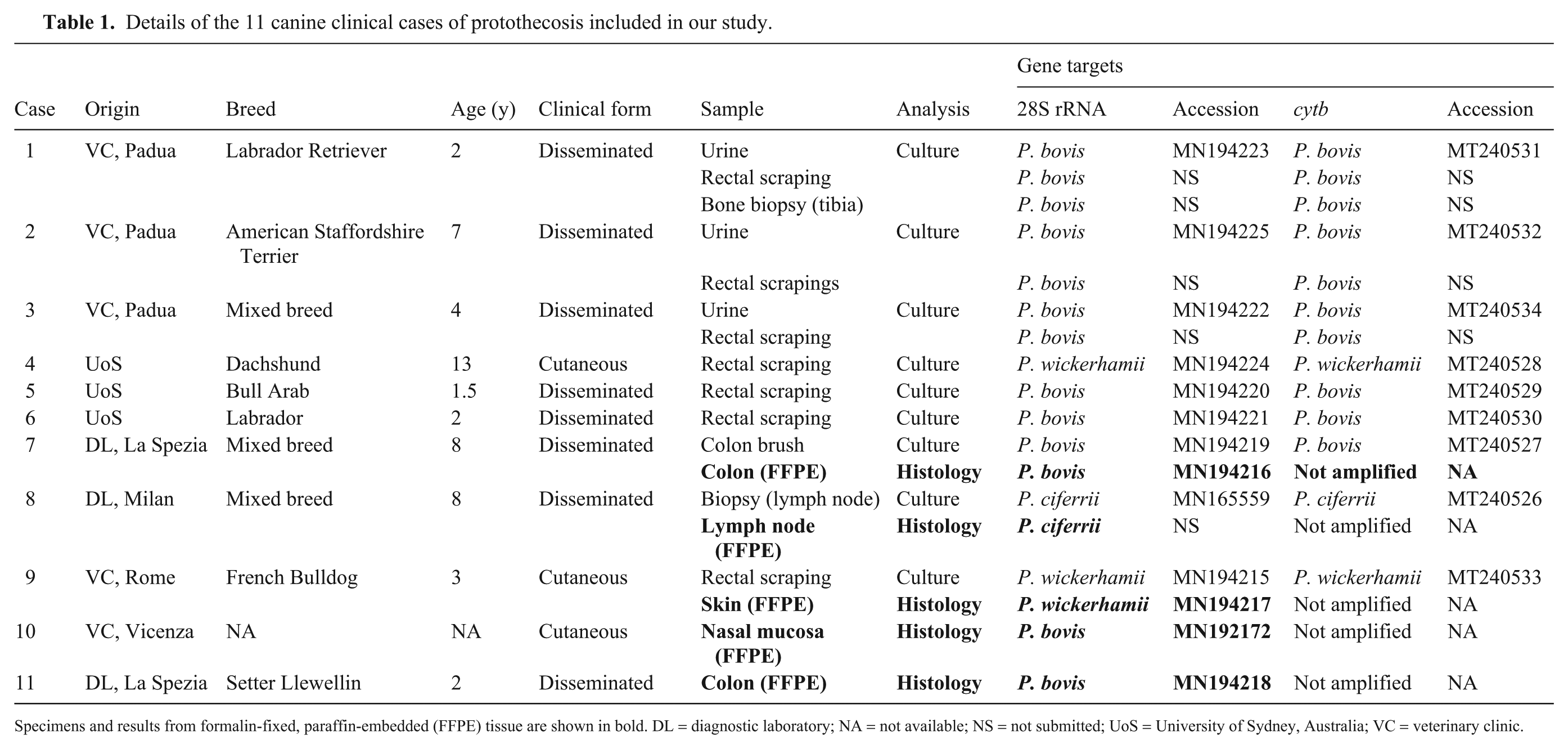
Specimens and results from formalin-fixed, paraffin-embedded (FFPE) tissue are shown in bold. DL = diagnostic laboratory; NA = not available; NS = not submitted; UoS = University of Sydney, Australia; VC = veterinary clinic.
Two hundred dogs were randomly selected to investigate the prevalence of asymptomatic alimentary carriage of Prototheca. Feces were collected from 104 German Shepherd dogs from a breeding facility (Perugia, Italy; group 1 [G1]); rectal scrapings were obtained from 96 stray dogs at the time of neutering at the municipal kennel of Padua province over the period 2018–2019 (G2). Stray dogs were from the countryside in the Veneto region where dairy farms are present, including some dairy facilities where cows with Prototheca mastitis had been diagnosed. 3 The approval by the Ethics Committee of the Istituto Zooprofilattico Sperimentale delle Venezie was obtained for our study (CE_IZSVE 8/2019).
All specimens from symptomatic canine patients and from G1 and G2 dogs were cultured on Prototheca isolation medium (PIM) 12 at 37°C for 7 d. DNA was extracted from Prototheca colonies (positive cultures), feces from G1 dogs, and from FFPE tissue, following protocols reported previously. 4 The only minor modification was that a section of canine tissue histologically negative for Prototheca and for fungi was included systematically in each series of DNA extractions as a negative control.
Prototheca DNA was amplified by a SYBR green rtPCR assay targeting 2 different genetic loci including the D1/D2 region of the 28S rRNA gene, using primers NL1 (5′-GCATATCAATAAGCGGAGGAAAAG-3′) and NL4 (5′-GGTCCGTGTTTCAAGACGG-3′) and the mitochondrial cytochrome b (cytb) gene with primers cytB_F1 (5′-GYGTWGAACAYATTATGAGAG-3′) and cytB_R2 (5′-WACCCATAARAARTACCATTCWGG-3′). The assay conditions were as reported previously, with a minor modification of annealing temperature (58°C for 28S rRNA; 54°C for cytb). 4
A negative control (sterile water) and a positive control (Prototheca bovis DNA, 18PAR/1160) were included in each PCR run, to monitor for contamination and correctness of the reaction, respectively. Part of the Felis catus interphotoreceptor retinoid-binding protein (IRBP) gene was amplified as an internal control for the adequacy of DNA extraction. 5 Fecal specimens (n = 35) were spiked with 3 µL of Prototheca DNA, and both the cytb and 28S PCR assays were also tested on those samples to exclude PCR inhibition.
All positive amplicons were sequenced, as described previously, 4 for taxonomic confirmation. Molecular phylogeny was performed using maximum likelihood on the 28S rRNA and cytb sequence data sets in MEGA v.6.0. 18 We added sequences of Prototheca spp. available from GenBank (https://www.ncbi.nlm.nih.gov/nucleotide), using Chlamydomonas reinhardtii as an outgroup (Suppl. Table 1). To compare the performance of the rtPCR assays for 28S rRNA and cytb on both culture isolates and FFPE tissues, all Prototheca DNA extracted from symptomatic dogs was tested using both PCR protocols.
Feces obtained from G1 dogs (n = 104) were screened exclusively using the cytb rtPCR because, unlike the 28S rRNA rtPCR assay, this assay was highly selective in discriminating Prototheca DNA from the DNA of commensal fungi, protozoa, and bacteria that are potentially present in samples from the intestinal tract. In validation work, the cytb rtPCR amplified Prototheca DNA exclusively and did not amplify DNA of yeasts (Candida albicans, C. tropicalis, Cryptococcus neoformans, Debaryomyces hansenii, Rhodotorula mucilaginosa, Trichosporon asteroides), filamentous fungi (Alternaria alternata, Aspergillus flavus, Aspergillus fumigatus, Cladosporium cladosporioides, Fusarium graminearum, Mucor circinelloides, Penicillium thomii), protozoa (Cryptosporidium spp., Giardia duodenalis, Toxoplasma gondii), or bacteria (Clostridium perfringens, Enterococcus spp., Escherichia coli) when tested as internal controls (data not shown).
Prototheca spp. grew on primary culture using PIM in 13 instances from 9 patients using rectal scrapings (n = 7), colonic brush preparations (n = 1), urine (n = 3), and fresh biopsy specimens (n = 2; 1 tibia and 1 lymph node; Table 1). Of the positive cultures, P. bovis was identified from 6 dogs, P. ciferrii from 1 dog, and P. wickerhamii from 2 dogs (Table 1). From FFPE tissue specimens, P. bovis sequences were obtained from 3 dogs; P. ciferrii (1 dog) and P. wickerhamii (1 dog) sequences were obtained from the other 2 patients. Prototheca identification from FFPE tissue (P. bovis, P. ciferrii, and P. wickerhamii) was concordant with the identification obtained from culture specimens from the same dog for each of the 3 patients (colon brush, lymph node biopsy, and rectal scraping from cases 7–9, respectively). For 2 dogs (cases 10, 11), FFPE tissue specimens alone were available, sourced from nasal mucosal and colonic biopsies, respectively, and no corresponding culture investigations were undertaken.
The 28S rtPCR successfully amplified DNA from 13 of 13 isolates with positive cultures and for 5 of 5 FFPE tissue specimens. The cytb rtPCR amplified DNA from all Prototheca cultures but from none of the FFPE tissue blocks. All amplicon sequences correctly identified P. bovis, P. ciferrii, and P. wickerhamii when blasted in the GenBank database (https://blast.ncbi.nlm.nih.gov/Blast.cgi). In the rooted trees constructed using 28S and cytb sequences, P. bovis, P. ciferrii, and P. wickerhamii identified from symptomatic animals are well separated from each other and from P. blaschkeae, P. moriformis (formerly P. ulmea), P. stagnora, and P. tumulicola (Figs. 1 and 2). Comparing the molecular protocols, the results of the 28S and the cytb rtPCR assays were congruent in terms of species identification, and both were well supported phylogenetically, confirming their high reproducibility (Figs. 1 and 2).
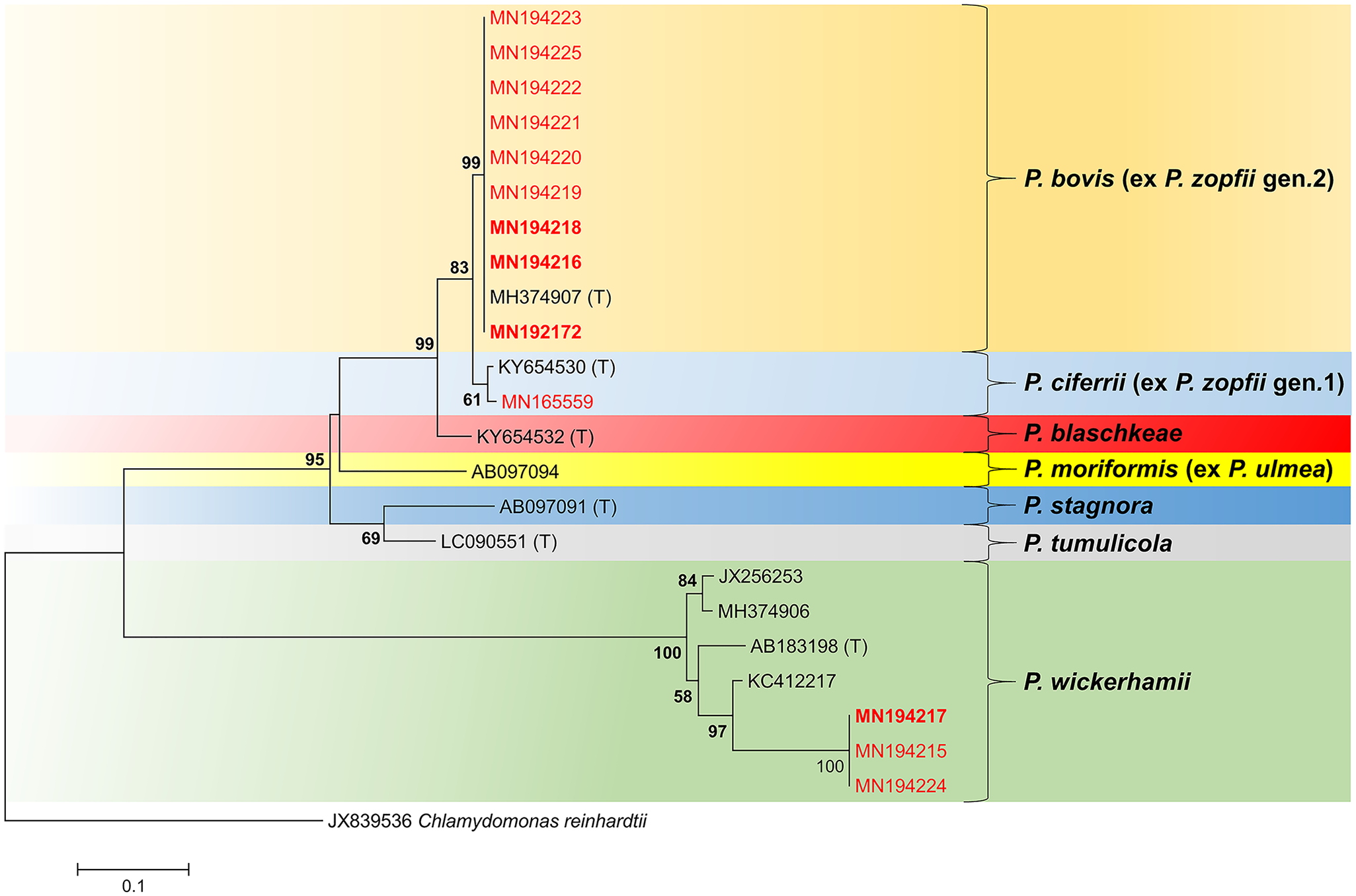
Phylogenetic tree based on the Prototheca 28S rRNA sequences. Prototheca sequences produced from our study are shown in red (from culture Prototheca isolates), and those from FFPE tissue are presented in red boldface. Chlamydomonas reinhardtii sequence was used as an outgroup. The tree was constructed using the maximum likelihood method. Bootstrap values shown at the main nodes represent the probabilities based on 1,000 replicates. (T) = type strains (color version of the figure is available online).
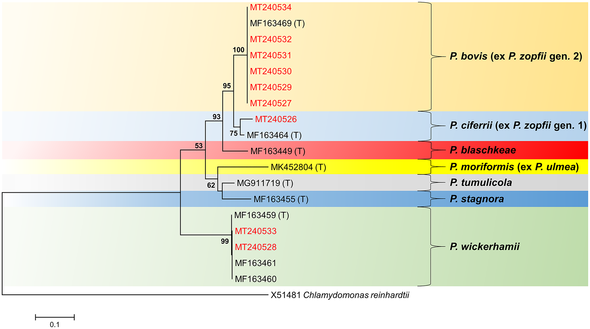
Phylogenetic tree based on the Prototheca cytb mitochondrial DNA sequences. Prototheca sequences produced from our study are shown in red. Chlamydomonas reinhardtii sequence was used as an outgroup. The tree was constructed using the maximum likelihood method. Bootstrap values shown at the main nodes represent the probabilities based on 1,000 replicates. (T) = type strains (color version of the figure is available online).
Among healthy dogs, all rectal scrapings (n = 96) and feces (n = 104) plated onto PIM were negative for Prototheca spp. Trichosporon asteroides was the only fungus cultured (>200 colony-forming units) from a single rectal scraping from a stray dog. Feces from asymptomatic dogs of G1 (n = 104) all tested negative for Prototheca with cytb rtPCR. PCR protocols performed on Prototheca-spiked samples all tested positive (n = 35), providing support that the negative results from the tested animals were indeed true negatives.
The results of the molecular protocols that we tested suggest that the 28S rRNA rtPCR is a reliable tool, suitable for detecting and categorizing Prototheca species in symptomatic animals. Indeed, Prototheca species were confirmed in 11 of 11 dogs with protothecosis. In addition, species identification is phylogenetically well supported by both the 28S and the cytb trees. It was interesting that analysis of the 28S rRNA P. wickerhamii sequences revealed higher heterogeneity than cytb mitochondrial DNA sequences. These results agree with previous studies that indicated a higher level of intra-strain polymorphism using rDNA markers (28S rRNA) compared with mitochondrial DNA markers (cytb). 6
Protothecosis is a rare disease in companion animals, with an insidious, often cryptic onset that can drive clinicians towards diagnostic possibilities other than that of an algal infection. Typically, an aspirate or biopsy sample is submitted for cytologic or histologic assessment without protothecosis having been considered as a diagnostic possibility. In such cases, the great benefit of the 28S rtPCR is the ability to detect Prototheca DNA even when specimens are not available for culture, and to use the amplicon sequence to determine the species responsible for the infection. Furthermore, identification based on sequence analysis enables the identification of other fungal pathogens distinct from Prototheca (mostly yeast forms) that may be mistaken with the alga morphology under the microscope (such as Blastomyces spp., Candida spp., Coccidioides spp., Cryptococcus spp., and Trichosporon spp.). 9
Considering that most canine protothecosis cases start as refractory colitis, the alimentary route is the most logical portal of entry for infective propagules. We would expect that stray dogs from the Veneto region, where protothecal mastitis is known to occur on dairy farms, 3 might be colonized, although most likely at a low prevalence. Our results, which are a single “snapshot” of these dogs at the time they were swabbed at the kennel, might have missed sporadic commensal carriage of Prototheca. Indeed, we may have had a better chance of detecting transient carriage had we sampled the dogs as soon as they entered the shelter. The number of clinical cases is probably too low to draw definitive conclusions concerning the source of infection and pathogenesis of protothecosis in dogs. Despite this, our data suggest that predisposing factors related to the host, such as host genetics, perhaps in relation to innate immunity or phagocytic capacity, are likely far more critical in the development of canine protothecosis than mere exposure to a contaminated environment.
Supplemental Material
sj-pdf-1-jvd-10.1177_1040638720976423 – Supplemental material for Molecular characterization of Prototheca in 11 symptomatic dogs
Supplemental material, sj-pdf-1-jvd-10.1177_1040638720976423 for Molecular characterization of Prototheca in 11 symptomatic dogs by Christian Falcaro, Tommaso Furlanello, Diana Binanti, Alessandra Fondati, Ugo Bonfanti, Mark Krockenberger, Richard Malik and Patrizia Danesi in Journal of Veterinary Diagnostic Investigation
Footnotes
Acknowledgements
We thank Dr. Carmen Losasso for sample contribution.
Declaration of conflicting interests
The authors declared no potential conflicts of interest with respect to the research, authorship, and/or publication of this article.
Funding
This work was partially funded by the Italian Ministry of Health, Directorate General of animal health and veterinary medicines (RC IZSVe 01/2015 and RC IZSVe 05/2018).
Supplementary material
Supplementary material for this article is available online.
References
Supplementary Material
Please find the following supplemental material available below.
For Open Access articles published under a Creative Commons License, all supplemental material carries the same license as the article it is associated with.
For non-Open Access articles published, all supplemental material carries a non-exclusive license, and permission requests for re-use of supplemental material or any part of supplemental material shall be sent directly to the copyright owner as specified in the copyright notice associated with the article.