
Abstract
Calving difficulty may lead to traumatic peripheral nerve injury. A male, 8-mo-old, Japanese Black calf with a history of secondary dystocia as a result of fetal gigantism had lameness and gait disturbance. At autopsy, multifocal dural thickening with adhesions to the adjacent spinal cord was observed at T12–13 and L4–5 vertebral levels. Microscopically, numerous traumatic neuroma-like fascicles of nerve twigs were embedded in the dura mater with abundant collagenous connective tissue. By immunohistochemistry, axons and Schwann cells were confirmed in each nerve fascicle. Our observations suggest that avulsion injuries in the preganglionic fibers of the spinal nerve roots, and secondary spinal cord compression, resulted in the development of neurologic signs.
Calving is a high-risk event for both cows and calves, and dystocia resulting from an oversized calf is common in beef cattle.9,13 When excessive or prolonged traction is applied during the delivery process, severe nerve damage could occur in both the cow and her calf. In cows, especially in first-calf heifers or dams with an oversized fetus, dystocia could result in compression of the obturator nerve as it courses ventrally on the medial shaft of the ilium. The sixth lumbar spinal nerve, which passes ventrally to the prominent ridge of the sacrum, is also vulnerable to compression and injury during dystocia. 3 Such nerve damage is referred to as calving paralysis.
Injury to the vertebral column, including vertebral fractures, myelomalacia, and spinal cord compression, are common in calves born following dystocia. 12 These types of injuries occur easily when the cow standing in a cattle stock suddenly becomes recumbent mid-parturition. 14 Femoral nerve paralysis is also observed frequently after dystocia, especially after a delivery that required forced traction as a result of hip-lock; the cause is considered to be over-extraction of the hip and tearing of the femoral nerve. 3 Furthermore, as in human neonates, a brachial plexus injury could be caused by severe laceration on the axilla, excessive traction on the forelimbs during dystocia, and severe abduction of a forelimb in calves.3,15 However, data pertaining to the lesions of intradural nerve-root avulsion injury in calves are very limited. We describe herein the pathologic features of multifocal injuries of thoracolumbar spinal nerve roots in a calf, which can resemble traumatic neuromas, and discuss the underlying mechanism.
A male, 8-mo-old, Japanese Black calf was examined. The calf was born following dystocia caused by his excessive size. The cow was standing during the assisted parturition, and she fell suddenly when the calf’s lumbar region protruded. Although the calf showed clinical signs of inability to rise after birth, he eventually stood on his own feet and drank milk with support. By the age of 6 mo, the calf started to collapse when turning left or right or when pressure was applied to the lumbosacral region. The calf received physiotherapy and was administered dexamethasone, vitamin B1, and non-steroidal anti-inflammatory drugs. However, clinical signs did not improve despite these treatments, and euthanasia was performed given that the prognosis was judged to be poor.
At autopsy, the dura mater was thickened and adherent to the spinal cord at the level of T12–13 (Fig. 1) and L4–5 vertebrae. The femoral and dorsal lumbar muscles were decreased in size, and the longissimus muscle at the T12–13 vertebrae was partially discolored and measured 6 × 6 cm.
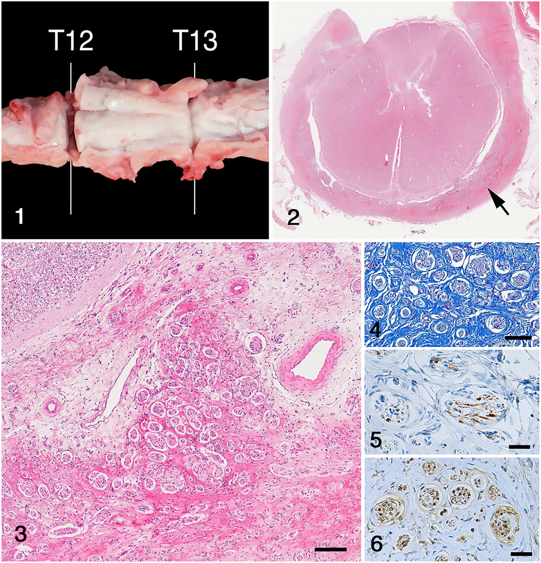
Calving-related intradural avulsion injuries of the thoracolumbar spinal nerve roots in a calf.
Major organs, including liver, spleen, kidneys, heart, lungs, skeletal muscles, spinal cord, and brain, were fixed in 10% neutral-buffered formalin. Tissue samples were processed routinely, and tissue sections were stained with hematoxylin and eosin; selected sections were stained with Masson trichrome.
Histologically, the dura mater at the level of T12–13 and L4–5 was thickened by abundant collagen deposition (Fig. 2). The dura mater contained numerous variably sized nerve fascicles, consisting of axons, swollen Schwann cells, fibroblasts, and perineurial cells (Fig. 3). Mild axonal injury was observed particularly in the dorsal and lateral funiculi in the white matter at the level of T12–13. The proliferating cells comprising the nerve fascicles showed no atypia or polymorphism. Masson trichrome staining revealed fibroplasia and collagen deposition (Fig. 4). Dural thickening was more pronounced at T12–13 than at L4–5. The grossly discolored longissimus muscles exhibited moderate atrophy of the skeletal myofibers with fat infiltration and mild edema.
Immunostaining using the labeled streptavidin–biotin method (LSAB2 system-HRP; Dako) was performed. The primary antibodies used (Dako) were a rabbit anti-S100 polyclonal antibody at a 1:5,000 dilution, a mouse anti-human neuron-specific enolase (NSE) monoclonal antibody at a 1:2,000 dilution, and a mouse anti-human Ki67 monoclonal antibody at a 1:1,000 dilution. Positive and negative controls were included in the protocols. Axons in the nerve fascicles were immunopositive for NSE (Fig. 5); hypertrophied Schwann cells surrounding axons were immunoreactive for S100 (Fig. 6). Based on Ki67 expression (< 1%), axons and Schwann cells showed no significant proliferative activity.
A diagnosis of multifocal intradural injury of the spinal nerve roots was made. The histologic features of this case closely resembled those of traumatic (amputation) neuroma, which is morphologically characterized by a reactive, non-neoplastic proliferation of axons, Schwann cells, perineurial cells, and fibroblasts at the proximal end of an injured peripheral nerve. 17 The hyperplastic lesions of peripheral nerves occur in response to injury, including surgery. 19 In the veterinary field, traumatic neuroma is well-known as a consequence of tail docking in dogs, 5 lambs, 4 and pigs.11,16 The disease has also occurred as a result of repair processes linked to cervical spinal cord injury in a dog, 8 and as a post-surgical complication following castration in horses. 1
Although nerve amputation would be expected to produce a localized nodular neuroma, amputation of spinal nerve roots would result in the proliferation of numerous small nerve bundles throughout the affected tissue. 5 Amputation neuromas develop over 1–12 mo after nerve injury. 7 The lesion would have been expected to become larger and the clinical signs worse. In the absence of nodular growth, it is speculated that, in our case, the intradural spinal nerve roots were avulsed, and multiple nerve stumps were produced in and around the dura mater, attended by marked fibroplasia. As the lesions progressively enlarged, the spinal canal gradually became stenosed, eventually applying pressure to the spinal cord, resulting in lameness and gait disturbance when the calf was 6 mo old. When the calf turned left or right, the spinal canal would be further compromised, resulting in recumbency.
The calf in our case had a history of calving accident, in which the cow suddenly became recumbent during assisted traction. It is postulated that, as a consequence, pressure and traction were applied to the lumbar region. Although the most frequent consequence of forced extraction is a fracture, followed by peripheral nerve damage and joint luxation, 10 an acute spinal cord injury also occurs, associated with vertebral fracture and luxation. Common sites of vertebral fracture in calves at calving are the C2–4, T10–13, and L3–6 vertebrae. 6 This lesion distribution is consistent with the affected sites in our case; however, no signs of fracture or luxation were found.
Damage to the femoral nerve, arising from the L4–6 nerve roots, is also commonly seen in calves following forced extraction. 18 In the only other case of spinal nerve root avulsion reported, the calf had no vertebral fracture 2 ; it had a history of dystocia requiring manual assistance, and autopsy revealed avulsion of the L4 and L5 spinal nerve roots on the right side. Although our case also showed nerve root injury at L4–5, the lesion was milder than at T12–13, and the femoral and sciatic nerves were intact. As such, paralysis was not present.
Footnotes
Declaration of conflicting interests
The authors declared no potential conflicts of interest with respect to the research, authorship, and/or publication of this article.
Funding
The authors declared that they received no financial support for their research and/or authorship of this article.