
Abstract
Ocular diseases are an important category in equine medicine; however, most articles regarding histologic ocular lesions in horses are exclusive to a specific condition and do not provide a complete review of clinically significant ocular disease frequency in a diagnostic laboratory. We reviewed sections of equine eyes from 140 cases (98 enucleations [biopsies] and 42 autopsies) with clinically relevant ocular alterations at 2 diagnostic centers in the United States. The most common primary conditions were non-traumatic keratitis (36), equine recurrent uveitis (ERU; 31), traumatic injuries (22), ocular and periocular neoplasms (19), and uveitis and/or endophthalmitis resulting from sepsis (18). Congenital anomalies (3) and retinal atrophy and detachment alone (3) were infrequent. Non-traumatic keratitis was frequently accompanied by anterior uveitis (22), corneal rupture (16), pre-iridal fibrovascular membrane formation (13), and secondary mycotic infection (11). ERU was the second and third most prevalent disease in autopsies and enucleations, respectively. This condition was commonly associated with glaucoma (15). Glaucoma (25) and cataract (20) were the most prevalent secondary alterations in the evaluated cases. Keratitis (20) and corneal rupture (16) were among the most prevalent consequences of trauma. Information presented herein may guide clinicians and pathologists, contributing to the early diagnosis of potentially vision-impairing conditions and raising the chances of successful treatment and cure.
Introduction
Ocular conditions are an important disease category in equine medicine.10,44 Depending on the cause and evolution of the ocular condition, ocular conditions could even lead to blindness and eventually culminate in euthanasia of the affected horse. Despite the importance of these conditions, most articles regarding histologic ocular lesions in horses are exclusive to a specific disease or alteration, such as corneal stromal abscesses, 21 keratomycosis, 4 recurrent uveitis, 12 corneal squamous cell carcinomas,11,25 retinal detachment, 39 or glaucoma.9,22,45
To our knowledge, there are no reports in the equine literature that provide a complete and general histologic review of clinically significant equine ocular diseases that led to enucleation or were associated with the cause of death or euthanasia. This type of investigation allows for a comparison of occurrence of clinically relevant ocular conditions affecting this species, which can guide clinicians and pathologists in the diagnosis of future cases. Furthermore, ocular histopathology may provide a more detailed analysis of lesions that cannot be fully characterized in clinical surveys. Several ocular conditions require histologic confirmation, and thus may be underrepresented in clinical surveys that do not involve enucleation and histologic examination of the globe. Therefore, we investigated the occurrence of primary and secondary ocular conditions leading to enucleation or to death or euthanasia in horses submitted for surgery or autopsy, in which the eyes were evaluated histologically at 2 diagnostic centers in the United States.
Materials and methods
We conducted a retrospective study of equine globes submitted to and archived at the Pennsylvania Animal Diagnostic Laboratory System (PADLS; Philadelphia, PA; 1997–2011) and the Louisiana Animal Disease Diagnostic Laboratory (LADDL; Baton Rouge, LA; 2003–2015). The autopsy and biopsy files from both institutions were reviewed, and equine cases for which the ocular histologic slides were available were analyzed. For biopsy cases, only ocular conditions leading to enucleation were included. For autopsy cases, inclusion criteria were as follows: 1) primary ocular diseases leading to death or euthanasia; 2) primary ocular diseases contributing to the decision for euthanasia; or 3) systemic diseases that led to death or euthanasia and that directly and significantly affected one or both eyes to the extent that ocular signs were reported in the clinical history, or the macroscopic appearance of the eye at autopsy led the pathologist to collect and analyze the eyes histologically.
The reports from the selected cases were evaluated. and information regarding breed, sex, age, clinical history, and gross and histologic findings were obtained. The histology slides from the included cases were reviewed by 2 pathologists (I.M. Langohr, M.M. Flores). When slides of periocular tissue were present, they were also analyzed; however, our study did not primarily assess periocular lesions. The ocular lesions were categorized according to 1) the primary pathologic process and 2) the secondary lesions accompanying the primary process. When 2 primary pathologic processes were present, the most severe and clinically important one was chosen.
Results
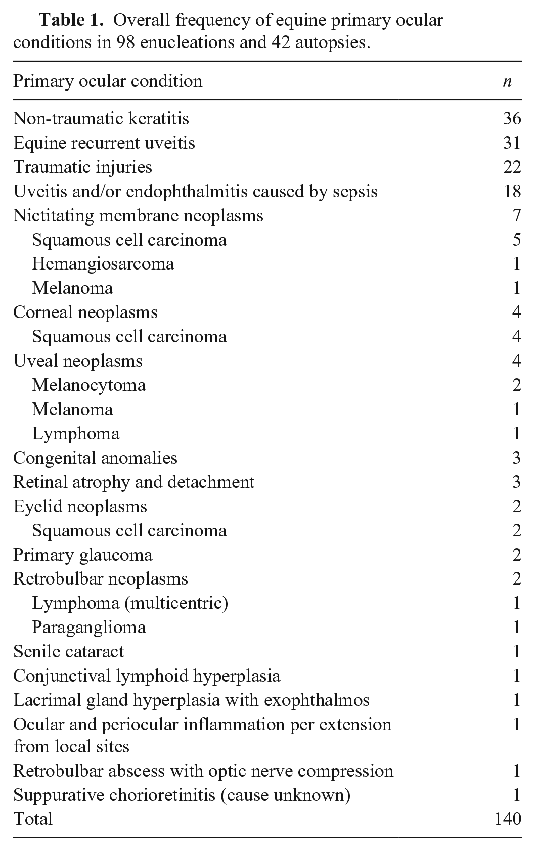
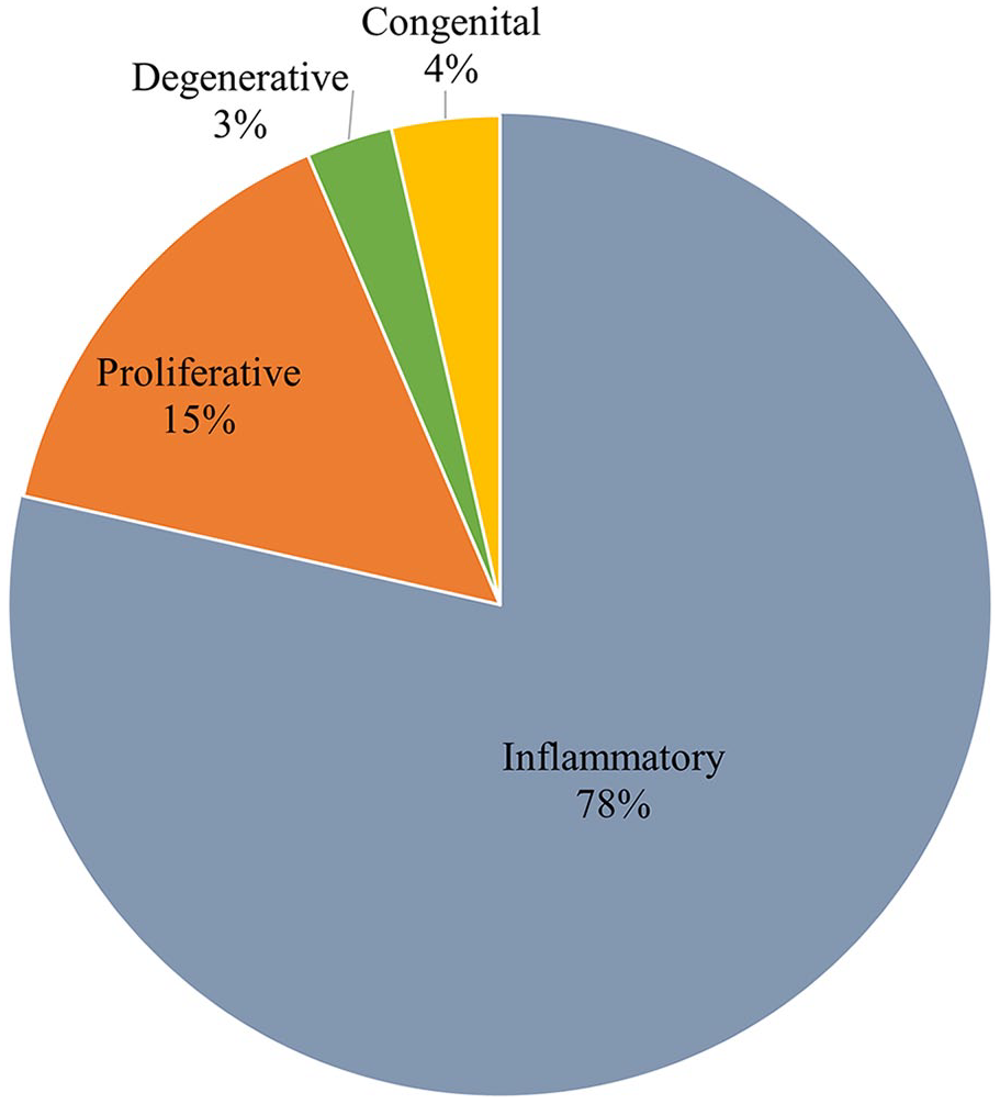
We included 140 horses with ocular diseases, of which 98 were enucleations and 42 were autopsies (Table 1; Fig. 1). Enucleation cases had only one available globe for histologic examination. Sixteen autopsy cases had slides corresponding to both globes, and the remaining cases had only one slide available for histologic review. In 14 of 42 autopsy cases, the primary ocular disease was the main cause of death or euthanasia. Five horses had a primary ocular condition contributing to the decision for euthanasia. The remaining 23 horses had systemic diseases that significantly affected one or both eyes, leading to sampling during autopsy and later microscopic analysis of the eyes.
Overall frequency of equine primary ocular conditions in 98 enucleations and 42 autopsies.
Overall frequency of equine primary ocular cases in 98 biopsy and 42 autopsy cases.
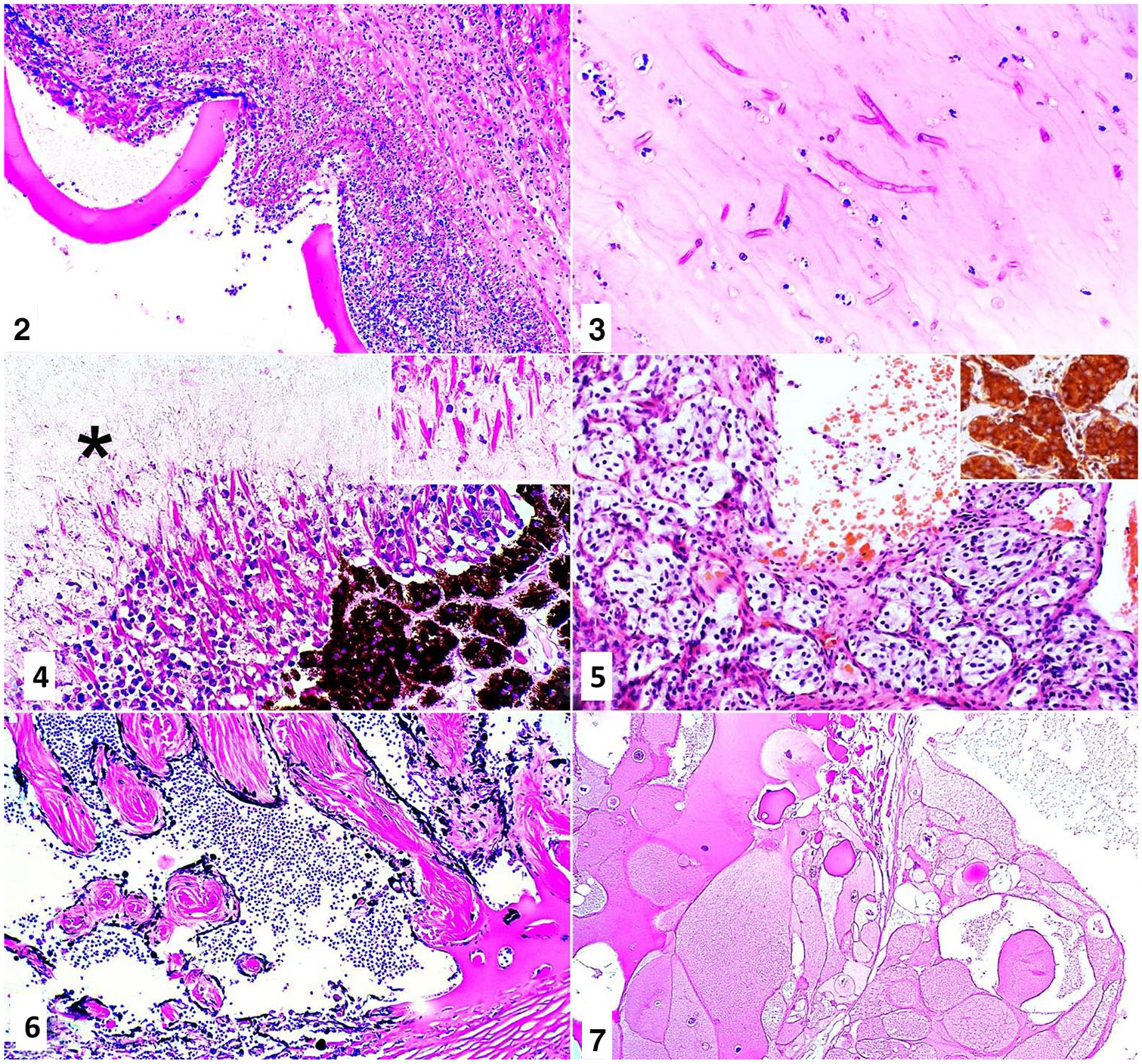
Thirty-six horses (33 enucleations and 3 autopsies) were diagnosed with keratitis, 34 of which were ulcerative. Only one case was reportedly bilateral. The primary cause of this corneal lesion was determined in only one case (corneal desiccation following previous eyelid surgery). None of the cases had any reported history of ocular trauma. The ocular alterations accompanying keratitis consisted of anterior uveitis (22), corneal rupture (16) with secondary iris prolapse (9) and lens rupture (3), keratomalacia (13), pre-iridal fibrovascular membrane formation (13), hyphema (7), Descemet membrane rupture (5; Fig. 2), retinal detachment (4), mild panuveitis (3), cataract (3), and corneal stromal abscess (2). Fungal hyphae were commonly present within the inflamed cornea (11; Fig. 3), often also deep within the cornea, at the level of the Descemet membrane (7), or even within the eye, specifically within the lens capsule (2), likely predisposing to the lens rupture in these cases. Most cases had non-pigmented hyphae with parallel walls, frequent septation, and acute-angle branching (morphologically consistent with Aspergillus and Fusarium). Only in one case did the fungi have brown-pigmented walls.
Histologic lesions in equine globes.
The diagnosis of equine recurrent uveitis (ERU) was made in 31 cases, representing the second most common condition in autopsy cases (11 of 31) and the third most common in biopsy cases (20 of 31). In 6 of 31 cases, the lesions were bilateral and, in 19 of 31 cases, the clinical history suggested that they were unilateral. ERU was histologically characterized by the expected lymphoplasmacytic inflammation of the anterior uveal tract associated with multiple eosinophilic crystals within the non-pigmented ciliary body epithelium (28 of 31) and/or amyloid-like material within the apical cytoplasm of the inner non-pigmented ciliary epithelium (18 of 31; Fig. 4). Pre-iridal fibrovascular membrane formation was a common secondary lesion (14 of 31; Fig. 5), occasionally accompanied by peripheral anterior synechiae (5 of 31). Additional histopathologic findings included inner retinal atrophy consistent with glaucoma (15 of 31) and cataract (10 of 31). In some cases (11 of 31), ERU was associated with another concomitant or preceding primary process. Three horses affected by ERU also had uveitis and/or endophthalmitis as a result of sepsis, including a 5-mo-old foal with Rhodococcus equi infection; 2 horses with ERU also had an ocular neoplasm (iridal melanoma and retrobulbar multicentric lymphoma). No infectious agents were identified histologically within the eyes affected by ERU, even in those cases of infectious diseases such as R. equi sepsis.
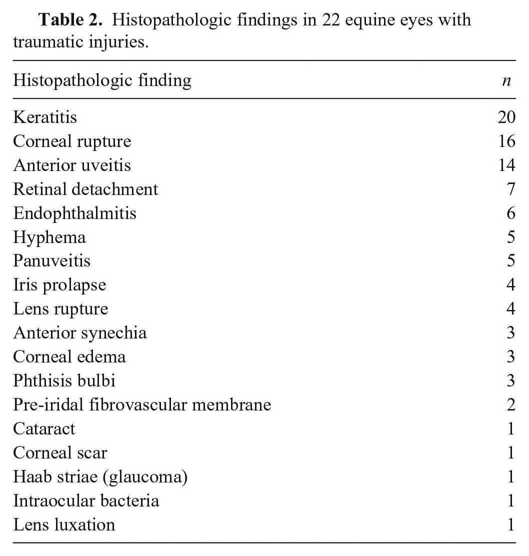
Cases with a clinical history of “trauma” were classified as “traumatic injuries” and were all enucleations. Such cases were characterized by various lesions in different ocular structures (Table 2). In 14 of 22 cases, the ocular trauma was reportedly a primary event. In one case, the ocular trauma was considered secondary to local irritation by a silver nitrate–treated recurring sarcoid in the medial canthus. In 7 of 22 cases, the clinical history did not provide enough information to indicate if the trauma was a primary event or if it was secondary to another condition.
Histopathologic findings in 22 equine eyes with traumatic injuries.
In 18 cases (all autopsies), the clinical history and/or autopsy findings were indicative of sepsis, and the ocular changes were secondary to this systemic condition. Of these, 7 were reportedly bilateral. Of the other 11 cases, only 1 eye was available for retrospective evaluation, which precluded the characterization of the case as unilateral or bilateral, although 2 cases had a clinical history indicating unilateral eye involvement. Inflammatory cells were present within the anterior chamber (Fig. 6), posterior chamber, and/or vitreous body in all cases included in this category. Intraocular bacteria were not seen in any of these cases. Most of the affected animals (13 of 18) were < 1 y old, and 7 of these were ≤ 1 mo old. Systemic R. equi infection was diagnosed in 8 of 18 cases. The other cases did not have any indication of the bacteria involved in the sepsis. Secondary ocular changes included keratitis (7), pre-iridal fibrovascular membrane (3), and serous retinal detachment (2).
Nineteen ocular or periocular neoplasms were diagnosed, of which 17 were in biopsy and 2 were in autopsy cases. Only one of the cases of retrobulbar (multicentric) lymphoma was reportedly bilateral. Half of the horses (10 of 19) were > 10 y old. The most common diagnosis was squamous cell carcinoma (SCC; 11 of 19), which affected 3 different sites: nictitating membrane (5 of 11), cornea (4 of 11), and eyelids (2 of 11). Uveal melanocytic neoplasms (2 melanocytomas and 1 melanoma) were the second most common diagnosis. Lymphoma was diagnosed in the retrobulbar space in 2 cases, 1 of which was classified under the concurrent process of ERU, and in the uvea in 1 case. Additionally, one case each was also diagnosed with the following neoplasms: retrobulbar paraganglioma, nictitating membrane hemangiosarcoma, and nictitating membrane melanoma.
Three foals were diagnosed with congenital anomalies, all of which were submitted to autopsy. These animals had congenital bilateral ocular conditions that led to euthanasia, consisting of cataract (1), retinal detachment (1), and microphthalmia (1). Only in the case of the foal with retinal detachment was there information available regarding the breed; it was an Arabian foal that was “blind since birth.”
Glaucoma and cataract were common secondary conditions in our study. Twenty-five cases of glaucoma were included, of which 23 were classified as secondary and 2 as primary. These glaucoma cases were diagnosed based on histopathologic findings, in particular inner retinal atrophy (23 cases). ERU accounted for 15 of 23 of the secondary glaucoma cases, followed by the uveal melanocytic tumors (3 of 23). Other causes of secondary glaucoma were uveitis and/or endophthalmitis caused by sepsis (2 of 23), non-traumatic keratitis (1 of 23), traumatic injury (1 of 23), and nictitating SCC (1 of 23) resulting in corneal invasion, corneal rupture, and panophthalmitis. In one case of secondary glaucoma, the primary cause was undetermined. Glaucomatous eyes had inner retinal atrophy (23), corneal striae (9), cataract (7), optic disc cupping (6), corneal edema (4), and keratitis (4, corresponding to mild-to-moderate secondary inflammation and edema, presumably caused by corneal desiccation because of failure of the lids to cover the enlarged globe and/or corneal trauma secondary to blindness).
Cataract was the most frequent secondary ocular alteration following glaucoma. Cataract was diagnosed in 20 cases (Fig. 7) and was generally secondary to other ocular lesions such as ERU (10 of 20), non-traumatic keratitis (3 of 20), uveitis and/or endophthalmitis secondary to sepsis (2 of 20), primary glaucoma (1 of 20), and traumatic injuries (1 of 20). Nonetheless, age-related primary cataract could not be completely ruled out because most cases affected horses > 10 y old. The only younger horse (6-y-old) with acquired cataract had primary glaucoma. Another juvenile horse (< 1-y-old) had bilateral congenital cataract.
Discussion
Keratitis (traumatic and non-traumatic), ERU, and neoplasms were among the most common diseases found in our retrospective evaluation of histologic ocular slides of 140 horses sampled between 1997 and 2015. Previously published studies regarding equine ocular pathology have generally not indicated the overall prevalence of these conditions. Among the few articles concerning the subject, a retrospective North American Veterinary Medical database survey noted uveitis (19%), corneal ulcers (18%), and ocular SCC (11%) as the most common problems. 17 A survey of ocular diseases in domestic animals in India listed “corneal opacity” (28%) and trauma (7%) among the most frequent clinical findings in horses treated for ocular problems in a referral clinic. 40 Another clinical study from India of the prevalence of ocular diseases in 500 apparently healthy army horses found 100 animals with at least 1 ocular condition 42 ; keratitis (30%), uveitis (24%), and conjunctivitis (12%) were frequent among these. A similar clinical investigation of 204 healthy Thoroughbred racehorses in Australia noted peripapillary chorioretinal lesions (52.5%) and cataract (17.2%) to be most frequent conditions. 24 Histopathology was not performed in either of these clinical surveys, however. Regardless of the differences concerning methodology, inclusion criteria, and disease prevalence encountered by these authors, in agreement with our results, these studies mention corneal disease, trauma, uveitis, and ocular neoplasms among the most common ophthalmic conditions in horses.
We interpreted all cases classified as “non-traumatic keratitis” as primary corneal lesions based on our histologic examination. The cornea can also become secondarily inflamed in several conditions. Therefore, differentiating primary from secondary corneal inflammation can be a challenge. 44 The importance of equine corneal diseases, particularly keratitis, is well established and has motivated several studies regarding clinical findings, treatment, and outcome.4,21,29,35 Bacterial, fungal, and even viral agents may be involved.1,26,29,31 If left untreated, keratitis may progress and ultimately result in enucleation. 10 Keratomalacia, stromal abscess associated with mycotic infection, corneal rupture with secondary iris prolapse, anterior uveitis, and pre-iridal fibrovascular membrane, as seen in our cases, are common consequences of keratitis.5,21,29,44 Corneal fungal infection (mycotic keratitis) is a classic entity of horses that represents a secondary fungal infection in an already injured cornea.1,4,14,23,35,44 Aspergillus and Fusarium, both with similar histologic appearance, are frequently incriminated in these cases.1,4,14,35 Unfortunately, only routinely stained histologic sections were available for most cases included in our study, which prevented further characterization of the causative agent. This, however, would not represent a major clinical problem given that the treatment and outcome of these 2 infections is reportedly similar. 38 In 2 horses in our study, fungal hyphae were also noted within the lens capsule, indicating secondary intraocular fungal invasion. Thus, the prognosis of mycotic keratitis may be guarded, occasionally leading to blindness and enucleation.1,35
ERU is an important cause of blindness in horses in the United States and other countries.9,10,16,42,44 It is characterized by recurrent episodes of anterior uveitis with increasing severity. As the inflammatory episodes get worse, sequelae such as cataract, lens luxation, synechiae, retinal separation, and interstitial keratitis may develop.10,44 ERU is considered by some to be one of the most common causes of glaucoma in the horse, 9 which our findings support. Several studies have attempted to investigate the etiology and pathogenesis of ERU; however, these remain controversial. Infection by Leptospira interrogans, autoimmunity,10,12,13,44 and breakdown of the ocular-blood barrier 6 are reportedly involved. Additionally, Appaloosas seem to be genetically predisposed.10,13 The eosinophilic material that typically accumulates along the surface of the ciliary body in ERU cases and that is frequently referred to as “amyloid-like” 44 has been confirmed as amyloid, specifically as amyloid AA, in an ultrastructural and immunohistochemical study. 32 No infectious agents were identified within the globes affected by ERU in our study; however, some cases occurred concurrently with other diseases, such as sepsis (including R. equi infection), ocular neoplasms, and anterior segment dysgenesis. Systemic R. equi infections in foals can trigger intraocular immune-mediated inflammation, occasionally causing ocular inflammation in the absence of the bacteria. 36 This may have triggered ERU. Whether ocular neoplasms and congenital defects play any role in the pathogenesis of ERU is not reported, to our knowledge. It is therefore undetermined if the concurrent occurrence of these conditions with ERU in our study was incidental or if there was any causative relationship between them.
Horses are highly susceptible to ocular trauma, which was reflected in our study and has also been observed by others.5,34,40 This can be attributed to several reasons, including the ocular anatomy of this species, characterized by large and prominent eyes. 28 The cause of most cases of ocular enucleation in horses was previously attributed to ocular trauma. 34 It is important to emphasize, however, that not all equine traumatic injuries culminate in enucleation or autopsy, which are the worst outcomes for a horse undergoing ocular trauma. Therefore, traumatic ocular conditions, although commonly reported, are likely underrepresented in a histologic study such as ours.
Thirteen of the 18 horses with uveitis and/or endophthalmitis secondary to sepsis were < 1 y old in our study, 7 of which were ≤ 1 mo old. Uveitis is a relatively common consequence of sepsis in foals, and generally a less common manifestation in older horses.30,41 When present in septic foals, it is a predictor of poor prognosis. 30 A bilateral presentation is most common.36,41 Although information regarding the involvement of one or both globes was not present in all of our cases, at least 7 of 18 were reportedly bilateral. Ocular lesions secondary to sepsis are caused directly, by the presence of bacteria within the uveal tract or ocular chambers, or indirectly, by the effect of bacterial endotoxins on the ocular structures or by immune-mediated mechanisms.30,36 However, most studies regarding equine ocular manifestations of sepsis do not explore these mechanisms in more detail and therefore do not allow the classification of the ocular lesions as directly or indirectly related to the bacterial infection.30,41 Intraocular bacteria were not seen in any case of uveitis and/or endophthalmitis caused by sepsis in our study; however, one cannot eliminate this possibility based exclusively on routine histologic examination. Apart from the 8 cases associated with systemic R. equi infection, other bacteria involved in these septic horses were not identified. In addition to R. equi,36,41 some of the bacteria reportedly associated with uveitis secondary to sepsis in horses include Escherichia coli 30 and Streptococcus equi. 3
The eyes can be the site of a variety of primary and metastatic neoplasms. SCC was the most prevalent eye tumor in our series, consistent with what is described in the literature.2,18,20,44 Most cases are associated with chronic exposure to ultraviolet (UV) radiation. 44 A frequent site for this tumor is the nictitating membrane,11,27,44 which was the most commonly affected structure in our study. If surgically excised early in the course of disease, SCCs of the nictitating membrane may not necessitate enucleation.11,33 Therefore, it is important that owners and veterinarians are aware of this entity in order to identify preneoplastic lesions early in the process, allowing surgery to be done in a reasonable time. SCCs at this site have an apparent decreased likelihood of recurrence when compared to tumors involving the eyelids, which is attributed to the treatment modality adopted based on the affected location. 11 Hemangiosarcoma is another tumor that might be induced by UV radiation and occasionally affects the nictitating membrane, as seen in one horse of our study. 44 A SCC and a hemangiosarcoma have been described affecting the same nictitating membrane in a horse. 15 This is a possibility in poorly pigmented periocular tissues. It is important to note that, because our study did not primarily assess periocular lesions and was confined to whole globes that also happened to have periocular lesions, neoplasms such as SCC and hemangiosarcoma were likely underreported herein.
Three of the 140 cases included in our study were diagnosed with congenital ocular anomalies, leading to euthanasia. Cataract and microphthalmia are the most frequent congenital anomalies reported in the horse,37,43 followed by other conditions including persistent pupillary membrane, retinal dysplasia,8,37 and rarely retinal detachment. 39 Although Arabian horses seem predisposed to some congenital ocular problems, such as cataract and microphthalmia, retinal detachment as seen in an Arabian foal in our study is not one of them. 37
Our study included a relatively high number of glaucoma cases. In the past, this condition was considered rare in horses and an uncommon consequence of ERU in this species. 45 With the availability of reliable tonometers, glaucoma in horses has started to be recognized as a common consequence of uveitis, 7 specifically of ERU, 9 as we observed in our study. Diffuse and persistent corneal edema may be the first clinical sign in a horse with glaucoma. 22 Corneal vascularization (83%), corneal striae (70%), corneal and anterior uveal inflammation (75%), descemetization of pectinate ligaments (83%), retinal atrophy (89%), and optic nerve hypercellularity (93%) are common histologic lesions in cases of equine glaucoma, 9 and were seen in several cases of glaucoma included in our study. Optic disc cupping was also occasionally observed in our study, as described by others.9,45 This histologic finding may be variable between studies, however, because it depends on correct trimming and inclusion of the optic nerve in the histologic section. Additionally, Descemet membrane detachment was recently described in 5 horses affected by glaucoma 22 ; ~ 75% of the Descemet membrane was detached in these enucleated globes, with spindle cell proliferation covering the overlying and denuded corneal stroma. A lesion previously described as “absence of Descemet membrane” in equine glaucomatous eyes 19 appears to hold some resemblance to the changes described as Descemet membrane detachment. 22 This seems to be an unusual feature of glaucoma, however, which has not been mentioned by others9,45 or seen in our series.
An important limitation of our histologic retrospective study was the access solely to routinely stained slides in many cases. This precluded further investigation of cases of suspected infectious etiology through histochemical, immunohistochemical, and PCR techniques. Considering that infectious agents (fungi and bacteria) are not necessarily readily visible with routine histologic stains, the presence of these agents might have been underdiagnosed in our study. Our survey nonetheless highlights the importance of ocular diseases in horses and describes the prevalence of each condition in 2 diagnostic centers in the United States. Information presented herein may guide clinicians and pathologists, contributing to early diagnosis and raising the chances of successful treatment and cure.
Footnotes
Acknowledgements
We thank the pathologists and residents who contributed to the work-up of the cases submitted to the pathology service at the Louisiana Animal Disease Diagnostic Laboratory and the Pennsylvania Animal Diagnostic Laboratory over the period of this study. We thank Suzanne Hindman and Jaqueline Ferracone, research assistants at New Bolton Center PADLS, for their invaluable help in retrieving the retrospective materials for our study.
Declaration of conflicting interests
The authors declared no potential conflicts of interest with respect to the research, authorship, and/or publication of this article.
Funding
The authors received no financial support for the research, authorship, and/or publication of this article.