
Abstract
Extraintestinal pathogenic Escherichia coli (ExPEC) strains carrying distinct virulence attributes are known to cause diseases in humans and animals and infect organs other than the gastrointestinal tract. A fatal case of bronchopneumonia in a 12-year-old female Quarterhorse was investigated. Following postmortem examination, E. coli, Enterococcus sp., and Klebsiella pneumonia were isolated from the lungs, which contained multifocal intra-alveolar accumulations of neutrophils and macrophages with edema, hemorrhage, and fibrin. The strain of E. coli belonged to O2:H21 and carried virulence genes cnfl, sfa, foc, fimA, and papG allele I that are known to be associated with ExPEC strains. The strain was resistant to several antimicrobials including clindamycin, erythromycin, oxacillin, penicillin, and rifampin. This is the first report, to the authors' knowledge, in which ExPEC O2:H21 has been associated with fatal bronchopneumonia in a horse.
Keywords
Extraintestinal pathogenic Escherichia coli (ExPEC) strains have functionally similar virulence profiles and phylogenetic backgrounds that are distinct from commensal or intestinal E. coli. 16 Typically, ExPEC strains have been implicated in causing urinary tract infection (UTI), pneumonia, meningitis, and other diseases in humans. In animals, these strains have been reported to cause UTIs 5,11 and hemorrhagic pneumonia in dogs 3,7 and cats. 17 The ExPEC strains carry genes encoding for virulence attributes such as cytotoxic necrotizing factor (Cnf-1), type 1 fimbriae, and P fimbriae adhesins. 6,8,11 Escherichia coli isolated from the present fatal case of bronchopneumonia with pulmonary edema and hemorrhage in a horse exhibited traits that were typical of ExPEC strains. To the authors' knowledge, this is the first report in which an ExPEC strain was associated with a fatal case of pneumonia in a horse.
A 12-year-old female Quarterhorse in late gestation was presented to a local veterinarian for mucopurulent nasal discharge. The mare's vaccination status was uncertain, although no vaccines had been given during the past 1.5 years. The mare was maintained on a small pasture with several other horses adjacent to a pasture that contained beef cattle. The horses had access to feed and shelter in a small barn that also housed approximately 20 Newfoundland dogs that were kenneled as part of a breeding operation. The mare's owner initiated penicillin treatment 2 days prior to the initial veterinary examination. Upon examination the mare was depressed, lethargic, anorexic, drooling, mildly ataxic, and had bilateral mucopurulent nasal discharge. Cranial nerve function was normal, and there was a low-grade fever (39°C). The mare was treated with supportive care and the owner was advised to switch from penicillin to an oral sulfonamide antimicrobial compound. By day 3, the horse was reluctant to stand and had difficulty swallowing. The owner was unable to administer oral medication because of the mare's inability to swallow and instead continued treatment with penicillin. On the fourth day after the onset of illness, the owner and veterinarian elected to perform a terminal hysterotomy in an effort to salvage the foal 11 days prior to the calculated date of parturition. The mare was euthanized immediately following the procedure. The foal received colostrum replacement and supportive care but died at approximately 2 weeks of age. No further diagnostics were performed on the foal.
One day following euthanasia, the mare was submitted for postmortem examination to the Animal Diagnostic Laboratory of The Pennsylvania State University (University Park, PR). Upon gross examination, the carcass was in good nutritional condition with mild postmortem autolysis. An open abdominal surgical incision was present on the ventral midline and through the uterus, consistent with terminal hysterotomy. There was foamy white material at both nares and throughout the nasal cavity bilaterally. The right lung was markedly reddened and edematous. There were extensive multifocal and coalescing linear ulcers in the esophageal mucosa and round to irregular ulcers in the gastric mucosa, both in the squamous and glandular portions. The liver displayed an enhanced lobular pattern. Selected tissue samples from all organs were fixed in 10% neutral buffered formalin and processed routinely, embedded in paraffin, sectioned at 5 μm, and stained using hematoxylin and eosin. Additional paraffin-embedded sections of lung were later sectioned at 5 μm and stained using the Gram method for tissue. a Additional tissues were submitted for fluorescent antibody assay, virus isolation, and bacteriological, parasitological, and toxicological testing.
Histologically there was marked pulmonary edema characterized by abundant proteinaceous fluid within alveolar spaces and interlobular septa (Fig. 1). Multifocally, the lung contained numerous intra-alveolar and fewer intrabronchiolar neutrophils admixed with macro-phages, fibrin, and acute hemorrhage (Fig. 2). There was mild multifocal necrosis of alveolar septa. There were numerous colonies of coccobacilli both free in alveoli and within the cytoplasm of many neutrophils and macrophages (Fig. 3). Gram-stained sections of lung revealed numerous Gram-negative bacilli and fewer Gram-positive bacteria (Fig. 4). Within the liver there was marked centrilobular microvesicular hepatocellular vacuolation. Multifocal esophageal and gastric mucosal erosions and ulcerations were present with infiltrations of neutrophils and deposition of fibrin in the subjacent connective tissue. Multiple fibrin thrombi were present within submucosal blood vessels in affected areas of the esophagus. No significant microscopic changes were detected in the other organs examined. No viruses, significant parasites, or toxic substances (except pentobarbital, consistent with the reported method of euthanasia) were detected from any of the evaluated tissues.
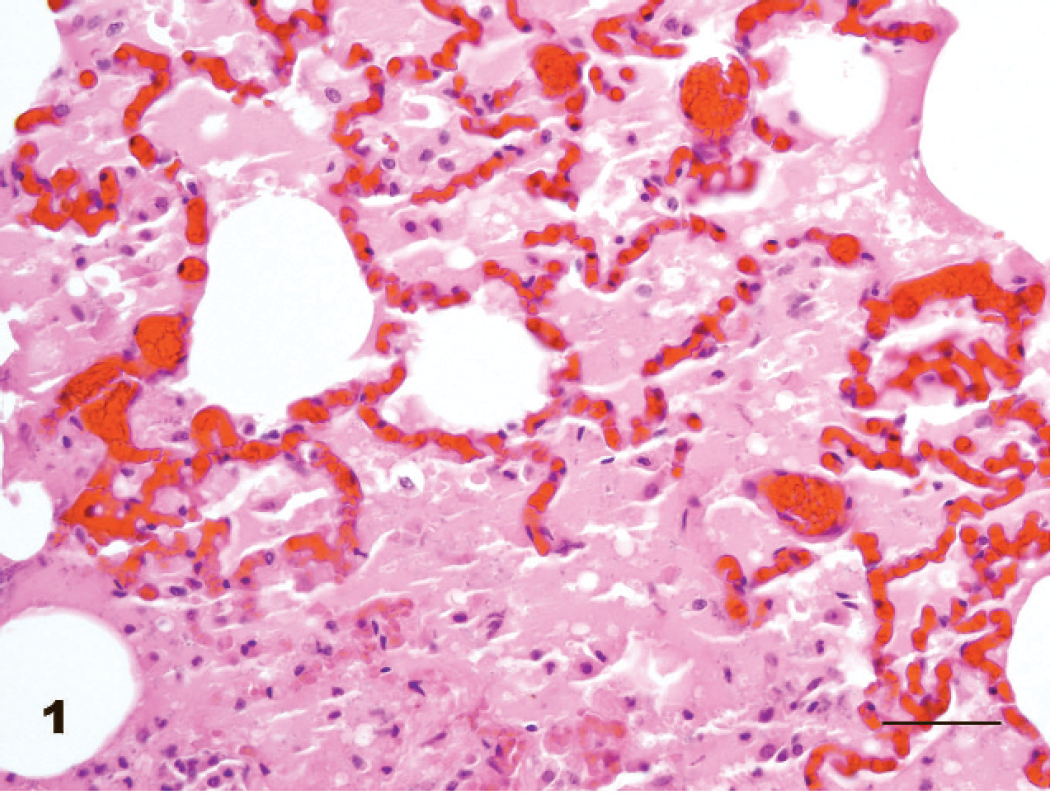
Lung; horse. Proteinaceous fluid with scattered macrophages and neutrophils within alveolar spaces. Hematoxylin and eosin. Bar = 50 μm.
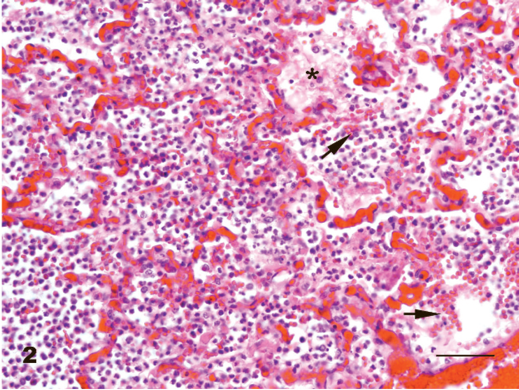
Lung; horse. Large numbers of intra-alveolar neutrophils admixed with macrophages, fibrin (asterisk), and hemorrhage (arrows). Hematoxylin and eosin. Bar = 50 μm.
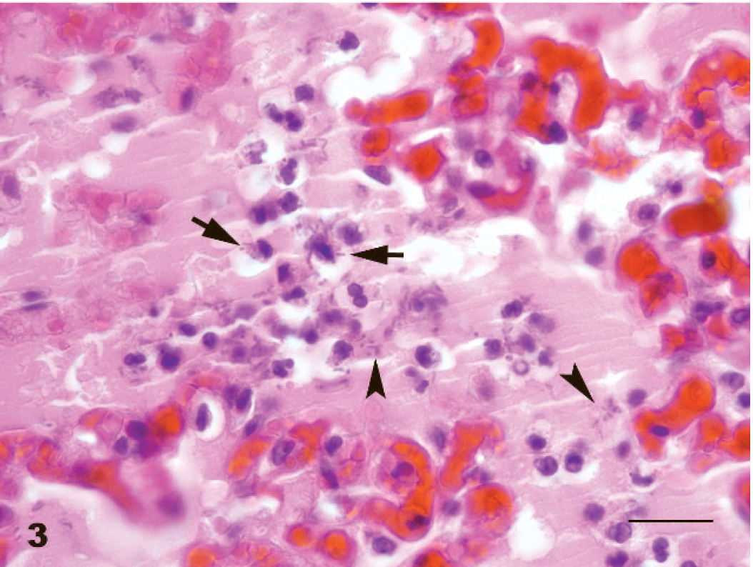
Lung; horse. Large numbers of rod-shaped bacteria both free in alveoli (arrowheads) and within the cytoplasm (arrows) of macrophages and neutrophils. Hematoxylin and eosin. Bar = 20 μm.
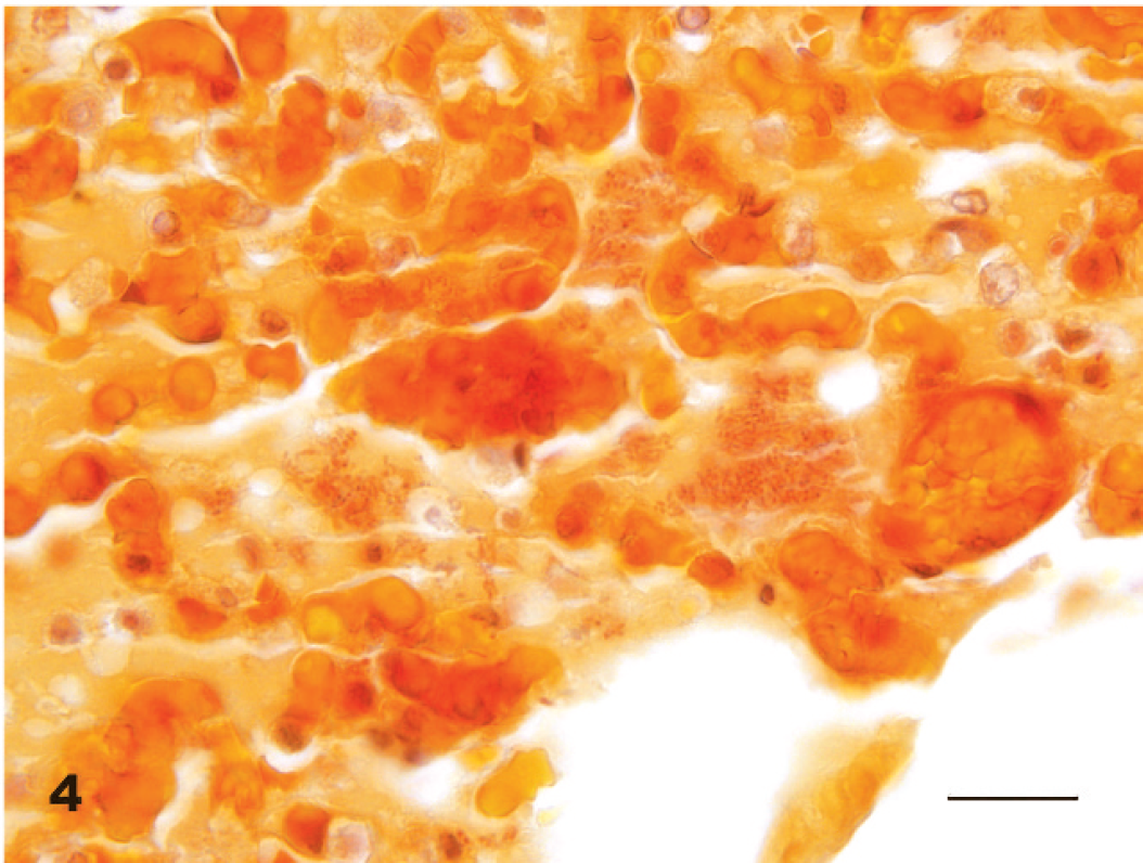
Lung; horse. Numerous free and intracytoplasmic Gram-negative bacilli within alveoli. Gram stain. Bar = 20 μm.
While light growths of E. coli, Enterococcus sp., Proteus sp., and Klebsiella pneumoniae were obtained from the brain and liver tissues, heavy growth was observed for E. coli, Enterococcus sp., and K. pneumoniae cultured from the lung tissues. Further characterization of E. coli isolate from the lung determined that it belonged to serogroup O2, as determined by standard method, 13 and serogroup H21, as determined by polymerase chain reaction–restriction fragment length polymorphism (PCR-RFLP) analysis of the fliC gene. 12 To determine if the strain was pathogenic, the virulence profile was determined. DNA was extracted from overnight culture, and genes encoding for virulence factors such as heat-stable toxins (STa, STb), heat-labile toxin (LT), shiga-toxins (Stx-I and Stx-II), cytotoxic necrotizing factors (Cnf-1, Cnf-2), and intimin (Eae) were detected by PCR, as described. 4 Once it was established that the strains carried genes encoding for Cnf-1, further testing for the presence of genes for ExPEC strains, sfa and foc 14 type 1 fimbriae, fim, 15 and papG alleles I and III 10 was performed by PCR following the conditions described in earlier reports. These genes were selected based on common virulence genes found to be associated with E. coli O4:H5 ExPEC strains in dogs. 11 Polymerase chain reaction was performed in a RapidCycler b following a specific Rapid-Cycle DNA amplification protocol. 19 The amplification products were subjected to electrophoresis in 1% agarose gels at 200 V for 30 min for all assays. The gels were stained with ethidium bromide and examined under ultraviolet light. Positive samples were identified based on the presence of bands of expected sizes compared to results with positive control strains J96 for sfa/focG, papG allele I, and fim genes; BUTI-3-1-4 for papG allele III. Escherichia coli K12 was used as a negative control strain.
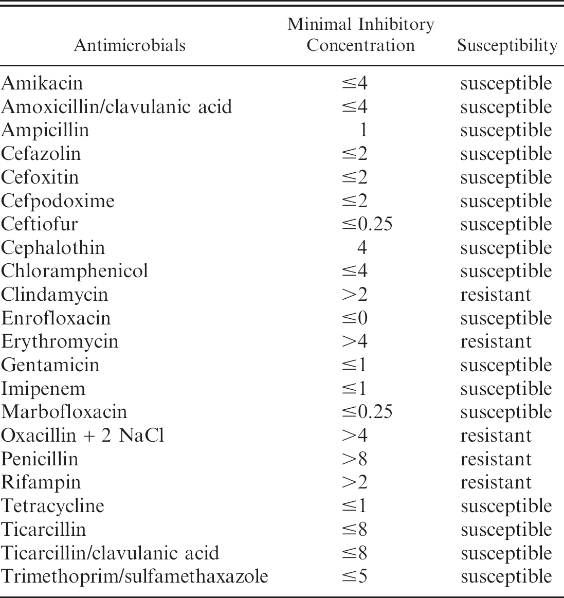
Susceptibility of Escherichia coli strain to different antimicrobials.
The E. coli isolate belonged to the O2:H21 serotype and carried genes encoding for virulence factors S and F1C fimbriae (sfa and foc), P fimbriae (PapG allele I), type I fimbriae (fim), and cytotoxic necrotizing factor (cnf-1) that are typically associated with ExPEC strains. Extraintestinal pathogenic E. coli strains belonging to O2 have been reported. While the serogroups O4 and O6 have been implicated in causing hemorrhagic pneumonia in dogs 3,7 and cats, 18 this is the first report to document the strain O2:H21 associated with bronchopneumonia in a horse. Escherichia coli O2 from cattle have been classified as Shiga-toxin-producing E. coli. 2 However, in this case the strain did not carry genes that encode Shiga toxins 1 or 2 (Stx), heat-stable (STa or STb) or heat-labile toxins (LT), or Cnf-2 and other genes associated with intestinal pathogenic E. coli.
Antimicrobial susceptibilities of the strain against 22 antimicrobials were tested using sensititer assay, as depicted in Table 1. The E. coli strain was resistant to clindamycin, erythromycin, oxacillin, penicillin, and rifampin. Gram-negative bacteria are inherently resistant to penicillin or exhibit a higher minimal inhibitory concentration, and, therefore, penicillin is not a drug of choice for treating Gram-negative infection in animals and humans. The actual effect of the penicillin therapy in this horse is not certain; however, the authors propose that it had little effect on the Gram-negative organisms and may have reduced the Gram-positive bacterial population. Since there was no clinical resolution of disease following penicillin treatment, this may indicate that the role of Gram-positive bacteria may have been minor or that their presence established the conditions that allowed ExPEC to proliferate and potentially cause pulmonary lesions similar to those reported in other ExPEC pneumonia cases. Thus, while the role of other bacteria in the pathogenesis of this horse's lung lesions cannot be excluded, it appears that ExPEC played a role in the disease process.
While the E. coli strains implicated in cases of hemorrhagic pneumonia in dogs 7 and cats 18 harbored most of the virulence genes, as observed in the isolate from horse, the ones from dogs and cats carried the papG allele III, which encodes a variant of the P fimbrial adhesin molecule PapG that is now known to be epidemiologically associated with human cystitis. 9 The O2:H21 strain carried adhesin PapG allele I and did not carry PapG allele III. The ExPEC strains carry many virulence attributes, and some of them are encoded in pathogenicity-associated islands. The production of disease requires bacterial adherence to host cells by P fimbriae, which expresses adherence molecules encoded by papG alleles I and/or III. Much work has been done on ExPEC strains that cause UTI in humans and animals. Type 1 fimbriae were found to be the most highly expressed adhesin during UTI. DNA microarray analysis demonstrated that expression of type 1 fimbriae coordinately affects the expression of P fimbriae in an inverse manner. 17 While type 1 fimbriae is involved in adherence, P fimbriae serves as an independent virulence factor in asymptomatic bacteria and is the cause of symptoms and tissue damage during UTI. 1 However, the role of P fimbriae or other types of fimbriae has not been established during cases of pneumonia.
In conclusion, the current study represents the first reported case of fatal bronchopneumonia associated with ExPEC in a horse. The E. coli isolate shared many virulence genes with E. coli isolates associated with hemorrhagic pneumonia in dogs and cats. Although this horse was housed in close proximity to a dog kennel, there is no known association (at the time of this publication) between the dogs and the E. coli from this mare. Additional studies are needed to determine if any association exists.
Acknowledgements. The authors wish to thank Drs. Kristin Abderhalden and Nancy Mauer (Spruce Run Equine Veterinary Associates, Somerset, PA) for their contribution to this case.
Footnotes
a.
Sigma-Aldrich Corp., St. Louis, MO.
b.
Idaho Technologies, Salt Lake City, UT.