
Abstract
Many of the sample matrices typically used for veterinary molecular testing contain inhibitory factors that can potentially reduce analytic sensitivity or produce false-negative results by masking the signal produced by the nucleic acid target. Inclusion of internal controls in PCR-based assays is a valuable strategy not only for monitoring for PCR inhibitors, but also for monitoring nucleic acid extraction efficiency, and for identifying technology errors that may interfere with the ability of an assay to detect the intended target. The Laboratory Technology Committee of the American Association of Veterinary Laboratory Diagnosticians reviewed the different types of internal controls related to monitoring inhibition of PCR-based assays, and provides information here to encourage veterinary diagnostic laboratories to incorporate PCR internal control strategies as a routine quality management component of their molecular testing.
Introduction
Sample matrices commonly used in veterinary molecular-based testing, including feces,15,29 milk,5,35 semen, 47 oral fluids, 7 and environmental swabs, 9 are prone to inhibit PCR and nucleic acid sequencing. There can additionally be considerable variability in the concentration of the inhibitors found in these matrices, as well as variability associated with different animal species. Substances capable of causing inhibition in a PCR assay are often intrinsic to the specimen type, such as complex polysaccharides, bilirubin, and bile salts found in stool2,24,29,49; proteases and calcium found in milk5,38; or hemoglobin, heparin, and hormones found in blood and tissues.38,50 Inhibitors can also be introduced inadvertently via contamination from the environment, 50 or intentionally introduced during specimen collection and transport (e.g., gel media9,12 and anticoagulants).11,28,51 Ethylenediamine tetra-acetic acid (EDTA), which is a common component in many blood collection, transport, and nucleic acid elution buffers, is known to have an inhibitory effect on downstream PCR applications,38,50 although EDTA is typically diluted or removed during the nucleic acid extraction process. 19
The mechanisms of action of common inhibitors include degradation or interference with PCR-critical proteases, degradation or interference with nucleic acids, competition with the nucleic acid template, and reduction in primer specificity.38,50 Commonly encountered examples of inhibition in biologic samples include the ability of heme in blood to block the DNA polymerase active site,1,38 and endogenous proteinases to degrade assay-critical polymerases. 35 For environmental samples, humic substances and components of soil and sediments such as iron have the potential to inhibit polymerase activity and primer binding.44,45,50 Reduced analytic sensitivity caused by inhibitors in environmental samples has been described extensively, and is particularly problematic in the surveillance of amphibian diseases. 23
Mitigation strategies for dealing with common inhibitors include thorough washing during nucleic acid extraction and screening for inhibitor-resistant polymerases. 44 Immunomagnetic 49 separation and immunocapture 38 have been described as being efficient in removal of PCR inhibitors for selected pathogens, particularly enterics, because the PCR target is specifically separated from the sample matrix and thus from sample-associated inhibitors. Sample dilution is another readily available option for attenuating the impact of inhibitory substances, with the caveat that dilution also reduces analytic sensitivity for detecting the target. Addition of substances to the PCR mixture to counter or bind inhibitors is also a common strategy, and may include bovine serum albumin, dimethyl sulfoxide, non-ionic detergents, and proteinase inhibitors.38,50 Commercial kits for nucleic acid purification and PCR amplification are generally designed to remove PCR inhibitors; however, the effectiveness of the individual commercial product is dependent on the sample matrix, and commercial products are often not optimized for the range of species tested in veterinary laboratories.
Despite efforts to remove inhibitors, absolute assurance that a negative PCR test result represents a true negative in the sample requires that the presence of inhibitors, as well as extraction failure and technical or reagent error associated with the individual test sample, be ruled out. External controls routinely used during PCR-based assays monitor the reagents and technical steps associated with extraction, amplification, and cross-contamination at the run level, but not at the individual sample level. Internal controls (ICs), which by definition are tested as a component of the sample containing the assay target, serve the purpose of ensuring that individual samples are effectively extracted, amplified, and importantly that inhibitory substances do not mask the intended target. The combination of external and internal controls during PCR-based testing ensures that inhibition, in addition to technical or reagent failures, is not responsible for causing false-negative and false-positive test results.
ICs can be categorized into endogenous ICs (EICs) and exogenous ICs (XICs). EICs are found naturally in the test specimen, for example as a sequence of the host genome such as beta-actin21,30,46,53 or beta-2-microglobulin,4,14,53 or alternatively are found in the specimen as a genome sequence from the host’s microflora (e.g., 16S rRNA). XICs are spiked into samples prior to processing for testing, during nucleic acid extraction, or prior to the amplification steps of the PCR assay.
Endogenous internal controls
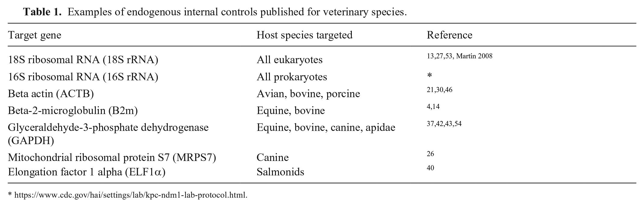
Ribosomal genes are a common choice for EICs (Table 1). The eukaryotic 18S rRNA gene has the versatility to be used as an EIC for multiple animal species when designed properly, but, because the gene occurs in all eukaryotes, it cannot be used to verify that a particular sample comes from the stated species. The conserved portion of the16S rRNA gene can also be used as an EIC in samples such as feces, in which bacteria are always present (Centers for Disease Control and Prevention [CDC]. Multiplex real-time PCR detection of Klebsiella pneumoniae KPC carbapenemase (NDM-1) and New Delhi metallo-β-lactamase genes. Available from: https://www.cdc.gov/hai/settings/lab/kpc-ndm1-lab-protocol.html). Because 16S rRNA is found in all prokaryotes, bacterial DNA, including that from a recombinant Taq expression host, will be detected by a 16S rRNA EIC if present in a diagnostic or research sample. Further, extra caution is warranted to prevent reagents from becoming contaminated with bacteria, given that contaminating bacteria will also be detected by a 16S rRNA EIC. The use of ribosomal genes for EIC has obvious advantages, including the presence of multiple copies in genomes, making ribosomal genes more readily detectable than single-copy genes. The practical disadvantage of employing ribosomal genes as EICs is that they are highly conserved and occur in relatively high copy number, which may facilitate assay cross-contamination (e.g., aerosol-related contamination from a strong-positive IC resulting in a false-positive result for the negative PCR amplification or no-template controls). In this scenario, a real-time PCR (rtPCR) cycle threshold (Ct) related to EIC cross-contamination would typically show a weak signal (e.g., Ct 33–38) compared to the expected Ct from true-positive samples (e.g., 15–25). Routine monitoring of the rtPCR Ct levels of EICs allows cross-contamination issues to be readily detected, traced, and resolved.
Examples of endogenous internal controls published for veterinary species.
Host-specific EICs have historically been used in the less quantitative molecular techniques, for instance many reference genes (housekeeping genes, conserved genes) used in northern blots are also commonly targeted in rtPCR (e.g., beta-actin). Many studies on human as well as animal species have suggested specific reference genes as potential candidates for EICs.41–43,48,53,54 The criteria for selection of appropriate host genes to be used as rtPCR EICs have been reviewed. 36 A number of software programs are available to assist with selection and validation of EICs (e.g., NormFinder, 3 BestKeeper, 34 and geNorm). 48 It should be noted that the software programs cited target identification of better reference genes for gene expression studies and may not be equally applicable for selecting reference genes to be used as EICs for clinical testing purposes.
EIC targets can be difficult to design; however, it is very likely that an EIC target designed for a PCR assay detecting a specific pathogen or target in a given matrix and species will additionally work for alternative targets in the same species and matrix. It is important to note, however, that each PCR target and the assay parameters must be optimized individually with the selected EIC to ensure that no adverse interaction occurs between the primer and probe sequences of the target and the EIC, and that competition for PCR components does not alter the assay limit of detection.
Exogenous internal controls
XICs are spiked into the test sample in a defined concentration or copy number. Compared to use of EICs that can vary with the health status of the animal, adding a known amount of XIC into the lysis buffer prior to extraction provides a more stable, easily standardized, and more readily implemented control for quantitative molecular applications. 20 Additionally, spiking a standard concentration of an XIC into all test samples can serve to normalize data and compare results across studies.10,20,31
XICs can be designed to be homologous (i.e., competitive) or heterologous (i.e., noncompetitive). Homologous XICs are artificial templates constructed to use the same primer binding sites as the assay target sequence and a different internal sequence for the XIC so that the two can be distinguished by amplicon size or by use of specific probes.18,38 Selecting the appropriate concentration of homologous XICs used in PCR reactions is critical to the detection limit of the assay 18 because of the possibility of competition for PCR reagents (e.g., oligonucleotides, DNA polymerase) between the assay target and XIC. An overabundance of an XIC can result in amplification being inhibited for one or both targets depending on the molar ratio, and the length, sequence, and secondary structure of the DNA fragments. 18
Heterologous XICs are designed using primers and probes unique to the XIC.18,38 The noncompetitive XIC design still requires that the concentration of the control is carefully managed in order to limit competition for oligonucleotides and DNA polymerase during the PCR reaction. 18 Heterologous XICs are considered very efficient for deployment in veterinary laboratories based on their ability to be used universally for different animal species and matrices.
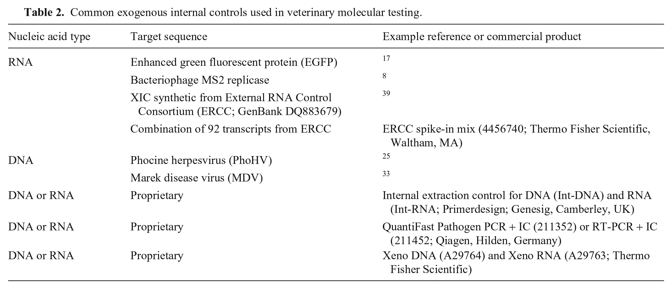
A number of synthetic sequences have been developed by academia and industry, including by the National Institute of Standards and Technology (NIST) as a part of the External RNA Controls Consortium (ERCC; https://www.nist.gov/programs-projects/external-rna-controls-consortium), an ad-hoc group with ~ 70 members from private, public, and academic organizations. The ERCC assembled a library of 176 DNA sequences that could be transcribed into RNA for use as XICs. Additionally, encapsulated Escherichia coli phage MS2 has been reported as a universal XIC, 8 having the benefit of not sharing homology with animal hosts or potential pathogen targets. Specific examples of XICs used for rtPCR in veterinary testing (Table 2) include an in vitro transcript of enhanced green fluorescent protein, 17 and herpesviruses including Marek disease virus 33 and phocine herpesvirus, 25 both used in assays targeting unrelated DNA pathogens. As seen with the herpesvirus example, an intact virus can be an effective XIC for monitoring extraction efficiency and subsequent PCR amplification steps used for the detection of either RNA or DNA targets. Compared to DNA-based XICs, the innate low stability of RNA and the ubiquitous nature of RNases make RNA-based XICs more susceptible to template degradation. Armored RNA, composed of RNA sequences artificially encapsulated in a protein coat to protect them from RNase digestion, was initially developed to provide assay controls and standards used in testing for the human immunodeficiency virus, 32 and has since been adopted for use as an XIC or surrogate for additional human and animal RNA targets.16,52 Armored or protected RNA formulations, and encapsulated RNA XICs such as MS2, are available commercially.
Common exogenous internal controls used in veterinary molecular testing.
Quality control
Assays using ICs must be developed, validated, and managed to ensure that the IC does not interfere with the assay limit of detection by competing with the assay target for essential PCR assay components. XICs, whether produced in-house or obtained commercially, must be titrated to the lowest possible concentration such that, when used with samples free of inhibitors, the assay target remains consistently within the detection limit of the assay and the IC is detected consistently. 38 For most commonly used rtPCR platforms available in veterinary diagnostic laboratories (VDLs), the XIC would typically be titrated for results in the 30–35 Ct range. Commercially available XICs often have a manufacturer-recommended range lower than 30–35 Ct; however, to ensure the optimal limits of detection for the assay, re-titrating the XIC to the lowest concentration that still allows the IC to be detected is warranted. An example showing how to properly titrate a heterologous (noncompetitive) XIC is provided (Fig. 1).
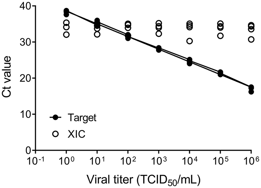
Example of a properly titrated exogenous internal control (XIC), provided by the Laboratory Technology Committee of the AAVLD. The XIC (open circles) maintains a consistent cycle threshold (Ct) value over a wide target concentration range without compromising the limit of detection of the assay. Serial dilutions of equine herpesvirus 1 cell culture lysates were tested with a DNA XIC spiked into lysis buffer. Four replicates were tested at each viral concentration.
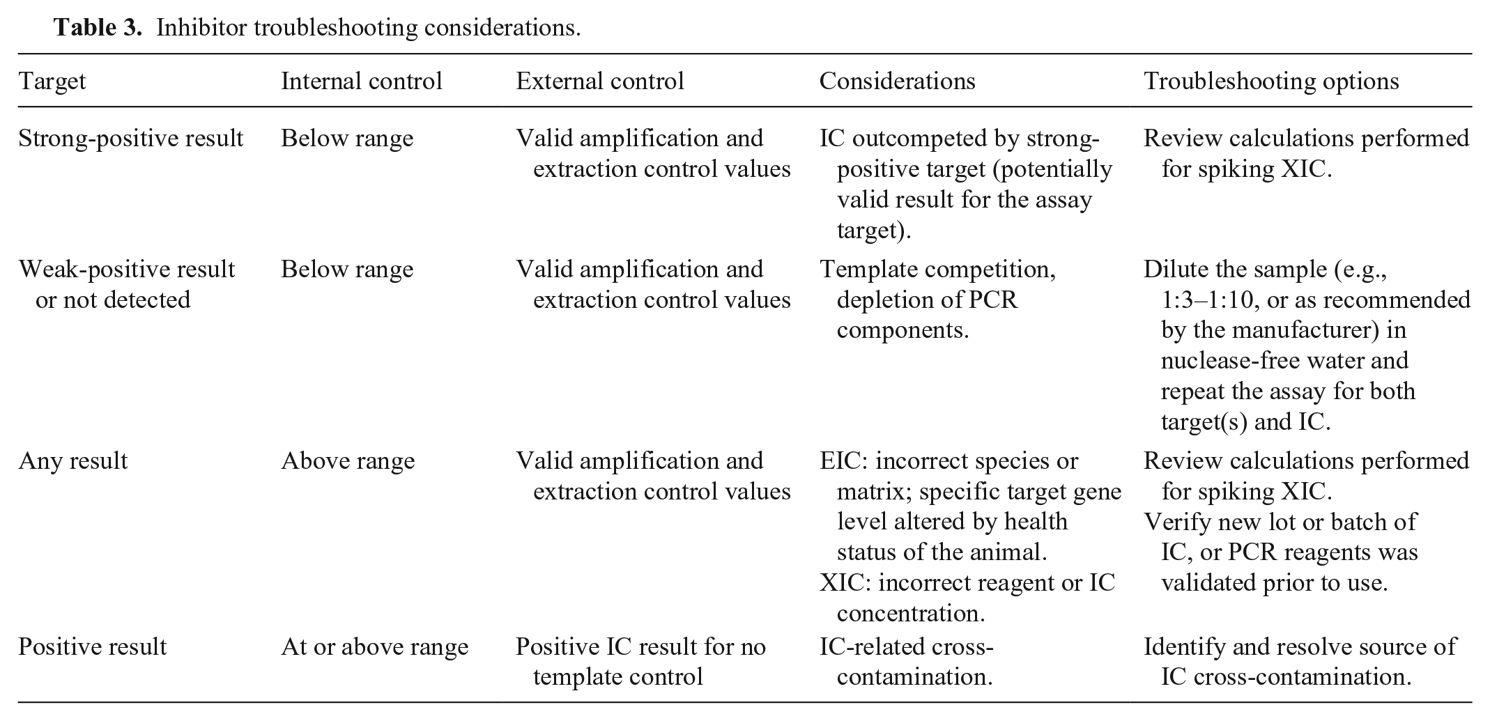
Inhibition of amplification can be detected using ICs; however, ICs cannot differentiate between inhibition and failures in PCR amplification caused by human error, faulty equipment, or reagent issues. Effective control of all steps of the PCR assay requires that external controls (i.e., positive amplification controls, extraction controls, and reverse-transcription controls where applicable) be used in conjunction with ICs. Because competition for PCR components cannot be avoided when multiple targets are amplified in the same reaction vessel, it is critical that the IC be titrated to not compete with assay target(s) for the critical reagents during PCR amplification. A higher than expected result value for an IC signals the potential for target competition and assay failure because of reduced or failed assay target detection in the sample. The potential for a strong-positive assay target to outcompete an IC additionally exists, as recognized by a lower than expected or negative IC response. Provided that other PCR system controls are valid, the result would in most cases be considered a valid test result 6 (Table 3).
Inhibitor troubleshooting considerations.
Control charting to monitor the performance of ICs allows laboratories to quickly initiate investigations when a deviation that could be the result of issues such as presence of inhibitors, defective reagent lot, failure of equipment performance, or human error is observed. In order to make the most effective use of ICs, quality control (QC) performance ranges must be established prior to their routine use. The IC performance range established should be specific to each detection scheme (i.e., single or multiplex assay), reaction conditions, thermocycling program, and thermocycler platform. IC performance ranges are based on a specified number of independent runs performed under the conditions established for a given assay, noting that the performance range for ICs used in multiplex assays must be re-established following any change in the number of multiplexed targets. In order to develop performance criteria for RNA EICs, representative samples from animals both with and without clinical disease are required in order to include variability resulting from differential gene regulation that occurs with different health conditions or disease states. 36 There is no established statistical rule for the number of repeats needed to establish an initial mean and standard deviation (SD) for a PCR IC. The Laboratory Technology Committee of the AAVLD, through consensus discussion and technical experience, considers 15 independently tested replicates sufficient to provide a reasonable balance in cost and other technical feasibility factors. The Clinical Laboratory Improvement Amendments (CLIA) Program, which sets standards for human clinical laboratory testing, recommends 20 separate determinations for establishing the initial mean and SDs, followed by monthly updates over a 3–6 mo period to establish a stable performance range. 6 The number of replicates for veterinary testing is ultimately determined by each laboratory and should be documented as part of their quality management system. EIC and XIC performance values should continue to be monitored throughout the use of the IC to ensure consistency of the assay and IC, 6 and in order to identify and manage trends that would signal assay performance changes. The performance range used by the laboratory for monitoring results may consist of 1, 2, or 3 SDs above and below the mean, dependent on the rejection criteria established by the individual laboratory’s quality management program. 22 An additional QC measure required of veterinary laboratories, beyond those of human clinical laboratories, is the need to establish and monitor IC performance specifically for the different animal species and the coinciding sample matrices routinely encountered in veterinary testing. It is strongly recommended that genome-level studies be performed or consulted using stability algorithms for statistical selection of appropriate targets. 48
Strategy for use of internal controls
ICs provide a means of monitoring PCR-based tests at the level of the sample, and therefore can be used to detect failures resulting from inhibitors in the sample, as well as failure in an assay variously caused by reagents, equipment, or human error. If the IC is included in the sample naturally (i.e., EIC), or is added prior to extraction (i.e., XIC), the IC can be used to verify effective extraction, reverse transcription where applicable, amplification, and lack of inhibitors in the sample.
Use of one or multiple host genes as EICs is a common method of inhibition monitoring. The principle advantage of the approach is provision of control for sample quality as well as for species of origin.36,37,41,43 Properly validated EICs are ideal for genetic assays requiring precise quantitation of copy-number variants, including testing situations and data handling benefiting from data normalization.3,4,36,46 The use of EICs as standards for gene expression studies in humans and animals is common 36 ; however, as a QC measure, this approach is less utilized in veterinary clinical testing given the need for workflow consolidation to efficiently accommodate the large number of host-pathogen combinations encountered. Disadvantages for use of EICs include innate variability because of inconsistent cellular counts in samples (e.g., nasal swabs), differences between sample matrices, and potentially the health status of the animal resulting in gene up- or down-regulation.30,46,48,53,54 Should the EIC concentration (i.e., copy number) be sufficiently higher than the assay target, the EIC can outcompete the assay target and thus not accurately detect the target at the detection limit of the assay, 38 potentially resulting in the serious consequence of a false-negative assay result.
XICs, whether added prior to or after extraction, also provide control for amplification and sample quality (i.e., inhibitors) in PCR assays, and, like EICs, must be developed and monitored to ensure that the XIC does not compete with the assay target for PCR components in order to prevent a decrease in assay detection limit or a potential false-negative result. XICs can be added into the PCR reaction vessel (i.e., vial or plate) at different stages of the PCR assay process, or can be spiked directly into the test sample prior to processing. When spiked into the lysis buffer prior to extraction, XICs provide the benefit of monitoring the effectiveness of the extraction step. Spiking XICs directly into the unprocessed biologic sample has the benefit of controlling at all stages of testing from sample handling and extraction through amplification and detection; however, directly spiking the sample may lead to degradation and reduced analytic sensitivity, especially for RNA XICs in samples with a natural abundance of proteases and RNases. It is possible to circumvent recovery and stability concerns associated with RNA by using DNA-based XICs in assays detecting RNA targets; however, control for the reverse-transcription step would have to be added to the overall assay control strategy.
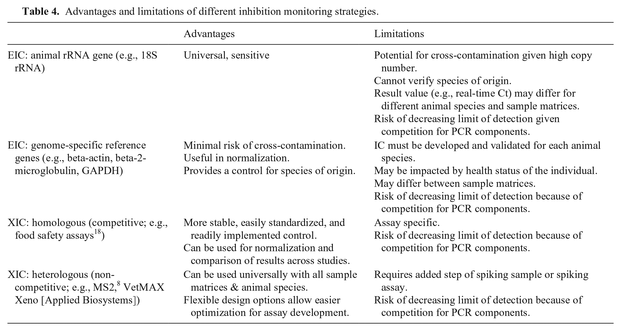
The use of ICs significantly enhances the reliability of PCR-based assays to provide meaningful results. Table 4 summarizes advantages and limitations of different inhibition monitoring strategies available to VDLs. The particular strategy employed for incorporation of ICs into a laboratory’s quality management scheme is the decision of individual laboratories and should take into account the cost versus benefit of each approach. An overview of common strategies is shown in Figure 2. XICs spiked prior to the extraction step can be used in conjunction with external PCR controls (i.e., positive amplification control, no-template control) to monitor inhibition, extraction efficiency, amplification, and cross-contamination. Alternatively, an assay-specific positive extraction control (PEC) can be used with an XIC spiked at the PCR setup stage for similarly monitoring all stages of the PCR assay. A third strategy is a combination of EIC and external controls (i.e., positive amplification, no-template control). The strategies defined are relevant for PCR, microarrays, and nucleic acid sequencing procedures.
Advantages and limitations of different inhibition monitoring strategies.
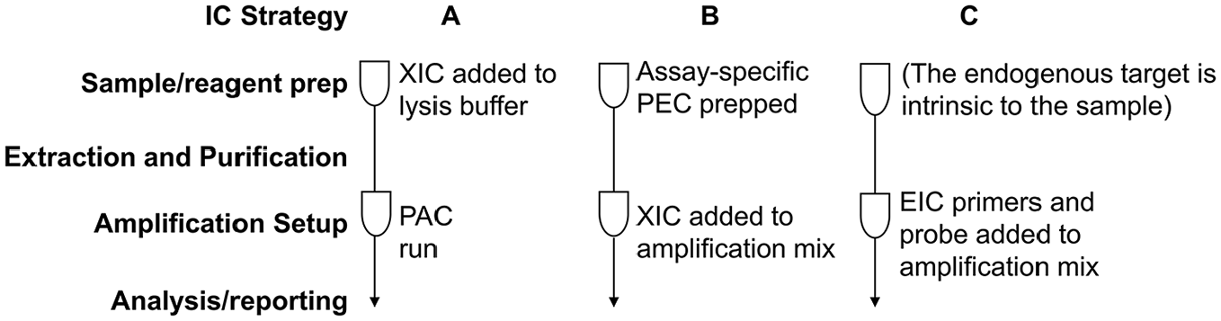
Flow chart for common inhibition monitoring strategies for PCR. The test tube symbols represent the stage at which specific controls or reagents are added. EIC = endogenous internal control; IC = internal control; PAC = positive amplification control; PEC = positive extraction control; XIC = exogenous internal control. Strategy
Conclusions
The volume of PCR-based tests performed in VDLs continues to grow at a rapid pace, as does the introduction of new PCR-based assays and methodologies aimed at ensuring high-quality testing for the entire range of animal species and matrices handled by most VDLs. Although not a recent innovation, the use of ICs has not yet become routine in PCR-based veterinary testing. We have reviewed IC options and strategies for use in controlling for sample-based inhibition, ultimately in order to improve the reliability of negative PCR test findings in which inhibition may be an issue. Veterinary molecular testing encompasses varied and complex sample types from a wide variety of animal species. The presence and impact of PCR inhibitors in the diverse sample set routinely handled by VDLs is not readily predictable, emphasizing the need for ICs at the sample level during PCR-based testing in order to validate individual test results. The Laboratory Technology Committee of the AAVLD has recommended to its membership that all new molecular assays being validated and implemented include an inhibition monitoring strategy based on internal validation for the host, target species, and sample matrix combination being tested. A component of the strategy to ensure that inhibition is not negating the value of the negative PCR result includes trend analysis during use of ICs. The information and discussion provided by the AAVLD Laboratory Technology Committee is intended to encourage more routine and standardized use of ICs to detect inhibitors in PCR assays utilized in VDLs.
Footnotes
Acknowledgements
We thank Monica Reising for contributing to the discussions and editing of the manuscript. For materials used to illustrate XIC titration, we thank Bettina Wagner for the equine herpesvirus 1 cell culture lysates and the Wisconsin Veterinary Diagnostic Laboratory for providing the XIC. Renee Anderson and Roopa Venugopalan provided technical support.
Authors’ contributions
All authors conceived and designed the study through regular participation in the AAVLD Laboratory Technology Committee. LB Goodman, L Yan, and RL Tallmadge drafted the manuscript. L Yan, KL Kurth, AL Glaser, and LB Goodman contributed to acquisition and interpretation of the data. All authors critically revised the manuscript and gave final approval.
Declaration of conflicting interests
The authors declared no potential conflicts of interest with respect to the research, authorship, and/or publication of this article.
Funding
The authors received no financial support for the research, authorship, and/or publication of this article.