
Abstract
We describe the pathologic, histochemical, and immunohistochemical findings associated with pulmonary and encephalitic cryptococcosis in a 3-y-old, mixed-breed, nanny goat from central-west Brazil. The goat had progressive neurologic signs over 30 d; cryptococcosis was diagnosed antemortem by cytologic evaluation of cerebrospinal fluid. Treatment was initiated, but the animal died spontaneously shortly thereafter. Grossly, there was a large space-occupying gelatinous mass (cryptococcoma) in the left lung and smaller masses in the cerebral temporal and frontal cortex, thalamus, hippocampus, basal nuclei, and mesencephalon with consequent internal hydrocephalus. Histologic evaluation revealed marked granulomatous cryptococcal pneumonia and meningoencephalitis. Intralesional narrow-necked budding cryptococcal yeasts were identified on special stains (Mayer mucicarmine and Grocott methenamine silver) in sections of lung and brain. Immunohistochemistry utilizing a panel of monoclonal antibodies that selectively label capsules of Cryptococcus spp. was consistent with C. neoformans var. grubii.
Cryptococcosis is a mycosis of humans and other animals that is caused by the Cryptococcus neoformans/C. gattii species complex of organisms. To date, this complex is divided into C. neoformans and C. gattii; C. neoformans has 5 known molecular types (VNI–IV, VNB), and 4 distinct lineages (VGI–IV) are identified in C. gattii.3,12 However, 2 major reorganizations of the Cryptococcus complex of organisms have been proposed. In the first proposal, C. neoformans var. grubii and C. neoformans var. neoformans are to be considered as separate species, with the inclusion of 5 species within C. gattii. 8 The second proposal suggested a simpler reorganization containing only groups of organisms: the Cryptococcus neoformans species complex (var. grubii and neoformans) and the C. gattii species complex. 12
Cryptococcosis is not often reported in goats, although cryptococcosis has been described in goats with meningitis, 1 pneumonia, 1 and mastitis. 20 In most of these cases, the species definition of Cryptococcus was not confirmed.7,21 C. neoformans var. gattii (C. gattii species complex) was identified in 5 goats with pulmonary, hepatic, and encephalitic disease in Spain. 1
Cryptococcomas (cryptococcal granulomas) are space-occupying granulomatous reactions within parenchymatous organs. 18 Cryptococcomas have been described in the cow,15,16 dog, 9 cat, 2 horse, 17 and sheep, 19 with a few descriptions in goats.14,21 We describe herein the pathologic, histochemical, and immunohistochemical findings associated with pulmonary and encephalitic cryptococcosis in a goat, and add to the documentation of encephalitic cryptococcomas in goats.
A 3-y-old, mixed-breed, nanny goat from the rural region of the city of Cuiabá, Mato Grosso, central-west Brazil, was presented to the Veterinary Teaching Hospital, Universidade de Cuiabá (VTH-UC). The owner reported that the goat demonstrated progressively abnormal behavioral changes during the 30 d prior to clinical evaluation. The animal was reared semi-extensively in a herd of 15 goats without a previous history of disease. No other goats were reported to be affected. Clinical and neurologic examination revealed anorexia, ruminal hypomobility, ataxia, muscle spasms, bruxism, nystagmus, anisocoria, spastic tetraparesis, and a semi-comatose state. A visit to the farm where the goat was maintained revealed that there were pigeons (Columba livia) feeding at the troughs and cohabiting with the goats.
Cytologic evaluation of cerebrospinal fluid revealed narrow-necked budding yeasts consistent with Cryptococcus spp. Neurologic impairment was inexorably progressive, and the animal died 4 d after hospitalization at the VTH-UC.
An autopsy was performed soon after the goat’s death. Specimens of brain, lungs, kidney, liver, myocardium, and intestine were collected, fixed in 10% buffered formalin solution, processed routinely, and stained with hematoxylin and eosin, Mayer mucicarmine, Grocott methenamine silver (GMS), and periodic acid–Schiff (PAS) stains. Masson trichrome stain (MTS) was used to determine the presence of fibrous connective tissue in the dura matter and adjacent brain tissue.
Selected formalin-fixed, paraffin-embedded (FFPE) fragments of the brain were examined by immunohistochemistry (IHC) utilizing monoclonal antibodies (mAb471, mAb302, and mAbF10F5) to label the cryptococcal capsule as described previously. 11 FFPE sections of brain lesions were used in a PCR assay that targeted the internal transcribed spacer (ITS) regions 1 and 2. 9
At autopsy, the goat was severely emaciated; the principal pathologic findings were in the lungs and brain. An 8-cm diameter irregularly shaped mass in the caudal lobe of the left lung (Fig. 1A) consisted of an external fibrous connective tissue capsule with centrally located gelatinous material (cryptococcoma). There was marked flattening of cerebral gyri with narrowing of sulci. Similar 0.5–1.5-cm diameter gelatinous masses were identified in the lungs and randomly in several regions of the brain, including the temporal and frontal cortex, thalamus, hippocampus, optic chiasm, basal nuclei, and mesencephalon. Some cryptococcomas were in contact with the leptomeninges; others were located deep within the brain parenchyma. In addition, cryptococcomas were adjacent to and within the mesencephalic aqueduct (Fig. 1B), resulting in obstruction to the aqueduct with consequent internal hydrocephalus. Cryptococcomas were not observed in the cerebellum, and sagittal sections of the head did not reveal rhinitis or other lesions. Apart from marked serous atrophy of pericardial fat, significant pathologic findings were not observed grossly or microscopically in other organs.
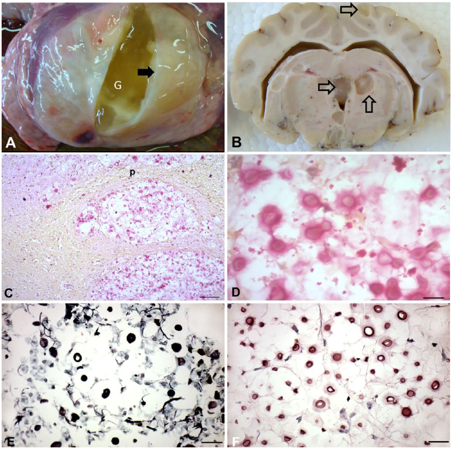
Gross anatomic findings and histochemical characterization of cryptococcosis in a goat.
Histologic evaluation of the left lung revealed a large central aggregation of narrow-necked budding, predominantly extracellular, encapsulated yeasts (4–12 µm diameter) admixed with mild accumulations of foamy macrophages in a background of mucoid-like matrix (Supplementary Fig. 1A). Multifocally, distant from the large lesion, there was marked pulmonary fibrosis with moderate numbers of cryptococcal organisms within alveoli (Supplementary Fig. 1B) and multiple foci of dilated and ruptured alveolar walls containing cryptococcal organisms (Supplementary Fig. 1C, 1D). Other pulmonary alterations included moderate bronchiolar and bronchial epithelial hyperplasia, scattered eosinophils, and a focal lymphocytic inflammatory infiltrate within the zone of pulmonary fibrosis.
Histologic evaluation of the brain revealed 2 distinct lesions: chronic cryptococcal leptomeningitis, and intracerebral cyst-like formations. Chronic cryptococcal leptomeningitis was characterized by severe swelling of the leptomeninges because of marked infiltration of macrophages, some lymphocytes, and congested blood vessels with vasculitis associated with numerous yeast cells (Supplementary Fig. 1E). However, these areas of chronic cryptococcal leptomeningitis were not in contact with the gelatinous masses observed grossly within the brain, which were comprised of large numbers of extracellular encapsulated yeasts forming cysts. Cryptococcomas were observed deep within the brain affecting gray and white matter, adjacent to the leptomeninges, or protruding into the lateral ventricles and without an associated severe inflammatory reaction. In some areas, cysts were adjacent to blood vessels. MTS did not reveal connective tissue around cysts regardless of their location within the brain (Supplementary Fig. 1F, 1G).
The capsule (3–5 µm) of the yeast was positively stained with Mayer mucicarmine stain, consistent with Cryptococcus spp. (Fig. 1C, 1D). GMS identified fungal cell walls but not the capsule of the intralesional organisms (Fig. 1E); PAS stained the fungal cell wall and variably stained components of the capsule (Fig. 1F).
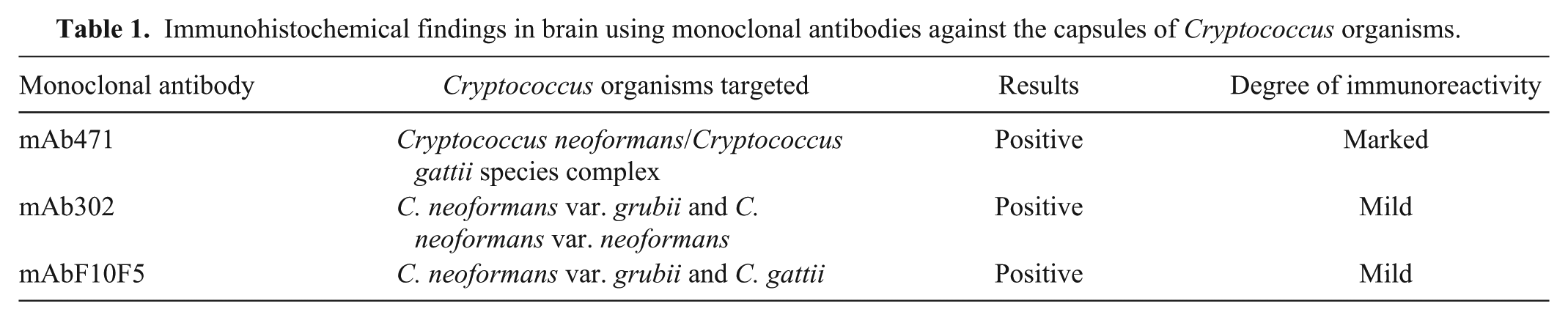
All mAbs used in IHC reacted positively with the capsule of the intralesional organisms identified from the FFPE brain fragments (Table 1; Fig. 2A–C); the negative control remained unstained (Fig. 2D). However, immunoreactivity and immunolabeling of the fungal capsule was more frequent with mAb471 compared with mAb302 and mAbF10F5. The results indicated that the intralesional cryptococcal organisms were in the C. neoformans species complex and more likely to be C. neoformans var. grubii. 11 Additionally, the PCR assay amplified the desired partial fragment, further confirming that the agent belonged to the Cryptococcus sp. complex. Sequencing was unsuccessful.
Immunohistochemical findings in brain using monoclonal antibodies against the capsules of Cryptococcus organisms.
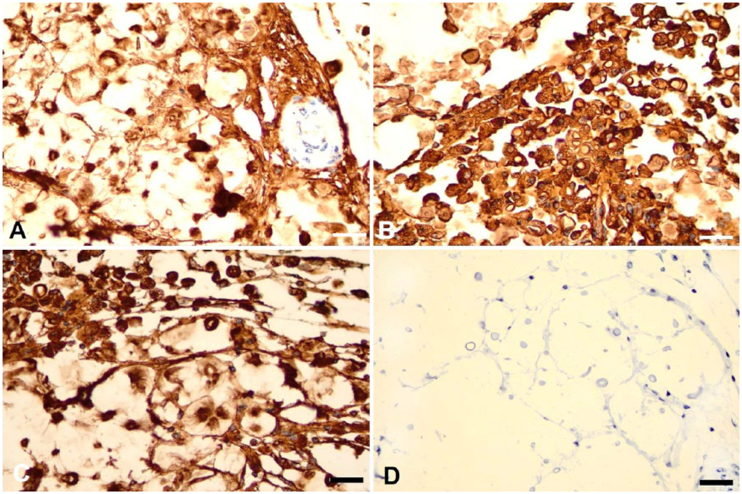
Immunohistochemical characterization of Cryptococcus complex organisms in a goat brain. Observe the specific immunostaining to the capsule of Cryptococcus species of organisms by monoclonal antibodies
Cryptococcal species identification to C. neoformans species complex (C. neoformans var. grubii) was achieved by immunohistochemical labeling of the cryptococcal capsule by a series of mAbs. This IHC method differentiates between the Cryptococcus species of organisms in FFPE tissue sections, 11 and has been used to characterize cryptococcal organisms in the cow.15,16 In our study, molecular testing using ITS1 and 2 primers amplified fungal DNA from the FFPE tissue sections, but the quality of the amplified fungal DNA was not adequate for sequencing; similar frustrating attempts to sequence cryptococcal DNA from archival FFPE tissues sections have been described.10,15 Molecular characterization would have provided additional support to the identification of the species of Cryptococcus in our goat, but was not successful. IHC serves as an excellent method for identification of cryptococcal organisms in retrospective studies from archival FFPE tissues in cases in which molecular methods fail to confirm the species of Cryptococcus within infected tissues.
Cryptococcomas similar to those in our goat have been described in cryptococcosis of companion animals,9,10 cows,15,16 and in a goat from São Paulo, Brazil. 14 In the case of caprine cryptococcosis from São Paulo, 14 pulmonary and encephalitic cryptococcomas were described (speciation was carried out by primers specifically targeting genus Cryptococcus), and there was evidence of a concomitant infection with Corynebacterium pseudotuberculosis. In our case, the species of Cryptococcus involved was identified by IHC, but histologic evidence of simultaneous disease was not observed. However, additional laboratory evaluations would have been required to exclude a concomitant disease in our goat. Cryptococcomas were not described in the cases of caprine cryptococcosis from Spain 1 and Portugal. 21 The intracerebral cryptococcomas observed grossly in our goat have been described in human histopathology4,6; we have described similar lesions in the kidney and brain of a dog with disseminated cryptococcosis. 9 Cerebral cryptococcomas associated with infection by Cryptococcus sp. have been ascribed to dissemination of intracerebral cryptococcal organisms along the swollen perivascular spaces of the brain, admixed with the mucoid-to-gelatinous secretion derived from the fungal capsule. 4 In human medicine, intracerebral cryptococcomas are considered to occur predominantly in the gray matter of the brain 6 ; in our case, both gray and white matter contained cryptococcomas.
We observed 2 unusual findings associated with cerebral cryptococcosis in domestic animals in our goat: cryptococcoma within the mesencephalic aqueduct, and internal hydrocephalus. Such lesions are not observed frequently in domestic animals with disseminated cryptococcosis or even in those with encephalitic cryptococcomas9,10,15; intraventricular cryptococcoma of the left lateral ventricle with ventricular dilation was described in a cow, 16 and there were pulmonary and encephalitic cryptococcomas in a goat from Brazil. 14 However, in our case, there was no evidence of a concomitant disease, and multiple cryptococcomas were observed within the brain; similar cases of intracerebral cryptococcomas without a predisposing infectious disease have been described in a dog9,10 and cow.15,16 Intracerebral cryptococcomas are associated more frequently with C. gattii than with C. neoformans in immunocompetent hosts. 3 However, in our case, infection was most likely caused by C. neoformans var. grubii, as occurred with cryptococcomas in the cow,15,16 without any underlying disease. Alternatively, C. gattii–induced cryptococcomas were described in the brain, lung, and kidneys of a dog 9 and in the nose of a cat, 5 both of which did not have any concomitant disease.
Our goat was maintained on a farm where pigeons were present; the excrement of pigeons is known to harbor C. neoformans and would have been the most likely source of Cryptococcus sp. Pigeon droppings were also associated with C. neoformans var. grubii infection in a cow from central-west Brazil, 15 and this species of Cryptococcus is considered prevalent within excreta of pigeons from several regions of Brazil. It is likely that infection began in the lungs with subsequent dissemination to the brain; inhalation of basidiospores, probably derived from pigeon feces, is the most frequent form of contamination in animals.3,13 Colonization within the brain has been related to the number of organisms inhaled and the pathogenicity of the associated strain. 13 Entry of the organisms into the brain in our case probably occurred hematogenously, resulting in chronic cryptococcal meningoencephalitis with the formation of intracerebral pseudocysts; similar entry into the brain has been described in other domestic animals.15,16,21
Supplemental Material
DS1_JVDI_10.1177_1040638718816358 – Supplemental material for Pathologic, histochemical, and immunohistochemical findings in pulmonary and encephalitic cryptococcosis in a goat
Supplemental material, DS1_JVDI_10.1177_1040638718816358 for Pathologic, histochemical, and immunohistochemical findings in pulmonary and encephalitic cryptococcosis in a goat by Selwyn A. Headley, Luciano A. Pimentel, Mariana Z. Michelazzo, Hugo S. Toma, Lucienne G. Pretto-Giordano, Rogério A. Marcasso, Alexandre M. Amude, Thalita E. Oliveira, Marcelo D. Santos and Mark Krockenberger in Journal of Veterinary Diagnostic Investigation
Footnotes
Acknowledgements
SA Headley and TE Oliveira are recipients of the National Council for Scientific and Technological Development (CNPq; Brazil) fellowships and grants. MZ Michelazzo is a recipient of a Coordination for the Improvement of Higher Education Personnel (CAPES; Brazil) fellowship. The antibodies utilized for immunohistochemical characterization of the fungi were kindly provided by Professor Tom Kozel, University of Nevada, Reno. The technical assistance of Ms. Elaine Chew, Mrs. Karen Barnes, and Mr. Huy Tan in the Veterinary Pathology Diagnostic Services, The University of Sydney, is gratefully acknowledged.
Declaration of conflicting interests
The authors declared no potential conflicts of interest with respect to the research, authorship, and/or publication of this article.
Funding
This study was partially funded by The National Council for Scientific and Technological Development (CNPq; Brazil).
References
Supplementary Material
Please find the following supplemental material available below.
For Open Access articles published under a Creative Commons License, all supplemental material carries the same license as the article it is associated with.
For non-Open Access articles published, all supplemental material carries a non-exclusive license, and permission requests for re-use of supplemental material or any part of supplemental material shall be sent directly to the copyright owner as specified in the copyright notice associated with the article.