
Abstract
Pythiosis is characterized most commonly by ulcerative dermatitis, mainly in the limbs of sheep and occasionally of goats. In sheep, Pythium insidiosum is also responsible for necrotizing rhinitis characterized by marked enlargement and deformity of the nasal region, severe respiratory difficulty, and bloody nasal discharge. A third form of pythiosis in sheep affects the digestive tract, involving ulceration of the esophagus, forestomachs, and abomasum. Pythiosis in sheep and goats has been reported only in Brazil where it occurs mainly in the semiarid region of the country, when animals congregate and stay for longer periods of time within or around water reservoirs. However, it has been reported as well in areas of humid environments, such as the Pantanal of Mato Grosso and in the Brazilian Cerrado. The diagnosis of the different presentations of pythiosis is based on gross and microscopic findings, coupled with detection of the agent by immunohistochemical, molecular, and/or culture-based methods.
Introduction
Pythiosis is an infectious disease of animals and humans 9 first observed in 1884 affecting horses in India. 2 It is common in horses14,16,23 but may also affect cattle,8,10,20 dogs,11,19 cats, 24 several zoo animal species,9,14 birds,18,29 sheep,3,7,17,21,22,25,27,28,30,31 and goats. 6 All cases of ovine pythiosis have been reported in Brazil, as was the only case of the disease described in a goat. 6 We review herein the main features of pythiosis in these 2 species, with emphasis on diagnostic features.
Etiology and epidemiology
Pythiosis is caused by Pythium insidiosum (class Oomycetes, order Pythiales, family Pythiaceae), a fungus-like microorganism. 21 Three forms of pythiosis have been described in sheep, namely cutaneous, rhinofacial, and digestive.
In the cutaneous form of pythiosis, it is believed that biflagellate mobile zoospores, which are the infective forms of P. insidiosum present in water, enter the body through skin injuries. However, there is evidence that the zoospores may also penetrate intact skin through hair follicles. 26 It has been suggested that the zoospores are attracted by chemotaxis to hair. 14 The morbidity of cutaneous pythiosis is 7–33%, with a lethality close to 100%. 30
Ovine rhinofacial pythiosis, also known as “bull nose,” has been reported in the northeastern and western regions of Brazil.7,17,22,25,27,31 This form of the disease may occur sporadically but can also be epizootic, with morbidity of 0.6–20% and lethality of 100%, and it is an important cause of sheep death and economic losses.3,7,17,22,25,27,31
Digestive tract pythiosis primarily affects the forestomachs, but sporadic infections in northeastern Brazil have also been reported involving the esophagus, abomasum, liver, and diaphragm. 21
In goats, only one individual case of cutaneous pythiosis has been reported. This case occurred in the semiarid region of northeastern Brazil in an 8-mo-old goat with daily access to an aquatic environment. 6
P. insidiosum requires an aquatic environment to survive. 16 It is well adapted to temperatures of 34–36°C, but can grow in temperatures as high as 45°C. 9 In the northeastern semiarid region of Brazil, where equine pythiosis is highly prevalent, the 3 forms of ovine pythiosis have been reported in sheep flocks that had daily access to ponds.1,17,21,25,30 The Brazilian semiarid region has a 3- to 5-mo rainy period with mean annual precipitation of 570–960 mm, and a mean temperature of 22–26°C. In this relatively semi-dry area, the animals congregate and stay for longer periods of time within or around water reservoirs.21,25,30 The construction of these reservoirs is an example of anthropogenic environmental changes that resulted in the expansion of the biological niche of P. insidiosum.21,25,30 A similar situation occurs in dry areas of California and Arizona, where pythiosis became frequent in dogs 4 and horses 33 after the development of large irrigated fields, mainly for rice production.4,33 Grazing around dams, the presence of abundant plant material, and grazing pressure are risk factors for the occurrence of rhinofacial pythiosis. 1
Cases of rhinofacial pythiosis have been also reported in the Pantanal of Mato Grosso 31 and in the Brazilian Cerrado17,31 regions, both in central-western Brazil. The Pantanal is the region of the world with the largest number of cases of equine pythiosis 14 ; it is characterized by swamps, and has a long, wet, and hot season (mean temperature 25°C, rainfall 1,200 mm/y). The Brazilian Cerrado is a vast tropical savanna with a wet season from October through April, rainfall of ~1,300 mm/y, and an average annual temperature of 19°C. Rhinofacial pythiosis in sheep has also been reported in the north of the Piaui state in northeastern Brazil, where the annual rainfall is 1,000–1,600 mm, the temperature is 19–36°C, and the relative humidity is 40–80% (F. Riet Correa, unpublished data). A unique case of rhinofacial pythiosis was reported in the state of Paraná, southern Brazil, in a flock of 30 sheep grazing in a paddock that has a weir as a source of drinking water. 3
The reason that all cases of ovine and caprine pythiosis have only been described in Brazil remains unknown. This is particularly puzzling because areas with similar topographic and climatic characteristics exist in other parts of the world where sheep and goats are raised.
Clinical signs
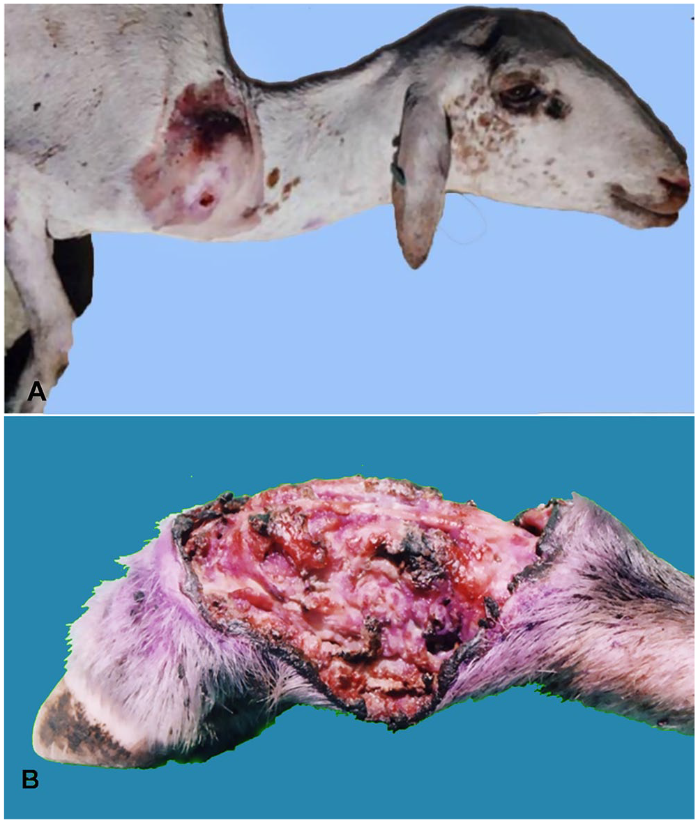
Clinically, cutaneous pythiosis in sheep is characterized by local swelling with progressive ulcerative dermatitis in the legs as well as abdominal and prescapular regions (Fig. 1A, 1B). Pain, anorexia, and lameness are also observed. Most animals with cutaneous pythiosis die.28,30 There is only one report of successful treatment of a sheep with potassium iodide (7 mg/kg, per os, for 7 d). 30 The single case of cutaneous pythiosis reported in a goat had similar characteristics described for sheep, except this animal was pruritic, 6 which has not been described in sheep. In this goat, surgical excision followed by topical treatment with chlorhexidine and insect repellents for 2 wk resulted in complete remission. 6
Rhinofacial pythiosis in sheep is clinically characterized by marked enlargement and deformity of the nasal region, severe respiratory difficulty, and bloody nasal discharge (Fig. 2). Oronasal-rostral fistulas with ulcers on the hard palate and enlargement of the retropharyngeal and parotid lymph nodes are commonly observed. The clinical course is highly variable, with a range of 15–90 d.25,31 Several cases of ovine rhinofacial pythiosis recovered after oral administration of 1 g/d of potassium iodide. 22
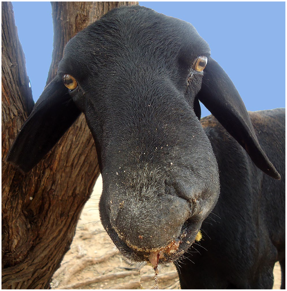
Pythiosis in sheep. Marked enlargement and deformity of the nasal region. Purulent exudate is observed in the mouth as a result of ulceration of the hard palate.
In digestive tract pythiosis in sheep, there is regurgitation, anorexia, and lethargy. 21 No cases of recovery have been reported in sheep with this form of pythiosis.
Gross pathology
Ulcerated areas of haired skin, ranging from 2 to 30 cm, which may be dry or wet and have a dark-red, brown, or black surface, are observed in the cutaneous form of pythiosis of sheep. The subcutaneous tissue in ulcerated areas is yellow or dark-brown and is surrounded by a thin, white, fibrous capsule. The surrounding tissues are usually edematous. The yellow coral-like firm masses, known as “kunkers,” which are considered the hallmark of the disease in horses, are not observed in sheep.6,28,30 Cutaneous lesions may expand to adjacent tissues including ligaments, tendons, nerves, and bones. Hematogenous spread may occur, in which case granulomas may be observed in the lungs, particularly in the caudal lobes, and in regional lymph nodes, mainly prescapular, which may also be markedly enlarged. 30 The gross lesions of cutaneous caprine pythiosis described in the one caprine case were similar to those described in sheep. 6
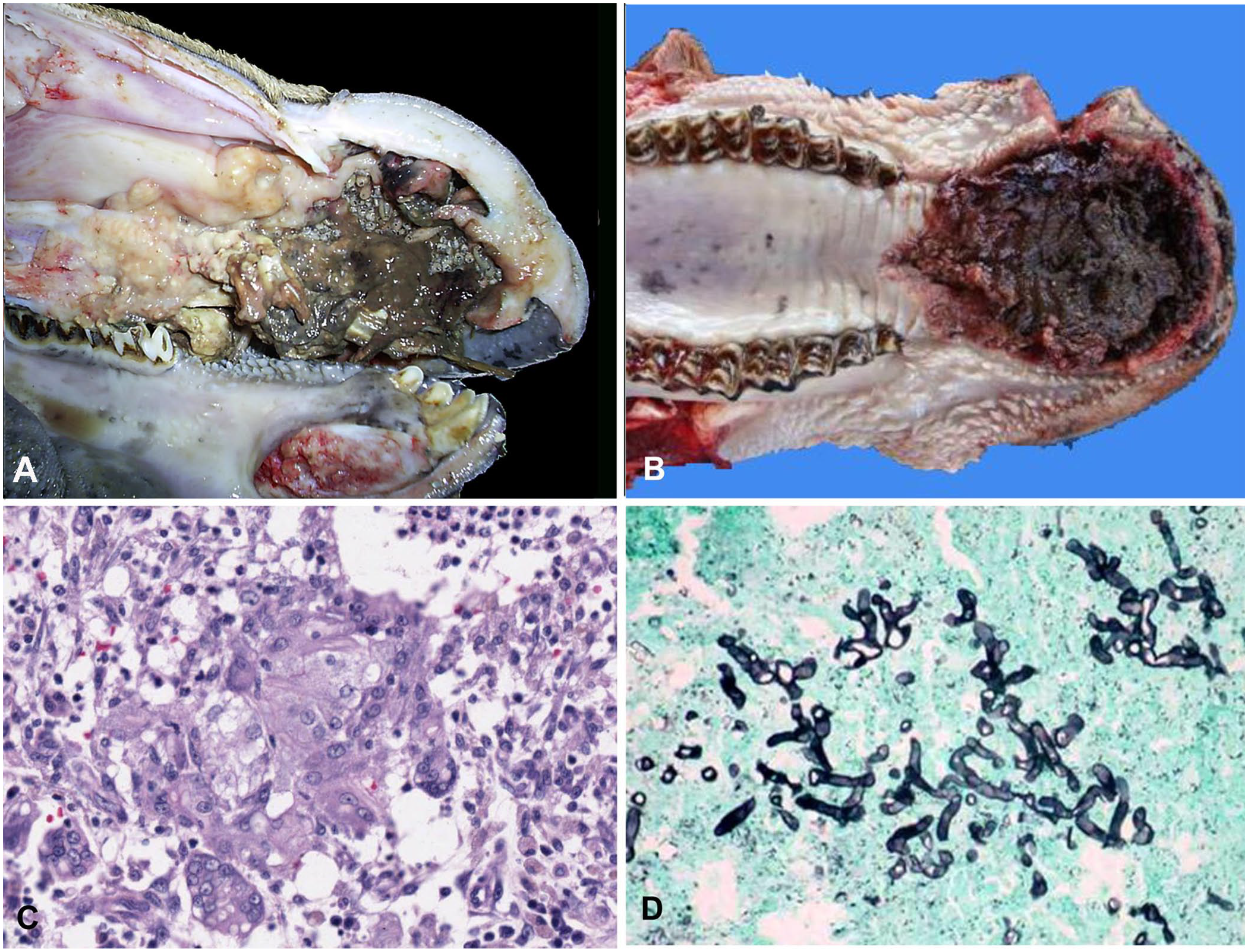
In rhinofacial pythiosis, the lesions usually extend from the mucocutaneous junction at the nares to the middle portion of the nasal cavity, and they may involve the nasal septum, hard palate, and maxilla (Fig. 3A, B). The lesions are characterized by caseous, yellow, and fetid exudate, associated with necrosis and probably secondary bacterial infection of the frontal bone and associated soft tissues.27,31 Rarely, the nasal infection caused by P. insidiosum in sheep extends to the rhinopharyngeal region 31 ; the submandibular and parotid lymph nodes are almost always involved.25,27,31 Occasionally, granulomas can also be observed in the lungs.25,27,31
Pythiosis in sheep.
In digestive tract pythiosis, ulcers covered with a fibrinous pseudomembrane are present in the esophagus, forestomachs, and abomasum. The forestomach and abomasal lesions may be transmural, resulting in perforation, peritonitis, and adhesions among abdominal organs. 21
Microscopic findings
Regardless of the form, pythiosis in sheep is characterized by pyogranulomas composed of central necrotic foci surrounded by moderate numbers of lymphocytes, plasma cells, eosinophils, neutrophils, regular and epithelioid macrophages, multinucleate giant cells, and fibrovascular tissue (Fig. 3C). Hyphae may be seen free in the necrotic center, in the cytoplasm of giant cells, or in the lumen of blood vessels associated with thrombi.6,17,25,26 Hyphae are 4–10 µm wide with non-parallel walls, irregular branches, and rare septa; they are often surrounded by radiating eosinophilic material (Splendore–Hoeppli reaction).6,21,30,31 The hyphae stain well with silver stains (e.g., Gomori methenamine silver; Fig. 3D) but not with periodic acid–Schiff. Vascular invasion by P. insidiosum and thrombosis of blood vessels is observed occasionally. 31 The microscopic lesions in the cutaneous case of caprine pythiosis 6 were similar to those described in sheep.
Diagnosis
A presumptive diagnosis of pythiosis can be made based on clinical signs as well as gross and microscopic lesions. Rhinofacial pythiosis should be distinguished from conidiobolomycosis, which is clinically and morphologically similar. Digestive tract pythiosis is clinically and pathologically similar to several fungal infections of the digestive tract, including aspergillosis and zygomycosis.
Although histology and examination of wet mounts in 10% KOH may help in the detection of this oomycete, neither technique allows differentiation of P. insidiosum from several pathogenic fungi, including zygomycetes, Conidiobolus spp., and Basidiobolus spp.15,16
Confirmation of the diagnosis of pythiosis should be based on identification of the agent by immunohistochemical, molecular, and/or culture-based methods.8,10,27,31 Immunohistochemistry has been used to identify P. insidiosum in formalin-fixed tissues of several animal species, 13 including sheep. 31
Culture can be performed on 2% dextrose Sabouraud agar plates incubated at 37°C for 24–48 h. P. insidiosum does not develop sporangia on routinely used culture media. Therefore, induction of a sporangium producing motile biflagellate zoospores is usually performed for identification purposes. All Pythium spp. develop zoospores in wet cultures with calcium and magnesium. However, because P. insidiosum is the only oomycete pathogenic for mammals, zoospore induction is helpful to establish a presumptive diagnosis of pythiosis. Further molecular identification of the culture by molecular methods is required for final identification of the organism. 9
Among several molecular tests used in the diagnosis of pythiosis in animals and humans, several authors have reported the use of PCR to detect the internal transcribed spacer of the rRNA locus of P. insidiosum as a specific test for detection of this microorganism.5,12,32 Serology is considered a valuable tool in the diagnosis of pythiosis in humans and some animal species (e.g., horses). 9 Antibodies to pythiosis were detected by an indirect ELISA in sheep with and without lesions of pythiosis. 7
Conclusion
Pythiosis is an important disease of sheep in Brazil. The high prevalence of the disease in this country seems to be related to the unique environmental conditions in which sheep are reared. It is possible, however, that pythiosis occurs with low frequency in other countries, but it may be misdiagnosed. Pythiosis should be considered among the differential diagnoses of granulomatous rhinofacial, cutaneous, and digestive tract diseases in sheep. The only way to prevent sheep pythiosis is to avoid grazing around water reservoirs, which is very difficult, mainly in the semiarid region during the dry season when pasture shortage is very marked.
Footnotes
Declaration of conflicting interests
The authors declared no potential conflicts of interest with respect to the research, authorship, and/or publication of this article.
Funding
The authors declared that they received no financial support for their research and/or authorship of this article.