
Abstract
A large, pedunculated cutaneous mass protruding from the left flank fold and an enlarged left prefemoral lymph node were found on examination of a 3-d-old crossbred Aberdeen Angus heifer. The calf was asymptomatic aside from peripheral lymphadenopathy, and the mass, along with the left prefemoral lymph node, was surgically excised. Histologic examination of the mass and the lymph node revealed a homogeneous population of neoplastic cells that stained positively with immunohistochemical stains S100 and melan A, supporting a diagnosis of congenital amelanotic melanoma with nodal metastasis. Two months later, the calf became acutely recumbent and was euthanized after clinical examination revealed widespread metastasis. Gross autopsy revealed widely disseminated metastases that involved vertebral bodies, spinal cord, heart, kidneys, lungs, oral mucosa, multiple lymph nodes, and the marrow cavity of several long bones. Our case serves as a reminder that, although rare, congenital neoplasms occur in bovids and have the potential for aggressive, metastatic behavior.
A 3-d-old black crossbred Aberdeen Angus heifer calf was presented for evaluation of a pedunculated cutaneous mass protruding from the left flank fold (Fig. 1). Calving had been unattended, and the mass had first been noticed the previous day. The owner reported that the dam appeared healthy and the calf had been nursing normally. Physical examination revealed normal mentation, adequate hydration, and vital signs within normal limits. The pedunculated mass was ~8 cm long and 6 cm in diameter at the widest point and was attached to the left flank fold by a stalk. The mass was soft-to-fluctuant with small firm nodules palpable internally. Skin overlying the proximal half was sparsely haired–to-alopecic and mildly abraded on the distal portion. Large vessels were palpable at the proximal end and within the stalk. The left prefemoral lymph node was markedly enlarged relative to the right, and there was moderate bilateral enlargement of the prescapular lymph nodes. The remainder of the physical examination was within normal limits. Ultrasound examination of the mass revealed irregular mixed echogenicity with multiple small hypoechoic nodules. The adjacent body wall appeared to be intact, and no abdominal structures were observed within the mass. The left prefemoral lymph node and mass were surgically removed that same day and submitted for histopathology. The calf became hyperthermic (41°C, normal 37.7–39.1°C) toward the end of the surgical procedure, but recovered normally with no other complications.
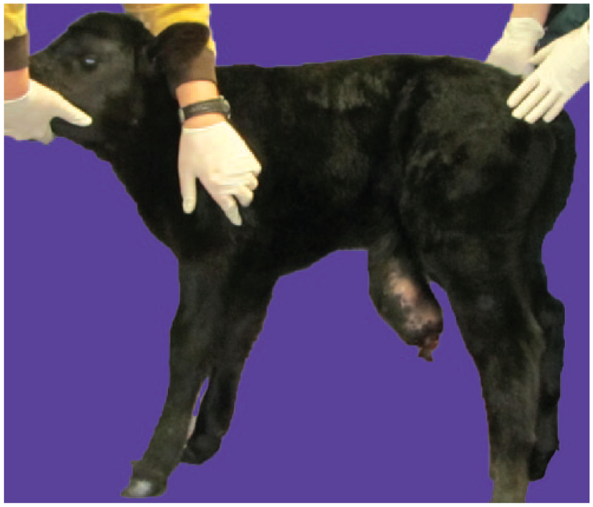
Initial presentation of the calf with a large, pedunculated, cutaneous mass protruding from the left flank fold.
Histologic evaluation of the biopsy specimen revealed the primary cutaneous mass to be a multinodular and infiltrative neoplasm. Morphologically similar neoplastic nodules regionally effaced and replaced the enlarged prefemoral lymph node. The neoplastic cells were regionally variable in their appearance, at least partially the result of trauma to the mass that caused ulceration and degeneration of portions of the neoplastic population. Nodules were composed of polygonal-to-spindle cells arranged in dense packets and cords separated by a fine fibrovascular stroma. Individual cells had a moderate nuclear-to-cytoplasmic ratio, with round-to-oval vesicular nuclei with finely stippled chromatin, a single, large nucleolus, moderate amounts of wispy eosinophilic cytoplasm, and distinct cell margins. No intracytoplasmic pigment was visible within neoplastic cells in biopsy specimens of the primary cutaneous mass or the regional lymph node.
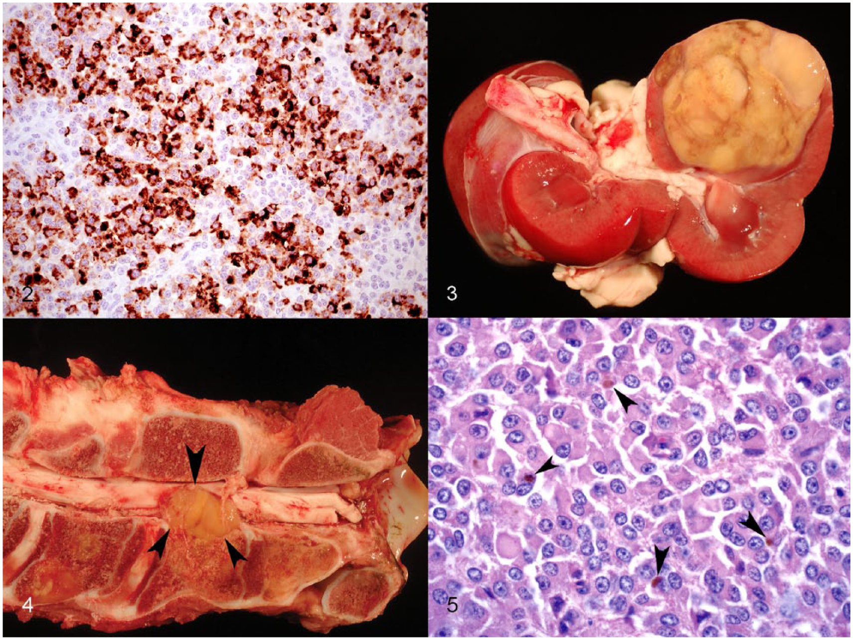
Specific neoplasms that were considered, given the marked pleomorphism of neoplastic masses and the young age of the calf, included amelanotic melanoma or poorly differentiated variants of neuroendocrine carcinoma, germ cell tumor, and round cell tumor. Fontana–Masson stain revealed small-to-moderate numbers of argyrophilic granules within the cytoplasm of scattered neoplastic cells in both the cutaneous mass and the lymph node. Immunohistochemical staining for S100 was moderately intense and widespread, with nearly all neoplastic cells exhibiting cytoplasmic positivity. Intracytoplasmic positivity for melan A (Fig. 2) was multifocal, with intense intracytoplasmic signaling in ~30% of neoplastic cells. Results of these immunohistochemical stains confirmed a diagnosis of congenital amelanotic malignant melanoma. Immunohistochemistry for bovine viral diarrhea virus was performed on an ear notch and was negative.
Congenital amelanotic melanoma in a calf.
Although evidence of metastasis was present, the owner elected to continue raising the calf. The owner was advised to monitor for any signs of recurrence or systemic illness in either the calf or the dam, and to call if noted.
At 2.5 mo of age, the calf was returned for evaluation because of generalized paresis and severe depression. She had been found recumbent and unable to rise the previous night with no improvement since that time. On physical examination, numerous 2–6 cm diameter, firm, fixed masses were palpable under the skin, the most prominent of which were along the ramus of the right mandible, at the eighth rib ventral to the right costochondral junction, and on the proximal and lateral aspects of the left tibia. Multiple small masses were present along the buccal surface of the molars in the upper arcades as well as within hard and soft palates. Neurologic abnormalities included recumbency with an inability to lift the head normally as well as a brief episode of horizontal nystagmus accompanied by a focal seizure of the upper lip. Preliminary blood work revealed that hematocrit, blood glucose, and electrolytes were within reference intervals. The calf was given supportive care overnight and was euthanized the following morning given the poor prognosis, and was submitted for autopsy.
At the time of euthanasia, the calf weighed 93.5 kg and was in good body condition. At autopsy, multiple pale-tan masses were found within the skin. Multiple 1–2 mm nodules were located at the lateral canthus of both eyes and spread in a small beaded pattern both dorsally and ventrally from this location. A 6 × 3 cm wide mass was also located in the subcutaneous adipose tissue on the lateral aspect of the left tibia and a 8 × 5 cm mass was palpable on the right lateral thorax immediately caudal to the right elbow. Multiple 1–6 cm diameter nodules were also found erupting from the oral mucosa, as well as within multiple subcutaneous lymph nodes, myocardium, lungs, and right kidney (Fig. 3). In addition to visceral metastasis, grossly similar nodules invaded the cortical bone and marrow cavity of multiple long bones, mandible, maxilla, and the second cervical vertebral body with extension into the vertebral canal and subsequent compression of the adjacent spinal cord (Fig. 4). Based on the homogeneous gross appearance of disseminated nodules and absence of evidence of inflammatory or infectious disease, differential diagnoses were limited to malignant neoplasia with widespread metastasis. Histopathology was performed on all involved tissues, revealing a neoplastic population identical to that seen on the biopsy; in addition, a small number of cells contained intracytoplasmic fine brown granular pigment (Fig. 5). The final diagnosis was congenital amelanotic melanoma with multi-organ metastasis.
Congenital neoplasia, defined as spontaneous tumors of fetuses and newborn animals up to 2 mo of age, is uncommon to rare. Given that production animals culled because of a failure to thrive may not receive a full postmortem examination, the true prevalence is unknown. In humans, the reported incidence of neonatal tumors ranges from 17 to 121 per million live births worldwide, with teratoma and neuroblastoma being the most common. 9 Congenital or hereditary tumors in pigs are among the best characterized examples of this type of disease in veterinary species, and multiple studies have demonstrated experimental reproducibility of malignant melanoma and lymphoma by selected matings. 2 Furthermore, occurrence of benign versus malignant melanocytic tumors varied widely between pig breeds and, in many cases, the tumors regressed spontaneously. 8
In our case, diagnosis was made more challenging by physical trauma to the mass that resulted in ulceration and significant degeneration of portions of the neoplastic population. Additionally, it was unexpected that a pedunculated mass would have such aggressive biological behavior with numerous metastases involving several organs and tissues. The malignant potential of the mass in our case differed markedly from a previous report of a pedunculated cutaneous malignant melanoma in a 2-mo-old calf that was excised with no evidence of metastasis present at slaughter 1 y later. 5
Melanocytic tumors account for ~6% of all bovine neoplasms and are seen most commonly in the skin 12 ; ~80% are benign melanocytomas. The tumors can occur in any breed and at any age, with an increased prevalence in the Aberdeen Angus breed. Congenital melanomas in calves are rare, 10 and the reported incidence of all congenital bovine neoplasia is 6 per 100,000 calves. 4 In one case study of 14 calves with congenital neoplasia, the most common types of tumors were malignant lymphoma (n = 3), mesothelioma (n = 3), and mixed mesodermal tumor (n = 3). 7 Only 1 case was found to be malignant melanoma, with masses present in the eyes, maxilla, kidneys, and liver. 7 Other reported congenital neoplasms in calves include granulosa cell tumor, 3 hemangioma, lymphangioma, ameloblastoma, and Sertoli cell tumor. 6 Most melanocytic tumors in neonates are benign, although melanoma in general is more prevalent in cattle with dark skin or hair such as the Aberdeen Angus breed. 1 There is no documented mode of inheritance in cattle.
Because most melanocytic tumors in cattle are benign, treatment is frequently not pursued. Surgical excision is the treatment of choice and is generally curative. Evaluation of prognosis is complicated by the fact that this is a production species; most cattle are not kept to the end of their natural lifespan. It is known that a greater degree of pigmentation is associated with increased survival time in dogs, 11 whereas, in humans, the prognosis is similar for pigmented and amelanotic melanoma. 4 Given the low incidence of melanocytic tumors in general, and especially the rarity of amelanotic melanoma in cattle, no comparable data exist for this species.
Our case serves as a reminder that, although rare, congenital neoplasms occur in bovids and have the potential for aggressive, metastatic behavior. The specific types of neoplasms that occur congenitally differ substantially from neoplasms typically seen in older cattle. In calves, mesodermal and hamartomatous lesions are seen far more frequently than in adult animals, in which epithelial tumors predominate. The hereditary or genetic character of the congenital tumor in this particular calf is unknown. However, at the time of this calf’s death, the dam had no history of melanoma or other neoplastic disease.
Footnotes
Declaration of conflicting interests
The authors declared no potential conflicts of interest with respect to the research, authorship, and/or publication of this article.
Funding
The authors received no financial support for the research, authorship, and/or publication of this article.