
Abstract
We investigated the effect of pyometra on kaolin-activated thromboelastography (TEG). Eighteen client-owned dogs with pyometra and 8 healthy spayed dogs were recruited. TEG parameters and packed cell volume were determined. Results from spayed females and from intact females with pyometra were compared using a Student t-test and Wilcoxon rank sum test. Bitches with pyometra were hypercoagulable compared to spayed bitches as evidenced by elevated maximum amplitude, G, and alpha angle. There were no significant group differences in R time, K time, or clot lysis at 30 or 60 min. Dogs with pyometra should be anticipated to have hypercoagulable TEG variables, and this should be addressed when planning surgical and medical therapy.
Pyometra is a common reproductive condition in bitches, and is typically associated with systemic inflammation, and potentially sepsis and disseminated intravascular coagulation (DIC). Although medical management is possible, most affected bitches are treated by surgical ovariohysterectomy, where knowledge of their hemostatic status would be useful. DIC may be associated with both bleeding and hypercoagulability. Systemic inflammation has been shown to have a procoagulant effect on primary and secondary hemostasis. Inflammation-mediated cytokine release results in increased release of platelets that have enhanced susceptibility to activation. Platelet activation in turn will further upregulate the inflammatory response. Tissue factor expression on the surface of endothelial cells and activated white blood cells triggers the formation of thrombin and ultimately fibrin. Thrombin is a potent platelet activator, leading to generalized hypercoagulability in response to inflammation. 6
Studies of bitches with pyometra have documented variable changes in hemostasis, based on measurements of prothrombin time (PT), activated partial thromboplastin time (aPTT), thrombin time (TT), antithrombin activity, platelet count, and fibrinogen and D-dimer concentrations.11,12 Given the role of the platelet surface and platelet reactivity on the speed and strength of clot formation, traditional plasma-based (platelet-poor) assays may under-evaluate the impact of inflammation on overall hemostatic balance.
Thromboelastography (TEG) is a whole blood assay that provides an overall assessment of clot formation (R time, clotting time [K time], α angle), clot strength (maximum amplitude [MA]), clot rigidity (G value), and fibrinolysis (clot lysis at 30 min [LY30] and 60 min [LY60]) and is useful to assess both hypo- and hypercoagulable states (Fig. 1). 8 TEG has been evaluated in other inflammatory conditions, including septic peritonitis, wherein evidence of accelerated clot formation and increased clot strength was documented in dogs that did not have DIC. 2 The use of TEG has not been reported in bitches with pyometra.
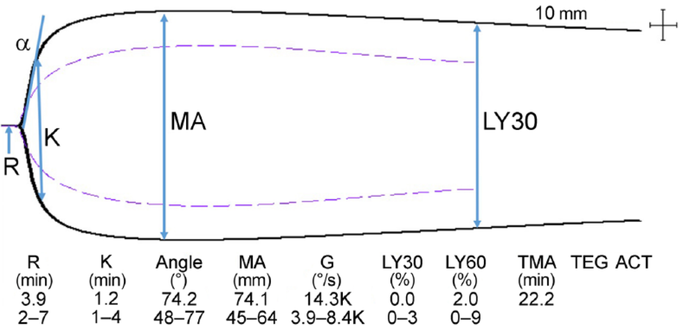
Normal thromboelastographic tracing showing the commonly assessed variables (with institutional reference intervals noted). The dotted line represents the mean of the reference values. Time to clot formation is described using the R time, K time, and α angle. The R time is measured as the time from the start of the assay to when the waveform reaches 2 mm above baseline. The K time indicates the time to achieve a specific clot strength (the time from 2 mm to 20 mm above baseline); α angle indicates the rate of clot formation (the slope of the line joining R and K). Clot strength is assessed using the maximum amplitude (MA, the maximum distance between the 2 lines). The G value is calculated using the MA. Clot lysis (LY) is a measure of fibrinolysis and is usually measured at 30 min (LY30) and 60 min (LY60). The dotted line represents the average values from healthy dogs used for determination of reference intervals, and the solid line is consistent with a hypercoagulable tracing with shortened R time and K time, and increased α and MA.
In pyometra, it is reasonable to expect TEG results consistent with hypercoagulability as a result of the presence of systemic inflammation as well as prolonged elevated serum progesterone levels accompanying diestrus. 7 Most centers utilizing TEG have established laboratory-specific reference intervals using healthy dogs, which likely consist of both males and females with a predominance of neutered dogs. The objective of our study was to use kaolin-activated TEG to characterize hemostatic changes in dogs with pyometra and to compare them to healthy spayed control bitches. We hypothesized that, given the marked impact of inflammation and the secondary impact of diestrus, bitches with pyometra would have TEG parameters consistent with hypercoagulability when compared to controls.
Our study was approved by our Institutional Animal Care and Use Committee. Dogs were recruited from the Emergency Service (ES) and from the students and staff of the Cummings School of Veterinary Medicine (North Grafton, MA). Bitches were characterized as spayed or as having pyometra. Pyometra was suspected based on clinical signs and ultrasonography of the uterus, and was confirmed surgically. Following signed owner consent, a 5-mL sample of blood was obtained from the jugular vein and placed in tubes containing 3.2% sodium citrate for kaolin-activated thromboelastography or sodium heparin for spun packed cell volume (PCV; 12,000 × g for 90 s). 10 Total plasma protein was measured by refractometry of plasma from the microhematocrit tube. The citrated tube was rested for 30 min prior to performing TEG (TEG5000 Thrombelastograph, Haemonetics, Braintree, MA), as described previously. 8 TEG variables recorded included R time, K time, angle, MA, G value, LY30, and LY60 (Table 1). A hypercoagulable TEG tracing was represented by a shortened R time and K time, steep angle, wide MA, and increased G. Delayed fibrinolysis was identified through decreased LY30 and LY60; increased LY30 and LY60 indicated accelerated fibrinolysis. A PCV ≤ 37% was defined as anemia. Only 4 of the bitches with pyometra had a complete blood count performed by our clinical pathology laboratory. Of 18 bitches, 16 were ovariohysterectomized soon after admission and 2 were euthanized prior to surgery given financial limitations; blood samples were collected at the time of presentation to the ES.
Age, thromboelastographic measures, packed cell volume, and total plasma protein in 8 healthy spayed bitches and 18 bitches with pyometra.
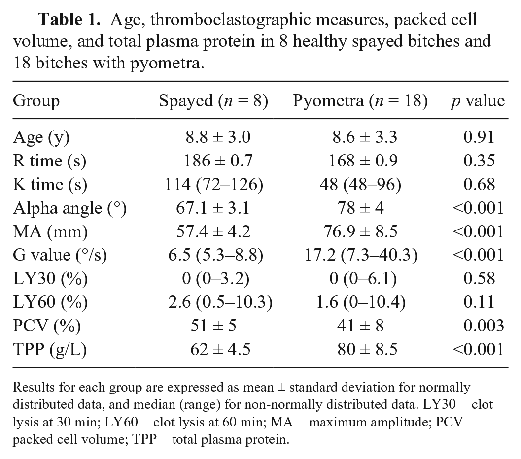
Results for each group are expressed as mean ± standard deviation for normally distributed data, and median (range) for non-normally distributed data. LY30 = clot lysis at 30 min; LY60 = clot lysis at 60 min; MA = maximum amplitude; PCV = packed cell volume; TPP = total plasma protein.
The distribution pattern of quantitative measures was determined by the Shapiro–Wilk test. We report mean and standard deviation for normally distributed variables, and median and range for non-normal distributions. Age, PCV, and TEG parameters were compared between groups using the Student t-test or the Wilcoxon rank sum test for normally and non-normally distributed measures. Linear regression models were used to analyze the association between PCV and the TEG parameters MA and G. In order to meet the model requirements, nonparametric variables were transformed, as necessary, prior to fitting the model. The proportion of bitches with anemia was compared between groups using the Fisher exact test. A multi-variable linear model was used to investigate the adjusted association of MA, G, and angle with pyometra by controlling for PCV. A p value of <0.05 was considered significant. All calculations were performed using commercial software (RStudio, Boston, MA)
Twenty-six dogs were enrolled, 18 with pyometra and 8 healthy spayed controls; 15 breeds were included. Spayed female dogs averaged 8.8 ± 3.0 y; dogs with pyometra were 8.6 ± 3.3 y (p = 0.91). The PCV was significantly lower in the pyometra group (42 ± 8%) compared with spayed females (51 ± 5%; p = 0.003). To further evaluate the clinical significance of the lower PCV in dogs with pyometra, the presence of anemia was evaluated between groups. Anemia (PCV ≤ 37%) was documented in 4 of 18 dogs with pyometra; no control dogs were anemic; this difference in proportion of anemia was not significant (p = 0.28).
Bitches with pyometra had significantly higher α angle, MA, and G value compared to the control group (p < 0.001 for all). There were no significant group differences in R (p = 0.35), K (p = 0.68), LY30 (p = 0.58), and LY60 (p = 0.11; Fig. 2). The association of MA, G, and α angle with pyometra remained significant after adjusting for PCV (p < 0.001, p = 0.011, p < 0.001, respectively). Bitches with pyometra were hypercoagulable when compared to spayed bitches. Specifically, the clot formation was more rapid, as evidenced by a steeper α angle, and clot firmness was much higher, as evidenced by markedly higher MA and G.
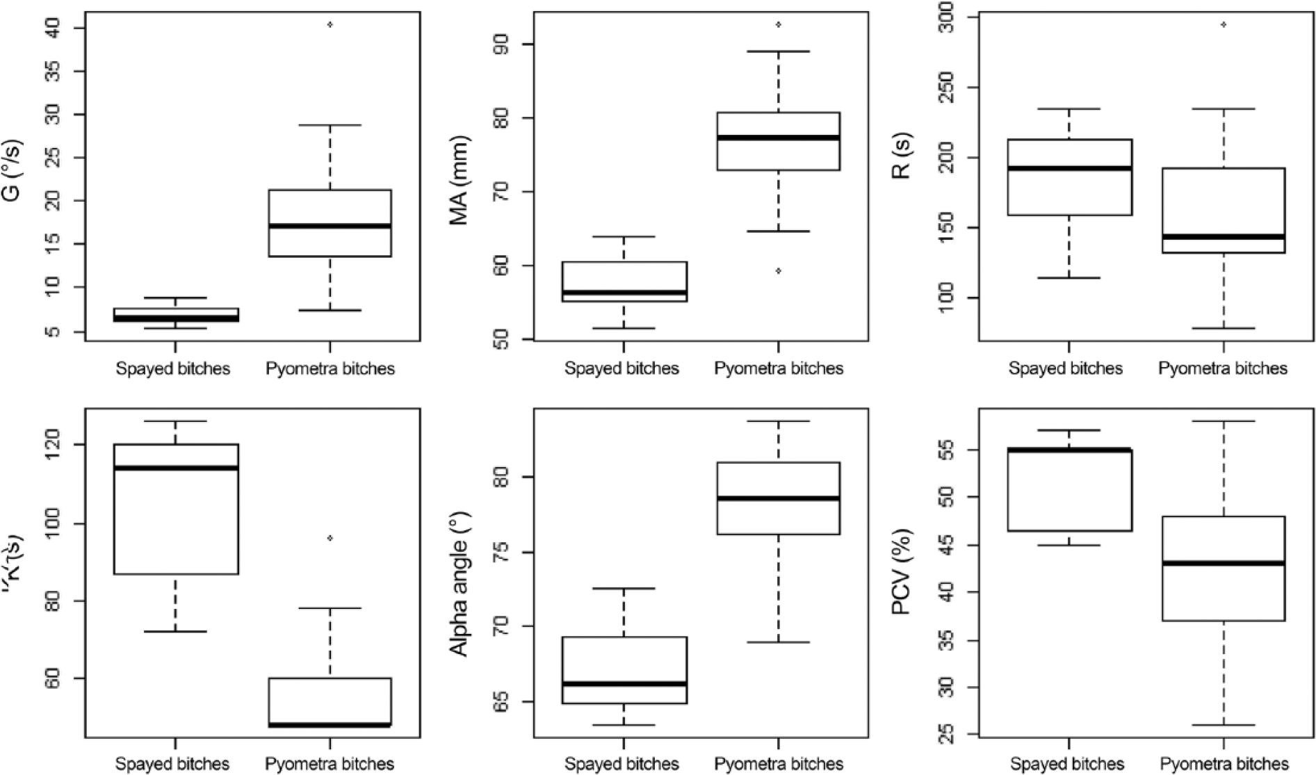
Box and whisker plots showing the thromboelastographic measures (G, MA, R time, K time, α angle) and packed cell volume (PCV) of healthy spayed bitches and bitches with pyometra. The asterisk (*) denotes outliers.
The hemostatic changes that occur in bitches with pyometra appear to be multifactorial. The mean PCV was significantly lower in dogs with pyometra. The impact of hematocrit on TEG is controversial. 2 Mild anemia is commonly associated with pregnancy in bitches. This relative anemia is the result of expanded plasma volume and proportionally decreased red blood cell mass, resulting in reduced blood viscosity, 9 and in bitches with pyometra, the anemia may be associated with blood loss 12 or systemic inflammation. 7 We found a significant negative correlation between PCV and MA and G, which is consistent with recent veterinary literature.3,5,8 The anemia in our sample of bitches was mild. Although PCV has been shown to impact TEG, 2 multivariate analysis confirmed that the statistical significance between pyometra and TEG parameters persisted after adjusting for PCV, implying that the findings of our study are not the result of anemia alone.
Hyperproteinemia was present in our bitches with pyometra (Table 1). Unfortunately, without determination of albumin and globulin, it is impossible to determine if this hyperproteinemia resulted from dehydration, hyperglobulinemia, or both. However, given that mild anemia was also present, we consider it less likely that dehydration was to blame, and more likely that the elevated globulins associated with inflammation were at play. Hypergammaglobulinemia in people is not believed to affect TEG tracings. 4
There are several significant limitations to our study, the first being a cohort of limited size, which weakens the power of the statistical analyses. Additionally, dogs with pyometra may develop DIC, which can manifest as hypercoagulability on TEG. 13 We did not specifically evaluate the presence of DIC in our study. Traditional hemostatic tests, including fibrinogen concentration and platelet count, were not evaluated. We sampled dogs once at the time of presentation to the ES and therefore any progression of changes or resolution of TEG changes was documented after ovariohysterectomy. In addition, the presence of hypercoagulability in bitches was not investigated in relation to surgical findings (i.e., hemostasis and bleeding) or the outcome. None of the surgically treated bitches bled abnormally. Future studies evaluating hemostatic alterations over time may confirm the progression of changes and the timeframe of hemostatic normalization.
The clinical relevance of hypercoagulability is unknown in bitches with pyometra, although thrombosis associated with pyometra appears rare in bitches. In a 2014 study, a Chihuahua was concurrently diagnosed with arterial thrombosis (bilateral external iliac) and pyometra, which were treated successfully with ovariohysterectomy and warfarin therapy. 1 Pyometra has a documented association with increased production of acute phase proteins (C-reactive protein) and inflammatory mediators secondary to endotoxin release (i.e., tumor necrosis factor-α, interleukin-6).7,12 Of the pro-inflammatory cytokines produced, platelet-activating factor plays a key role in promotion of procoagulant mechanisms. 12 These procoagulant mechanisms may in part increase the potential for sepsis-related thromboembolic complications. 12 The role of inflammation in the resulting TEG changes was not further investigated in our study.
Footnotes
Declaration of conflicting interests
The authors declared no potential conflicts of interest with respect to the research, authorship, and/or publication of this article.
Funding
The authors received no financial support for the research, authorship, and/or publication of this article.