
Abstract
Thromboelastography (TEG) is a point-of-care whole blood test of hemostasis. While TEG is becoming more widely used in veterinary medicine, few studies describe the use of TEG in cats. The objectives of the current study were to: 1) document the range of TEG variables produced in healthy cats using 3 sample types (citrated native, kaolin-activated, and tissue factor–activated), and 2) determine if there was a significant difference between 2 separate samples obtained from individual healthy cats on the same day. Jugular venipuncture was performed in 20 cats, and citrated blood collected for TEG. TEG analysis was performed on citrated native, kaolin-activated, and tissue factor–activated blood for each sample. Two hours later, the procedure was repeated from the opposite jugular vein, yielding a total of 120 analyses. Reaction time (R), alpha angle (α), kappa value (κ), and maximum amplitude (MA) were recorded from each tracing. No significant differences were found between TEG tracings from the first and second venipuncture samples. Significant differences were found between sample types for R, α, κ, and MA. Means for citrated native/kaolin-activated/tissue factor–activated methods were R = 4.1/3.7/0.6 min; κ = 2.5/1.8/2.2 min; α = 59.9/65.1/70.4 degrees; MA = 47.4/49.9/44.7 mm. A limitation of this study was the small number of cats used. Thromboelastography analysis may be a suitable method of evaluating hemostasis in cats.
Introduction
Thromboelastography (TEG) is a point-of-care measure of hemostasis, and a kinetic determination of thrombus development, allowing assessment of rate and strength of clot formation, as well as subsequent clot lysis. 3 Thromboelastography evaluates the entire hemostatic process whereas traditional tests of hemostasis such as prothrombin time (PT), activated partial thromboplastin time (aPTT), activated clotting time (ACT), and platelet aggregation only measure isolated portions of the hemostatic pathway. 13 Another advantage of TEG is its ability to detect both hyper- and hypocoagulable states. 3 Thromboelastography has been used to identify hypercoagulability in dogs with parvoviral infections, disseminated intravascular coagulation, neoplasia, and immune-mediated hemolytic anemia,7,10,11,15 and was a better predictor of bleeding in dogs compared to PT and aPTT. 17
Several variables describing clot formation are recorded during TEG analysis. The reaction time (R) measures the time to fibrin formation and represents enzymatic coagulation activity. The alpha angle (α) is an indicator of fibrinogen concentration and platelet function, measuring not only the gradual strengthening of the clot and but also how quickly it forms. The kappa value (κ) measures time for clot formation to a predetermined strength, and is influenced by many variables including factors II and VIII, platelet quantity and function, thrombin formation, fibrin precipitation, fibrinogen concentration, and hematocrit. The maximum amplitude (MA) measures overall clot strength and is an indicator of platelet function and aggregation as well as fibrin activity.3,8
Various sample types have been used for TEG analysis in veterinary and human medicine. The addition of an activator, such as tissue factor or kaolin, to whole blood has been reported to minimize variability and produce more rapid results in horses and human beings.4,5 Additionally, activated samples produced lower analytical variability compared to unactivated samples in a group of healthy cats. 9
While TEG is extensively used to detect hemostatic abnormalities in dogs,3,7,11,17few studies have reported the use of TEG in cats.1,9 The goals of the current study were to compare 3 TEG sample types in healthy cats and to determine the intraindividual variability of TEG measured at 2 different time points in individual cats.
Materials and methods
Twenty healthy cats, owned by students and staff at the Ontario Veterinary College, University of Guelph (Guelph, Ontario, Canada), were recruited for the study. Cats were not on any medications except parasite prophylaxis, and were determined to be healthy on the basis of a normal physical examination, complete blood cell count, serum biochemical profile, and coagulation profile. The study was designed in accordance with the standards of the Canadian Council on Animal Care and the Ontario Animals for Research Act and was approved by the University of Guelph Animal Care Committee.
A 5-ml blood sample was obtained from a single, atraumatic jugular venipuncture using a 22-gauge needle and a 6-ml syringe. Blood for TEG analysis was transferred into a 1.8-ml 3.2% sodium citrate tube, ensuring a 9:1 blood to citrate ratio. Tubes were gently inverted 5 times to allow mixing of the blood and citrate. Samples were allowed to equilibrate at room temperature for 30 min prior to TEG analysis. The procedure was repeated from the opposite jugular vein, approximately 2 hr after the first sample collection.
Thromboelastography analysis
Thromboelastography was performed using 2 thromboelastography analyzers a to allow for all 3 samples to be analyzed simultaneously. For every sample, 20 µl of calcium citrate was added to the pre-warmed (37°C) cup. b For the citrated native samples, 340 μl of citrated whole blood was then added to the cup. For kaolin-activated samples, 1 ml of citrated whole blood was poured into a kaolin-coated vial, c and 340 μl of this activated sample was then added to the cup. For tissue factor–activated samples, 20 μl of recombinant human tissue factor (1:50,000 dilution) stabilized in a HEPES (N-2-hydroxyethylpiperazone-N-2-ethanesulfonic acid) buffer with 2% bovine serum albumin d and 320 μl of citrated whole blood were added to the cup. Each sample was run for 75 min; R, κ, α, and MA were recorded from each channel simultaneously via an electronic transfer from the TEG analyzers to the computer. The procedure was repeated for the second blood sample.
Statistical analysis
Paired t-tests were used to assess the null hypothesis that there were no differences between the R, κ, α, and MA generated for the first and second samples. Data was assessed for normality using a Shapiro–Wilk test. If the data was not normally distributed, it was log transformed and re-tested for normality. The TEG variables generated by each sample type were compared using Spearman rank correlation coefficient. Values for R, κ, α, and MA of sample 1 and sample 2 from each cat was averaged, then 2-way analyses of variance accounting for cat and sample type effects were performed to determine differences between the 3 TEG sample types. A Tukey adjustment for multiple comparisons was performed post-hoc on all normally distributed data. A Levene test was used to assess equality of variances between the sample types. Standard deviations (SDs) and coefficients of variation (CVs) for the population were calculated for each variable. Intraindividual CVs were calculated between the 2 samples per cat for each TEG variable. Statistical analysis was performed using computer software programs.e,f
Results
The mean age of cats in the current study was 3.8 years (range: 1–10 years). Of the 20 cats, 19 were Domestic Short or Longhair cats and 1 was a Norwegian Forest Cat. Eleven were neutered males; 9 were spayed females. R, α, and MA values were normally distributed; κ was normally distributed after log transformation.
There were no significant differences between first and second samples for each TEG variable (p range: 0.307–0.993). The R (p = 0.0009), κ (p = 0.0126), α (p < 0.0001), and MA (p = 0.0435) values were significantly different among the 3 TEG sample types. The R-value was significantly shorter with tissue factor–activated activation (mean: 0.6 min) compared to citrated native (mean: 4.1 min; p = <0.0001) and kaolin-activated (mean: 3.7 min; p < 0.0001) samples; activation with kaolin also caused a shorter R-value compared to citrated native samples (p = 0.0074). The κ-value was significantly longer in citrated native samples (mean: 2.5 mm) compared to kaolin-activated (mean: 1.8 mm; p = 0.0071) and tissue factor–activated (mean: 2.2 mm; p = 0.0148) samples. The α-value was significantly smaller in citrated native samples (mean: 59.9°) compared to kaolin-activated (mean: 65.1°; p = 0.0118) and tissue factor–activated (mean: 70.4°; p < 0.0001) samples; kaolin activation resulted in significantly smaller α values compared to tissue factor activation (p = 0.0107). The MA-value was significantly larger when samples were activated with kaolin compared to tissue factor (mean: 49.9 vs. 44.7 mm; p = 0.0129).
Using a Levene test, there was significantly more variation for R with kaolin activation compared to citrated native (p = 0.0006) and tissue factor–activated (p = 0.029) samples. Variation between sample types was not significantly different for κ, MA, and α. Mean (± SD), range, and CV for each TEG variable are illustrated in Table 1. The mean intraindividual CV between the first and second sample per cat for each TEG parameter is illustrated in Table 2.
Influence of sample type on thromboelastography variables (n = 20 healthy cats).*
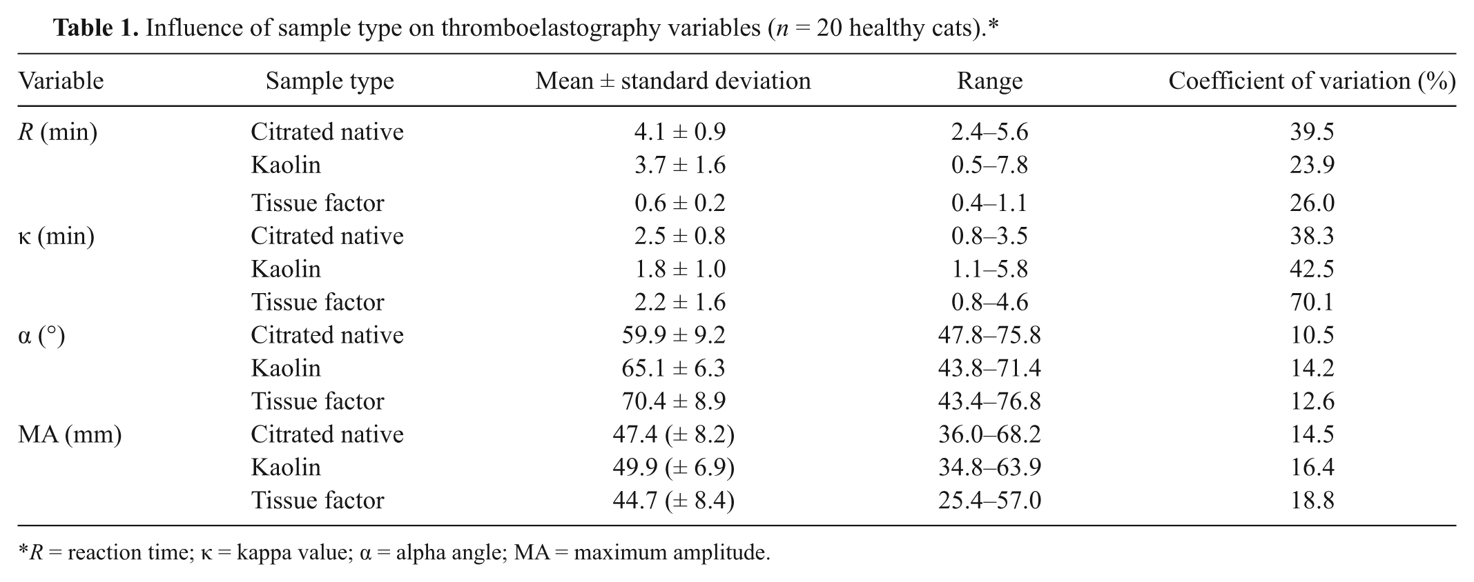
R = reaction time; κ = kappa value; α = alpha angle; MA = maximum amplitude.
Mean coefficients of variation between sample 1 and sample 2 per individual cat, for each thromboelastography variable and sample type (n = 20 healthy cats).*
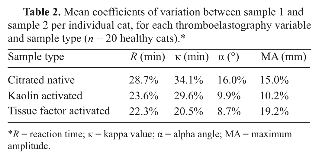
R = reaction time; κ = kappa value; α = alpha angle; MA = maximum amplitude.
Discussion
Three different sample types for TEG analysis (citrated native, kaolin-activated, and tissue factor–activated) have been described previously in dogs and cats.1,2,9,10,16 In the current study, the 3 TEG sample types were performed twice on each cat using 2 separate samples acquired and analyzed approximately 2 hr apart. There were no significant differences between first and second samples for each TEG parameter for any of the 3 sample types, suggesting there is minimal biological variability for TEG variables in cats. Additionally, within-cat variability between 2 samples obtained at separate times ranged from 8.7% to 34.1% for each TEG variable.
Activators such as kaolin and tissue factor are used in other species to reduce the variability in TEG tracing as well as accelerating initiation of the coagulation process, providing more uniform and faster TEG results.4,5 Kaolin (hydrated aluminum silicate) initiates coagulation via the contact activator pathway. Ready-to-use kaolin vials make the use of this activator convenient and consistent.2,5 Tissue factor is a stimulus of in vivo coagulation, making it the preferred activator in some laboratories. However, recombinant tissue factor must be diluted from stock solution, potentially introducing a source of variability in TEG analysis.4,5,16 The R and κ values are measures of the time to thrombus initiation. The R-value reflects the time it takes for the clot to start forming, while κ indicates the time to reach a defined clot strength.3,8 The current study showed that activators allowed a more rapid onset of the coagulation process, as indicated by the significantly shortened R-value when samples were activated with tissue factor, and the significantly shortened κ-value when samples were activated with tissue factor or kaolin. Additionally, the amplification phase of thrombus formation, as indicated by α, proceeded at a significantly faster rate when either activator was used compared to unactivated samples. The effects of activators observed in the present study are similar to those previously reported in cats and other species.4-6,9,18 Activating samples caused a mild increase in MA in human studies, but not in a previous study of healthy horses.4,6,18 Kaolin, but not tissue factor, activation resulted in significantly increased MA values in the present study, when compared to unactivated samples. This is similar to a previous feline study, in which kaolin activation produced significantly higher MA values compared to tissue factor–activated and citrated native samples. 9 While sample activation led to a more rapid onset and amplification of the coagulation process in the present study, the effects of sample activation on hypo- or hypercoagulable states is not known. It is possible that sample activation could mask states of altered hemostasis.3,8 Sample activation to allow faster TEG results may not be as important in cats as it is in other species such as horses, where the mean R-value without activation is 17 min compared to 6.6 min in tissue factor–activated samples. 4 Interoperator variability was not assessed in the current study as all TEG analyses were performed by a single author (AB). Similar to a previous study, 9 the current study revealed significant differences between R, κ, α, and MA values for all 3 TEG sample types tested, which suggests that unique reference intervals should be created for each sample type used.
Kaolin activation produced the most variability with respect to measurement of R values. However, the 3 sample types did not significantly differ in variability for the other TEG parameters measured. The variability observed in the current study differs from the conclusions of a previous study, where citrated native samples consistently provided the highest CVs for each TEG parameter measured while kaolin-activated samples produced the lowest CVs. 9 In the present study, one method did not consistently produce the highest or lowest CVs for each TEG parameter measured. This may be due to the differences in the cat populations and methodology between the studies. Specifically, the previous study had a wider breed variation. Additionally, the previous study performed TEG in duplicate by analyzing samples from a single blood draw in parallel, assessing intra-assay variability. In contrast, the present study analyzed 2 separate samples per cat to reflect intraindividual variability.
One limitation of the present study was the small number of cats used (20). It is recommended that samples from at least 40 individuals are obtained when generating a reference interval. 12 The current study used lower than optimal numbers for generation of a reference interval, but may still provide a useful interval for interpretation of clinical data. Most cats included in the study were Domestic Short and Longhair, and therefore may not be universally applicable to other breed populations. It has been shown that Greyhound dogs have significantly different TEG values from other breeds, 14 and the effects of breed on TEG should be investigated in cats.
No statistically significant differences were found for TEG variables of individual cats measured over 2 separate samples. Further research evaluating day-to-day variability of TEG, or repeatability between 3 or more samples should be performed to evaluate the consistency of TEG measurements in cats. The current study established TEG variables for R, α, κ, and MA values using 3 different sample types. Thromboelastography appears to be a repeatable test in healthy cats. Due to the significantly shorter R-value found with tissue factor activation, it may not be a suitable method for evaluating states of altered coagulation in cats.
Footnotes
Acknowledgements
The authors thank Gabrielle Monteith for her help with statistical analysis.
a.
TEG® 5000 Thrombelastograph Hemostasis Analyzer, Haemonetics Corp., Braintree, MA.
b.
TEG® Hemostasis System Cups and Pins, Haemonetics Corp., Braintree, MA.
c.
TEG® Hemostasis System Kaolin, Haemonetics Corp., Braintree, MA.
d.
Innovin®, Dade-Behring, Newark, DE.
e.
SAS version 9.1, SAS Inc., Cary, NC.
f.
GraphPad Prism version 5.0, GraphPad Software Inc., San Diego, CA.
The author(s) declared no potential conflicts of interest with respect to the research, authorship, and/or publication of this article.
This work was supported by the Ontario Veterinary College Pet Trust Fund (grant no. 49853).