
Abstract
Inherited forms of ichthyosis, or generalized scaling of the skin, have been reported in many animal species, including cattle, and are characterized by an autosomal recessive mode of inheritance. We investigated 2 calves affected with ichthyosis fetalis, a Polled Hereford and a Shorthorn. Both cases had hard white plaques on the skin consistent with excessive keratinization. This was confirmed by histopathology, which showed severe diffuse epidermal and follicular orthokeratotic hyperkeratosis. The known mutation (H1935R) in gene ABCA12, responsible for ichthyosis fetalis in Chianina cattle, was shown to be absent in both affected calves and their obligate heterozygous parents. These molecular findings indicate that allelic heterogeneity exists for this condition in cattle.
Ichthyosis comprises a heterogeneous group of inherited, congenital, and acquired skin disorders, which are characterized by fish-like scales on the skin as a result of excessive keratinization of the epidermis. Ichthyosis has been reported in humans and several animal species, including cattle. 1 In animal species, ichthyosis describes impaired formation of the stratum corneum, which is either congenital and/or heritable. 7
In cattle, there are 2 clinical forms of ichthyosis: ichthyosis fetalis and congenital ichthyosis. The more severe form is ichthyosis fetalis (also referred to as harlequin ichthyosis).3,5 Ichthyosis fetalis has been reported in several cattle breeds including the Norwegian Red Poll, Swiss breeds, Belgian White and Red breeds, Chianina, and Hanwoo.3,4,6,10 Cattle with this severe form are either stillborn or die shortly after birth. 8 They are generally hairless with extensive thick scaly skin, which forms folds and deep fissures along the wrinkle lines of the skin.
We report herein the occurrence of ichthyosis fetalis in 2 calves: a Polled Hereford and a Shorthorn. Gross and histopathologic findings are presented as well as a preliminary investigation into the genetic basis of this disease in these cattle breeds. We hypothesize that allelic heterogeneity exists for this condition in cattle and that the known mutation in Chianina cattle is not the cause in the Polled Hereford and Shorthorn breeds.
The Polled Hereford cattle investigated were from a property in Canbelego, New South Wales (NSW), Australia. Fresh and fixed skin tissues were collected from the affected Polled Hereford female calf, and tail hairs and blood cards were collected from the sire and dam. Hair and blood cards were collected from an additional sire and dam that had also produced an affected calf from the same property. The Shorthorn cattle were from a property in Nyngan, NSW, Australia. Similar samples were collected from the affected Shorthorn female calf and its sire and dam. Fixed tissues from the affected calves were submitted in 10% neutral-buffered formalin for histopathology. The fixed samples were processed into paraffin blocks, sectioned at 4 µm, stained with hematoxylin and eosin, and examined (BX-41 microscope, Olympus Optical, Center Valley, PA).
DNA was extracted from tail hair and tissue using standard techniques, and from blood cards by excising 5 × 1.2 mm discs (Harris Uni-Core, Sigma-Aldrich, St. Louis, MO) from the blood card and submerging them in 100 μL of sterile H2O. Samples were pulsed at 10,000 × g for 10–20 s and then incubated at ambient temperature for 15 min. The supernatant was discarded and 50 μL of buffer was added, comprised of 10 mM Tris-Cl, 50 mM KCl, 1.5 mM MgCl2 (pH 8.3), 0.1 mg/mL proteinase K (Roche, Basel, Switzerland), and 0.5% (v/v) Tween 20 (Sigma-Aldrich). Samples were centrifuged (Heraeus Pico, Thermo Fisher Scientific, Waltham, MA) again at 10,000 × g for 10–20 s and then incubated at 60°C for 30 min, followed by inactivation at 100°C for 10 min.
The ABCA12 gene (219 bp), including the causative mutation of ichthyosis fetalis in Chianina cattle, 2 was amplified by PCR using primers Itt-F (TACTGGGGAAACTGGTGCAT) and Itt-R (GTCGGGCAGCATCATATTTT). PCR was performed (AmpliTaq Gold DNA polymerase, Thermo Fisher Scientific) in a final volume of 30 μL, using 35 cycles with 56°C annealing temperature. Control samples for the 3 possible genotypes were co-amplified. Purified DNA fragments were sequenced (BigDye Terminator v.3.1 cycle sequencing kit, Thermo Fisher Scientific). Electrophoresis of purified sequencing reactions was performed (ABI PRISM 310 DNA analyzer, Thermo Fisher Scientific) and analyzed (Chromas Lite v.2.1.1, Technelysium, South Brisbane, Australia).
Four of 10 calves born at the Polled Hereford property showed evidence of disease. The calves were either stillborn or died shortly after birth. The herd history provided by the producer suggested that the herd is significantly inbred. A stud Polled Hereford bull was used by the producer several years ago. In subsequent breeding, male progeny of the stud bull were used as sires and joined to the female progeny. Therefore, the parents of the affected individuals are half-siblings. At the Shorthorn property, a previous case was observed, but not investigated; the same sire was implicated in both cases at that property.
The clinical presentation of the affected calves from both breeds was similar. The calves had hard white plaques over the entire body, in patterns formed along the wrinkle lines of the skin, making them appear like scales. There was very little hair on the animals. They had small ears, and their lips and eyelids were everted (Fig. 1). The parents of these calves showed no clinical signs of disease. Gross postmortem examination of the affected animals was otherwise normal.
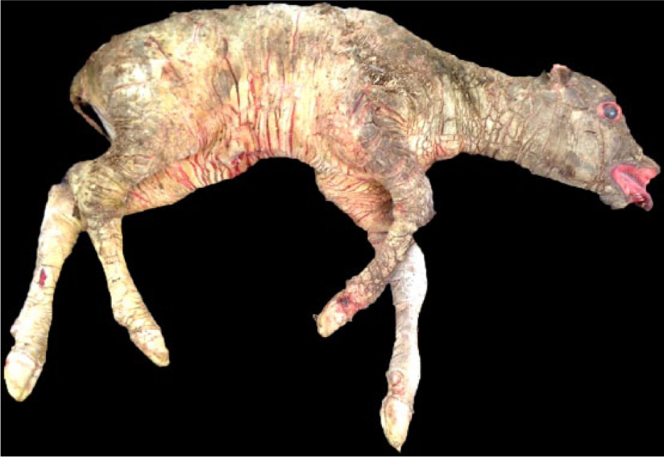
Shorthorn calf displaying characteristic features of ichthyosis fetalis including everted eyelids and lips, and hard plaques across the entire body.
Histologic examination of the skin revealed severe, diffuse, epidermal and follicular orthokeratotic hyperkeratosis (Fig. 2). The stratum corneum was markedly thickened by laminated anuclear keratin, ~3–5 mm thick, containing entrapped hair shafts. There was also hyperkeratosis of the dermal root sheath of the hair follicles. The stratum corneum was divided by deep fissures into plates of keratin (Fig. 2). Apocrine glands in the dermal layer were dilated, with mildly attenuated epithelium. There were a few perivascular and scattered lymphocytes and plasma cells within the dermis.
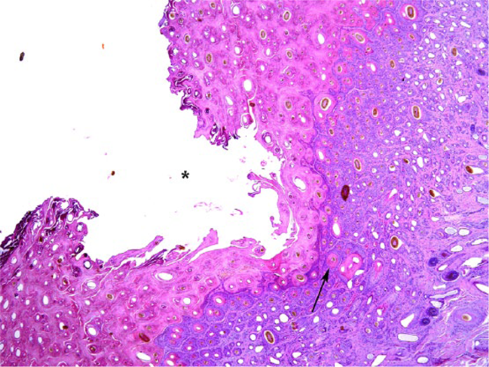
Severe follicular (arrow) and epidermal orthokeratotic hyperkeratosis in a calf with ichthyosis fetalis. The stratum corneum is divided by deep fissures (asterisk) into plates of keratin. H&E.
Genotyping for the known ABCA12 causative mutation (H1935R) 2 was performed on both affected individuals and the healthy obligate heterozygotes. Genotypes at the Chianina locus (H1935R) were consistent with the homozygous wild-type genotype. The expected genotype was observed for each of the controls. Genotyping showed that the Chianina mutation was not present in the affected calves or obligate heterozygotes. These genotyping results are not surprising given that there have been no reported breed improvement programs in Polled Hereford or Shorthorn involving the Chianina breed. Our results support the hypothesis that allelic heterogeneity exists for ichthyosis fetalis in all cattle.
Ichthyosis fetalis has not been reported previously in Australian cattle populations, to our knowledge. Moreover, we found no reports about this disease in the Polled Hereford or Shorthorn breeds worldwide. The gross and histologic findings were consistent with previous cases of ichthyosis fetalis in other cattle breeds and species.3,10 The disease history from each herd supports a genetic basis for the condition, with a recessive mode of inheritance. The sire of both affected calves was implicated in other cases on each property, and the sire and dam were healthy individuals with respect to this disease.
Seven genes have been implicated in humans and animals for the inherited forms of ichthyosis. The vast majority of harlequin ichthyosis cases in humans have mutations in ABCA12, 9 a lipid transporter responsible for cholesterol efflux from epidermal keratinocytes, which is also the locus harboring the Chianina mutation. 2 The ABCA12 gene is a suitable candidate for further investigation to identify the mutation responsible for this disease in Polled Hereford and Shorthorn cattle. Identifying the causative mutation will be critical for management of this inherited condition and to mitigate the amplification of these deleterious alleles in the wider population. Any new cases in either breed will provide valuable genetic material for future investigations. Until additional genetic data are available, examination of pedigree data may indicate whether the condition is present.
Footnotes
Acknowledgements
We thank the property owners for providing information and samples from their herds.
Declaration of conflicting interests
The authors declared no potential conflicts of interest with respect to the research, authorship, and/or publication of this article.
Funding
This work was partially funded by District Surveillance from NSW Department of Primary Industries.