
Abstract
A 7-year-old pregnant Angus cow was found dead in the field. At necropsy, the aortic valve was expanded by moderate fibrous connective tissue and acidophilic coagulum containing multifocal marked bacteria, mineral, neutrophils, and red blood cells. Numerous tiny grayish, opaque bacterial colonies were detected on blood agar plates at 7 days after inoculation with a swab of the heart valve of the cow. The bacterium was a Gram-negative, very small coccobacillus that was catalase, oxidase, and urease negative, and did not change litmus milk, triple sugar iron agar, and sulfide-indole-motility medium. The bacterium was negative for esculin hydrolysis, phenylalanine deaminase, nitrate reduction, and gelatin hydrolysis. The isolate did not produce acid from glycerol, inulin, lactose, maltose, mannose, raffinose, salicin, sorbitol, sucrose, trehalose, glycogen, ribose, or starch. Polymerase chain reaction tests for the gltA, ssrA, ftsZ, ribC, rpoB, and 16S ribosomal RNA genes of Bartonella species were positive for the isolate. Amplicons were sequenced, and the gltA, ribC, ssrA, and 16S ribosomal RNA gene sequences were found to have 100% homology to the type strain of Bartonella bovis, whereas the fts and rpoB sequences showed 99.9% and 99.6% homology, respectively, to the type strain of Bartonella bovis. Diagnosticians should be aware of slow-growing microorganisms, and culture media should be incubated beyond the standard period to enhance the recovery of Bartonella species.
Bacterial endocarditis is the most common endocardial disease in cattle, and the diagnosis of this condition remains challenging for practitioners and diagnosticians, particularly as the infection may be present with or without clinical signs. 2 Although the most frequent pathogens isolated from endocarditis valves or bloodstream of cattle are Trueperella pyogenes (also known as Arcanobacterium pyogenes), Streptococcus species, and Enterobacteriaceae, other organisms are occasionally implicated. 2
The genus Bartonella was named after A. L. Barton, who first described the intraerythrocytic bacterium Bartonella bacilliformis in 1909. 5 Bartonella species are members of the alphaproteobacteria within the order Rhizobiales and family Bartonellaceae, and most Bartonella species are vector-borne organisms.1,6 Bartonella species are fastidious, slow-growing, small Gram-negative rods and could be intraerythrocytic or erythrocyte-adherent bacilli. 5 Several new species have been discovered within the genus Bartonella. 5 There are now at least 20 species or subspecies, most of which are zoonotic agents. 9 Several polymerase chain reaction (PCR) and DNA sequencing assays have been developed for identification of Bartonella species.7,8,11-15
Bartonella species have been detected in endocarditis cases and blood specimens of bacteremic human beings and animals.1,5,10 Bartonella bovis has been detected in the diseased heart valves of cows by PCR and other molecular methods,3,10 but this organism has never been, to the authors’ knowledge, isolated from cows with endocarditis.
A 7-year-old pregnant Angus cow was found dead in the field, and a necropsy was performed at the University of Kentucky, Veterinary Diagnostic Laboratory (UKVDL; Lexington, Kentucky). Routine necropsy was performed, and slides from internal organs were prepared and stained with standard hematoxylin and eosin. Fluorescent antibody tests for Bovine respiratory syncytial virus, Bovine viral diarrhea virus, Bovine herpesvirus 1 (Infectious bovine rhinotracheitis virus), and Bovine parainfluenza virus 3 were performed. In addition, eye fluid was tested for general chemistry, calcium, magnesium, sodium, chloride, and creatinine, and heart blood was tested for cyanide. The swabs taken from affected heart valve were streaked onto blood agar, eosin methylene blue agar, and Columbia nalidixic acid with 10% horse blood. Because the pathologist suspected a bacterial endocarditis and possibly Bartonella spp. based on the necropsy findings, the plates were incubated under microaerophilic conditions at 37°C with 8% CO2 for 7–10 days at the UKVDL. The plates were examined daily for bacterial growth. Bacterial genomic DNA was prepared by heating a suspension of the bacterium for 10 min at 95°C. A series of PCR assays was performed using Bartonella-specific primers to amplify fragments in citrate synthase gene (gltA), the cell division protein gene (ftsZ), the riboflavin synthase gene (ribC), the RNA polymerase beta-subunit gene (rpoB), the transfer messenger RNA (ssrA), and the 16S ribosomal RNA (rRNA) gene (see Table 1). The PCR products from each examined gene were separated and visualized by 1.5% agarose gel electrophoresis with ethidium bromide staining, then purified and sequenced. The sequences were aligned with other Bartonella species available from GenBank to determine homology.
The primers and references used for each of the genes for polymerase chain reaction assays and sequencing of Bartonella bovis isolate.*
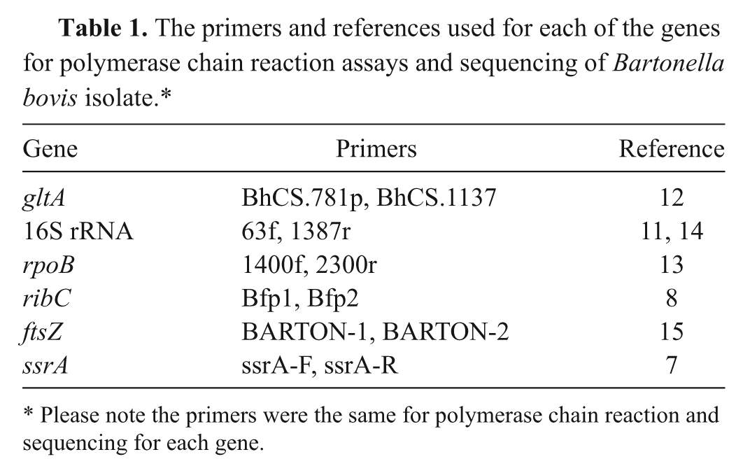
Please note the primers were the same for polymerase chain reaction and sequencing for each gene.
On gross examination, the cow presented in good flesh and in poor to fair postmortem preservation. There was an abscess in 1 spleen pole adhering to the diaphragm. The cow was pregnant with a 4-month male fetus having a crown rump length of 3.2 cm. There was no bloat line. The rumen content was fairly dry. A 2 cm × 2 cm × 3 cm hemorrhagic nodule with a laminated cut surface was present in an aortic valve cusp (Fig. 1). On histopathological examination, multifocal protozoan cysts containing bradyzoites were seen in the heart. Multifocal minimal portal to midzonal subacute inflammation was present in the liver. The kidneys showed minimal multifocal, cortical, interstitial subacute inflammation. The aortic valve cusp was expanded by an acidophilic coagulum containing multifocal mineral deposition, bacteria, neutrophils, and red blood cells. An adjacent valve cusp was expanded by moderate fibrous connective tissue with multifocal mild plasma cells, lymphocytes, macrophages, and neutrophils in the subendocardium. No microscopic changes were observed in the mammary gland, rumen, reticulum, adrenals, abomasum, and intestines.
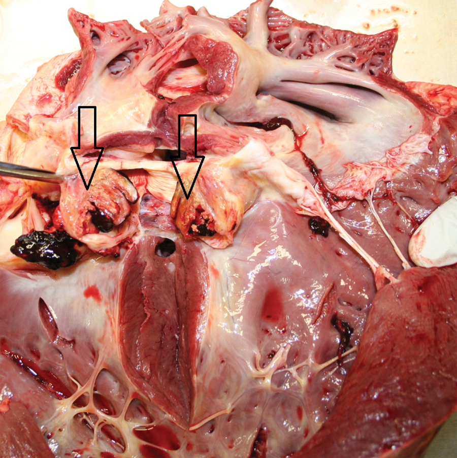
Valvular endocarditis in an Angus cow. Hemorrhage is present in the aortic valve cusp (arrows).
Fluorescent antibody tests for Bovine respiratory syncytial virus, Bovine viral diarrhea virus, Bovine herpesvirus 1, and Bovine parainfluenza virus 3 were negative. No cyanide was detected in the heart blood. Testing of eye fluid demonstrated that general chemistry, calcium, magnesium, sodium, chloride, and creatinine were within normal range, while blood urea nitrogen (19 mg/dl), phosphorus (1.8 mg/dl), and potassium (8.0 mg/dl) were higher than normal limits.
In microaerophilic culture, tiny colonies in numerous numbers were detected at 7 days postinoculation of the swab of the cow heart. The isolate was a Gram-negative, very tiny coccobacillus that did not change litmus milk and triple sugar iron agar. The isolate was negative for sulfide-indole-motility medium and also negative for esculin hydrolysis, phenylalanine deaminase, nitrate reduction, and gelatin hydrolysis. The isolate did not produce acid from glycerol, inulin, lactose, maltose, mannose, raffinose, salicin, sorbitol, sucrose, trehalose, glycogen, ribose, and starch. Because of the site of isolation, microaerophilic nature, and biochemical characteristics, the isolate was subjected to 6 different PCR tests targeting the gltA, ribC, ssrA, 16S rRNA, ftsZ, and rpoB genes of Bartonella spp. These PCR reactions showed that the isolate was Bartonella-positive by all 6 examined genes. Sequence analyses provided strong support that the isolate was B. bovis with 100% identity to the type strain of B. bovis in the gltA, ribC, ssrA, and 16S rRNA genes. The ftsZ and rpoB sequences also showed close relatedness to the type strain of B. bovis with 99.9% and 99.6% similarities, respectively.
Bacterial valvular endocarditis is often missed or misdiagnosed and usually not discovered till necropsy or slaughtering. 10 Bartonella species can cause endocarditis in some other animals and human beings.3-5 Cattle are thought to be the main reservoir for B. bovis. 9 The Angus cow described in the current report from a farm with 150 cattle; none of them showed any clinical signs. The cow was found dead in the field and brought to the UKVDL for necropsy. Because the organism is fastidious and difficult to culture, several enriched liquid and solid media have been reported. 5 However, the detection mostly relies on PCR and DNA sequencing methods targeting genes of gltA, 12 16SrRNA,11,14 rpoB, 13 ribC, 8 ftsZ, 15 and ssrA. 7 In the present study, blood agar media supported the growth of B. bovis. According to a 2006 study, the level of bacteremia was higher in pregnant cows than in nonpregnant cows (P = 0.05), and the level of bacteremia rose during the last two-thirds of gestation (P < 0.001). 9 Interestingly, the cow presented herein was also pregnant.
Bartonella bovis has been detected in cattle with endocarditis by PCR, DNA sequencing, and serology.3,10 However, in these prior studies, the organisms were not isolated in culture, a feature of the current study. Practitioners and pathologists should keep in mind the possibility of Bartonella involvement in endocarditis cases and alert microbiologists to the possibility of Bartonella infections. Microbiologists should also consider incubating culture media beyond the accepted standard period (2–3 days) to enhance the recovery of Bartonella species.
Footnotes
Acknowledgements
The authors thank the staff of the Pathology, Bacteriology, and Molecular Biology sections of the University of Kentucky, Veterinary Diagnostic Laboratory. Part of this article was presented as a poster at the 55th Annual Conference of American Association of Veterinary Laboratory Diagnosticians, Greensboro, NC, 2012.
Declaration of conflicting interests
The author(s) declared no potential conflicts of interest with respect to the research, authorship, and/or publication of this article.
Funding
The author(s) received no financial support for the research, authorship, and/or publication of this article.