
Abstract
Feline leukemia virus (FeLV) is an oncogenic retrovirus of cats. Immunoassays for the p27 core protein of FeLV aid in the detection of FeLV infections. Commercial microtiter-plate ELISAs have rapid protocols and visual result interpretation, limiting their usefulness in high-throughput situations. The purpose of our study was to validate the PetChek FeLV 15 ELISA, which is designed for the reference laboratory, and incorporates sequential, orthogonal screening and confirmatory protocols. A cutoff for the screening assay was established with 100% accuracy using 309 feline samples (244 negative, 65 positive) defined by the combined results of FeLV PCR and an independent reference p27 antigen ELISA. Precision of the screening assay was measured using a panel of 3 samples (negative, low-positive, and high-positive). The intra-assay coefficient of variation (CV) was 3.9–7.9%; the inter-assay CV was 6.0–8.6%. For the confirmatory assay, the intra-assay CV was 3.0–4.7%, and the inter-assay CV was 7.4–9.7%. The analytical sensitivity for p27 antigen was 3.7 ng/mL for inactivated whole FeLV and 1.2 ng/mL for purified recombinant FeLV p27. Analytical specificity was demonstrated based on the absence of cross-reactivity to related retroviruses. No interference was observed for samples containing added bilirubin, hemoglobin, or lipids. Based on these results, the new high-throughput design of the PetChek FeLV 15 ELISA makes it suitable for use in reference laboratory settings and maintains overall analytical performance.
Introduction
Species Feline leukemia virus (FeLV; family Retroviridae, subfamily Orthoretrovirinae, genus Gammaretrovirus) is considered to be the cause of one of the most serious infectious diseases of the cat.6–8,11,18 In a recent study, the overall prevalence of FeLV infection in cats in North America was 3.1%. In cats with clinical signs (oral disease, abscesses, respiratory signs, and other), the prevalence was 7.1% compared to 1.7% in healthy cats. 2 Transmission of FeLV typically occurs through friendly contact between cats in multi-cat households. The virus is shed in saliva, nasal secretions, feces, urine, and milk.5,13 If an infected cat does not mount an effective immune response to the virus when it is initially replicating within local lymph nodes, a persistent infection is likely, with integration of exogenous FeLV proviral DNA into the genome of the infected cells. 8
The integration of FeLV-specific nucleic acid can be detected by commercial PCR assays that are performed in specialized reference laboratories.10,18 However, because the p27 core protein of FeLV is produced in abundance in infected cells, it is the target antigen of choice for detection of infection by immunofluorescence assay (IFA) and a variety of in-clinic and reference laboratory format assays. Notwithstanding the value of PCR and IFA, immunoassays for the detection of p27 antigen have ultimately been adopted as the routine screening method for FeLV infection, primarily because of their ease of use and accuracy. 16 These immunoassays have been available in veterinary medicine for >20 y utilizing well-characterized monoclonal antibodies.14,15 The commercial assays are not well suited for the increased demands of high-volume reference laboratories because 1) they are designed for ease of use in testing individual or small numbers of samples; 2) use rapid, 5-min protocols for immediate test results; and 3) rely on visual inspection for result interpretation. 9 The purpose of our study was to validate a microtiter-plate ELISA, the PetChek FeLV 15 ELISA (IDEXX Laboratories, Westbrook, ME), for the detection of FeLV p27 antigen. This new test is specifically designed to maintain performance relative to established reference methods while incorporating improved operational protocols and objective, instrument-based interpretive criteria suitable for a reference laboratory setting.
Materials and methods
Measurement of FeLV p27 antigen
PetChek FeLV ELISA method
The PetChek FeLV 15 ELISA is a microtiter-plate ELISA that utilizes orthogonal assay protocols to screen and subsequently confirm the presence of FeLV p27 antigen in a serum or plasma sample. Both protocols employ microwells coated with anti-FeLV mouse monoclonal antibody (mAb), a second anti-FeLV mouse mAb conjugated to horseradish peroxidase (HRP), and positive and negative controls consisting of inactivated FeLV particles diluted in fetal bovine serum (FBS) and neat FBS, respectively. The term 15 used in the name of the product refers to the incubation times for the sample and conjugate mixture and the 3,3′,5,5′-tetramethylbenzidine (TMB) substrate, which are different from previous versions of the product.
In the screening protocol, 50 μL of each sample is added to coated microwells followed by the addition of 50 μL of the HRP conjugate. The sample and conjugate mixture is incubated on the plate for 15 min at room temperature and then washed. Subsequently, 100 μL of TMB substrate is added to each microwell and incubated on the plate for 15 min at room temperature. Stop solution (50 μL) is then added to each well, and the absorbance is measured at a wavelength of 650 nm (A650) using a plate-reader spectrophotometer. Samples with A650 values greater than or equal to the assay cutoff (A650 ≥ 0.300) are considered FeLV positive and can be confirmed using the confirmatory protocol.
In the confirmatory protocol, 50 μL of sample is added to each of 2 microdilution tubes. To one tube, 10 μL of diluent containing nonspecific protein and detergent (solution A) is added and to the second tube, 10 μL of neutralizing reagent (solution B) consisting of unlabeled anti-FeLV non-murine polyclonal antibody (pAb) is added. The tubes are incubated for 15 min at room temperature prior to transferring 50 μL each of solution A– and solution B–treated samples into adjacent coated microwells. The assay is completed as described above with the A650 measured using a plate-reader spectrophotometer. A sample is confirmed to be positive for FeLV p27 antigen when the sample in the solution B well is neutralized at least 50%:

For strong positive samples, if the A650 of the solution A–treated sample is >1.500 and the solution B–treated sample is not neutralized by at least 50%, then the sample is diluted 1:11 or 1:22 in sample diluent (solution A) and retested following the confirmatory protocol. If the A650 of the solution A–treated sample is ≤1.500, but the A650 of the solution B–treated sample is not neutralized by at least 50%, the sample is classified as a negative, non-confirming sample.
Reference FeLV antigen ELISA method
The reference microtiter-plate ELISA (ViraCHEK FeLV, Zoetis, Florham Park, NJ) was performed in accordance with the instructions in the kit insert. Briefly, 50 μL of sample is added to a single coated microwell followed by one drop of reagent 1 (HRP conjugate). After a 5-min incubation at room temperature, the plate is washed and then 2 drops of reagent 2 (TMB substrate) are added to each well followed by another 5-min room temperature incubation. After the second incubation, the operator visually interprets the results for each well. Positive samples are blue; negative samples are clear. To mitigate individual operator bias, visual interpretations for reference test results in our study were based on the consensus of 3 operators, each interpreting the test wells in a blinded and independent manner.
Real-time FeLV PCR
Real-time (rt)PCR assays (FeLV RealPCR, IDEXX Laboratories) were conducted at the IDEXX Reference Laboratory. The rtPCR detects and quantifies exogenous FeLV-encoded DNA in leukocytes obtained from peripheral blood. PCR was performed using whole blood samples submitted to the laboratory for testing. The rtPCR primers and probes, and the protocol for extraction of total nucleic acid from EDTA whole blood, were adapted from previously described methods.1,17 The assay was confirmed to detect exogenous FeLV DNA only. Briefly, 180 µL of whole blood was resuspended in lysis solution and incubated for 10 min. Nucleic acids were extracted, eluted into 150 µL of PCR-grade, nuclease-free water, and 5 µL amplified in subsequent rtPCR reactions. Analysis was performed on a LightCycler 480 (Roche Applied Science, Indianapolis, IN) and raw data analyzed to generate cycle thresholds (Ct). Real-time PCR for FeLV was run with 6 quality controls including 1) quantitative (q)PCR-positive controls, 2) PCR-negative controls, 3) negative extraction controls, 4) qDNA pre-analytical quality control targeting the host ssr ribosomal RNA gene complex, 5) an internal positive control spiked into the lysis solution, and 6) an environmental contamination monitoring control. These controls assessed the functionality of the PCR protocol (1 and 5), absence of contamination in the reagents (2) and laboratory (6), absence of cross-contamination during the extraction process (3), quality and integrity of the DNA and RNA as a measure of sample quality (4), rtPCR protocol (4), and absence of PCR inhibitory substances as a carryover from the sample matrix (5).
Validation criteria
Diagnostic accuracy and determination of screen cutoff
Feline plasma samples (n = 309) derived from whole blood previously submitted to IDEXX Reference Laboratories for retroviral PCR testing were collected, randomized, and blinded for testing by the reference FeLV antigen ELISA and the PetChek FeLV 15 ELISA. The method’s diagnostic accuracy was assessed against the concordant results obtained between FeLV PCR and the reference FeLV antigen ELISA. The A650 cutoff for the PetChek FeLV 15 ELISA screen protocol was determined using receiver operating characteristic (ROC) analysis (Prism v.6.05 for Windows, GraphPad Software, LaJolla, CA).
Precision
The precision of both the screening and confirmatory protocols was evaluated for negative, low-positive, and high-positive manufactured samples consisting of inactivated FeLV in FBS using described methods. 3 Intra-assay precision was estimated based on 20 replicates of each sample in a single test run. Inter-assay precision was estimated based on each sample run in singlet across 20 test runs over 5 d. For each sample and assay protocol, the A650 mean and standard deviation (SD) were determined, and the precision was reported as percent coefficient of variation (%CV) which was calculated as follows: %CV = (SD A650/mean A650) × 100%.
Interference in hemolyzed, icteric, or lipemic samples
The effect of elevated hemoglobin, lipids, or bilirubin on the PetChek FeLV 15 ELISA was evaluated based on the methods described. 4 Each substance was spiked into pooled sera from FeLV-negative and FeLV-positive cats at increasing concentrations and tested in both the screening and confirmatory protocols. Bovine hemoglobin (Sigma-Aldrich, St. Louis, MO) was tested at concentrations ranging from 0.30 to 5.50 g/L, bilirubin (ditaurobilirubin disodium salt, Scripps Laboratories, San Diego, CA) was tested at concentrations ranging from <0.001 to 0.1441 g/L, and lipid (Intralipid 30% IV solution, VWR International, Radnor, PA) was tested across optical density at 660nm (OD660) values ranging from 0.2 to 6.4. Each condition was tested across 5 wells, and results were analyzed by one-way analysis of variance using an alpha risk of 0.05.
Analytical sensitivity and specificity
Half log dilution series of recombinant p27 (ICL, Portland, OR) and inactivated FeLV (Advanced Biotechnologies, Eldersburg, MD) in FBS were prepared to span concentrations with a range of 0–10,000 ng/mL and 0–5,000 ng/mL (or 0–2.1 × 10⁷ virus particles/mL), respectively. Protein concentrations and viral particle counts for viral antigen stocks were based on certificates of analysis from the respective manufacturers. Dilutions were tested in triplicate in the screening protocol to determine analytical sensitivity based on the interpolated concentration at the A650 screen cutoff.
Half log dilution series of inactivated murine leukemia virus (MuLV; American Type Culture Collection [ATCC], Manassas, VA), and inactivated feline RD-114 virus (ATCC) diluted in FBS across concentration ranges of 0–5.28 × 10⁶ and 0–9.00 × 10⁶ virus particles/mL, respectively, were prepared. Protein concentrations and viral particle counts for viral antigen stocks were based on certificates of analysis from the respective manufacturers. The dilution series were tested in duplicate to assess the potential for cross-reactivity with other retroviruses known to contain p27-like antigens.
In addition, sera from 5 cats immunized with mouse IgG were tested to validate the specificity of the assays in samples with anti-mouse IgG activity. Feline anti-mouse serum was obtained by immunizing 5 naive cats (Liberty Research, Waverly, NY) with purified mouse IgG (Fitzgerald Industries International, Acton, MA). Antibody was mixed with adjuvant (TiterMax, Norcross, GA) and was administered multiple times over the course of 168 d at several sites by either intramuscular or subcutaneous injections. The immunizations were performed by Covance Research (Denver, PA) using protocol- and study-specific procedures that were in compliance with their animal welfare policy and with the Animal Welfare Act Regulations (9 CFR 3). Anti-mouse reactivity was demonstrated by ELISA using microtiter plates coated with mouse IgG and a commercial anti-cat IgG conjugate (Jackson Immunoresearch, West Grove, PA; data not shown).
Results
Diagnostic accuracy and determination of assay cutoff
Based on concordance between FeLV PCR and the reference FeLV antigen ELISA, 244 FeLV-negative and 65 FeLV-positive samples were tested on the PetChek FeLV 15 ELISA (Table 1; Fig. 1). In this population, the diagnostic accuracy was 100% when applying an A650 cutoff of 0.300 that was derived from a ROC analysis of the screen protocol results. All positive samples neutralized in the confirmatory protocol with a range of 56–97% (median = 95%; IQR = 92–96%).
Summary of ELISA results for feline leukemia virus (FeLV)-negative and FeLV-positive populations.
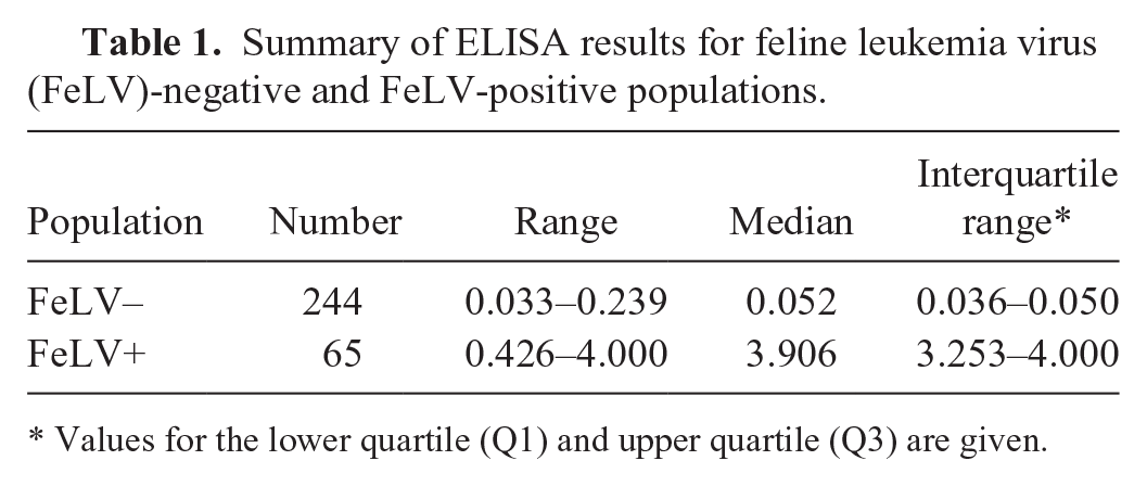
Values for the lower quartile (Q1) and upper quartile (Q3) are given.
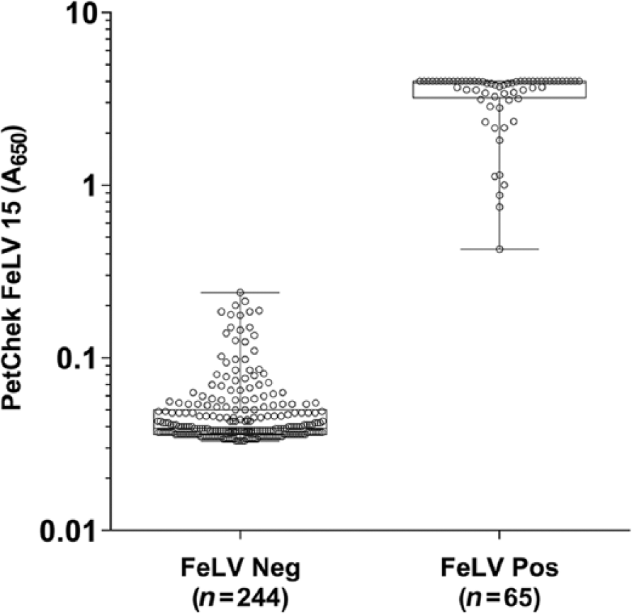
Distribution of PetChek FeLV 15 A650 results for feline leukemia virus (FeLV)-negative and -positive samples. Samples were defined as negative or positive on the basis of concordant results in the FeLV PCR assay and the reference FeLV antigen ELISA. The A650 cutoff for the PetChek FeLV 15 ELISA was 0.300.
Assay validation
Precision
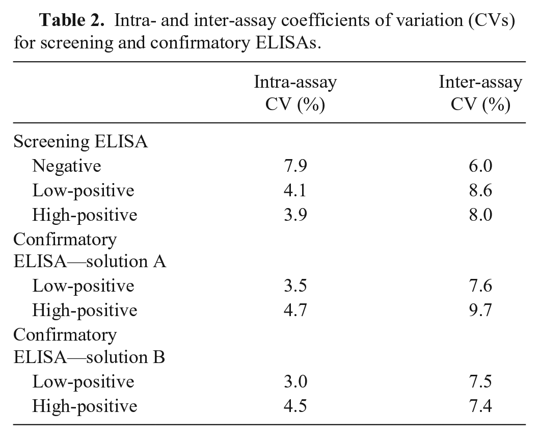
Screening assay precision was determined using 3 samples (negative, low-positive, and high-positive). The intra-assay (within-plate) CV was 3.9–7.9%, and the inter-assay (between-plate) CV was 6.0–8.6% (Table 2). For the confirmatory assay, the same low-positive and high-positive samples were used, demonstrating an intra-assay CV of 3.0–4.7% for the 2 wells and an inter-assay CV of 7.4–9.7% (Table 2).
Intra- and inter-assay coefficients of variation (CVs) for screening and confirmatory ELISAs.
Interference in hemolyzed, icteric, or lipemic samples
The analysis of interfering substances on the screening assay showed no statistically significant difference between negative or positive feline serum controls or when these samples were spiked with hemoglobin up to a concentration of 5.50 g/L (p = 0.181 for negative and p = 0.986 for positive), bilirubin up to a concentration of 0.1441 g/L (p = 0.479 for negative and p = 0.831 for positive), or lipids up to an OD660 of 6.4 (p = 0.136 for negative and p = 0.577 for positive). The same was true of the confirmatory assay when using negative or positive feline serum controls or when these samples were spiked with hemoglobin up to a concentration of 5.50 g/L (p = 0.988 for negative and p = 0.348 for positive), bilirubin up to a concentration of 0.1441 g/L (p = 0.372 for negative and p = 0.749 for positive), or lipids up to an OD660 of 6.4 (p = 0.379 for negative and p = 0.446 for positive).
Analytical sensitivity and specificity
Based on the dilution response of inactivated FeLV and recombinant FeLV p27, analytical sensitivity for p27 antigen was 3.7 ng/mL for inactivated whole FeLV and 1.2 ng/mL for purified recombinant FeLV p27 (Fig. 2). The PetChek FeLV 15 ELISA exhibited no cross-reactivity to related retrovirus antigen from MuLV and feline RD-114 (Fig. 3). Sera from 5 cats immunized with mouse IgG all tested negative (all were screen-positive, non-confirming negatives) in the PetChek FeLV 15 ELISA (Table 3).
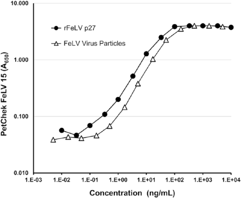
Comparison of PetChek FeLV 15 A650 results between recombinant feline leukemia virus (FeLV) p27 and inactivated FeLV whole virus particles. Dilutions of recombinant p27 antigen and whole virus were prepared in fetal bovine serum to span concentrations ranging from 0 to 10,000 ng/mL and tested using the PetChek FeLV 15 ELISA.
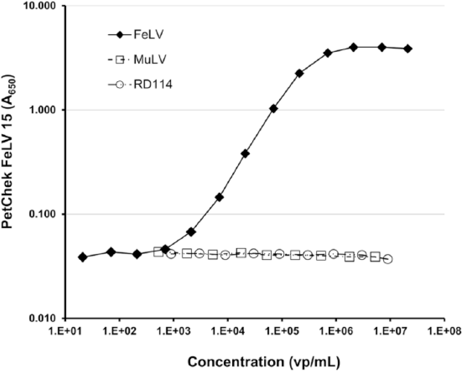
Comparison of PetChek FeLV 15 A650 results for samples containing inactivated RD-114, murine leukemia virus (MuLV), and feline leukemia virus (FeLV). Virus stocks were diluted in fetal bovine serum between concentrations of 0 and 9.00 × 10⁶ virus particles/mL and tested using the PetChek FeLV 15 ELISA.
Screening and confirmatory assay results for samples from feline leukemia virus (FeLV)-positive and FeLV-negative cats immunized with mouse IgG.
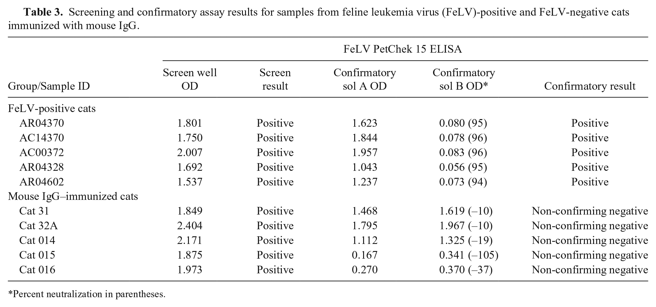
*Percent neutralization in parentheses.
Discussion
The PetChek FeLV 15 ELISA uses operational protocols and instrument-based interpretive criteria suitable for use in high-throughput volume reference laboratories. Our study provides data that validate the assay. The decision to use concordant results of the commercial p27 ELISA to detect soluble antigen and PCR to detect FeLV nucleic acid was done to obtain an unambiguous population of positive and negative samples. Maintaining alignment with both PCR and the commercial p27 ELISA helped to ensure that the appropriate cutoff was established for the PetChek FeLV 15 screening assay. Specificity of the PetChek FeLV 15 ELISA was maintained, in large part, because of the pair of mAbs used in the assay. These antibodies were developed to have high affinity for p27 and have previously been shown to be specific for the p27 of FeLV.14,15 The specificity of these reagents was not impacted by alterations to the assay protocol and was confirmed in our study by testing with closely related feline and murine retroviruses. The PetChek FeLV 15 ELISA also achieved high specificity through the use of the confirmatory assay that is performed on all positive samples identified in the screening portion of the protocol. The inherent burden of a confirmatory assay is to perform with high specificity. The derivation of the confirmatory assay cutoff is based on the previously established standard of a 50% reduction in color for any sample with a positive result in the screening assay (PetChek Feline Leukemia Virus Antigen Test Kit [Screening/Confirmatory], IDEXX Laboratories). As demonstrated with the positive population of field samples in our study, the median reduction in color was 95%, which conforms to the previously established cutoff. The confirmatory assay can also be useful in eliminating false-positives that might arise because of nonspecific interactions, as was the case for the feline samples that reacted with mouse IgG. This is a significant distinction between the PetChek FeLV 15 ELISA and other assays that detect FeLV p27 antigen, given that false-positive reactions associated with anti-mouse activity are known to occur in serotests for FeLV antigen. 12 False-positive reactions related to antibody in cat serum directed against mouse immunoglobulins were found in 10 of 2,830 (0.35%) of sera tested. 12
Operational efficiencies for screening assays are important for large-volume reference laboratories. The method validation of the PetChek FeLV 15 ELISA described in our study demonstrated that operational improvements could be made while maintaining performance consistent with established methods, such as PCR and a commercial ELISA, that have been characterized against the reference standard of virus isolation. The PetChek FeLV 15 ELISA incorporates objective interpretive criteria thereby eliminating operator-to-operator variability encountered with the visual interpretation of current ELISA methods. Additionally, utilization of stop solution and a plate reader allowed for immediate and simultaneous capture of numerical A650 results in a controlled fashion and provided continuous data for analysis. Finally, including a 15-min sample incubation time within the assay protocol allowed more samples to be added to each plate and preserve consistent performance between the first and last sample on the microtiter plate. Results of our study validate that the new microtiter-plate format ELISA is a highly specific, precise, and accurate method for detection of FeLV p27 antigen in feline patient samples and is appropriate for high-throughput applications in a reference laboratory setting.
Footnotes
Acknowledgements
We thank Marko Estrada for running the FeLV real-time PCR assay.
Declaration of conflicting interests
The authors declared the following potential conflicts of interest with respect to the research, authorship, and/or publication of this article: The authors are employees of IDEXX Laboratories, Westbrook, ME.
Funding
The authors are employees of IDEXX Laboratories, Westbrook, ME.
References
Supplementary Material
Please find the following supplemental material available below.
For Open Access articles published under a Creative Commons License, all supplemental material carries the same license as the article it is associated with.
For non-Open Access articles published, all supplemental material carries a non-exclusive license, and permission requests for re-use of supplemental material or any part of supplemental material shall be sent directly to the copyright owner as specified in the copyright notice associated with the article.