
Abstract
The current report describes a case of congenital subcutaneous and intramuscular tumors of the neck and tail base in a 4-week-old female Angus-Charolais crossbred calf. Results of clinical and ultrasound examination are summarized. Biopsy and necropsy findings indicated an infiltrative lipoma. Congenital lipomas are uncommon tumors in bovids. Clinical and morphologic differentials, as well as classification and the possible pathogenesis of congenital neoplasms, are discussed.
Congenital neoplasms are uncommon in animals, especially in bovids. Infiltrative lipoma is a rare variant of lipoma characterized by sheets of well-differentiated adipocytes that readily invade adjacent muscle and fascia. 8 Infiltrative lipomas have been reported most often in dogs and horses. They are locally invasive and have the capacity to infiltrate musculature, fascial structures, articular capsules, tendons, vessels, nerves, and even bones 9,11 but do not metastasize. 5,16 Lipomas are rare in cattle and usually are located in internal organs 7 ; however, 1 description of an infiltrative lipoma in the face of a calf has been published. 4
A 4-week-old female Angus-Charolais crossbred calf was presented at the Clinic for Ruminants and Swine (University of Giessen, Giessen, Germany) because of dyspnea and an enlarging mass on the right side of the neck. According to the owner, the mass was present at birth and had dramatically increased in size (Fig. 1). At the time of presentation, the mass extended from the right ear to the thoracic inlet and from the right jugular vein to the sagittal midline of the neck. The mass had a soft and elastic texture on palpation and was not painful. The overlying skin was intact and mobile. The large mass impaired movement of the neck to the right, and the calf had inspiratory dyspnea. Multiple smaller nodules of the same consistency were detected at the base of the tail.
Ultrasonographic examination of the mass and surrounding tissue revealed muscle, adipose tissue, and fluid accumulation. However, there was no capsule that demarcated the mass from the surrounding soft tissue. Surgical excision of the mass was requested by the owner. Intraoperatively, the mass had a white surface, displayed an elastic to fatty texture, and involved nearly the entire musculature of the right side of the neck extending from the masseter muscle to the scapula. Complete surgical excision of the mass was impossible. Histologic examination of the neoplastic tissue revealed sheets of well-differentiated adipocytes that infiltrated the adjacent connective tissue and musculature. The tentative histologic diagnosis was infiltrative lipoma.
After partial excision of the neoplasm, the general condition of the calf temporarily improved. However, rapid regrowth of the tumor occurred and dyspnea reoccurred. Euthanasia was performed because of the poor clinical prognosis.
At necropsy, an unencapsulated, soft, homogeneous, yellow to white mass was present in the subcutis of the right neck. The mass measured 25 cm × 16 cm × 9 cm; was composed of multiple irregularly sized nodules of adipose tissue separated by septa of connective tissue; and infiltrated the surrounding skeletal muscles, including the brachiocephalic, trapezius, biventer cervicis, longissimus capitis, and spinal muscles. In addition, the neoplasm extended through the sternohyoid and omohyoid muscles to the depth of the right side of the esophagus and trachea
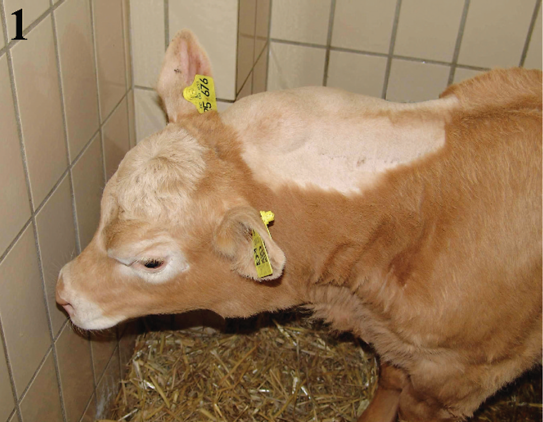
Cervical neoplasm in an Angus-Charolais crossbred calf.

Paramedian sagittal section of a congenital infiltrative lipoma in a calf. The tumor (T) in the subcutis of the right side of the neck is infiltrating the nuchal ligament (a), spinal muscle (b), multifidi muscles (c), thoracic hyoid muscle (d), and acromiohyoid muscle (e). The atlas (1), axis (2), and cervical vertebra (3) are not infiltrated.
(Fig. 2). A similar neoplastic mass was located at the base of the tail and infiltrated the sacrococcygeal musculature. There was no evidence of metastasis or abnormal findings in other internal organs.
Histologically, the morphology of both masses was identical to that of the original surgical biopsy. Both neoplasms were composed of mature adipocytes that were arranged in small lobules, which infiltrated the subcutis, skeletal muscle, and fascia (Figs. 3, 4). Mitoses were not observed. The definitive histologic diagnosis was infiltrative lipoma of the neck and sacrococcygeal region.
Infiltrative lipomas have been well described in dogs, horses, and humans. 5,9,11,12 They are regarded as benign tumors that originate from adipocytes in the subcutis but lack a connective tissue capsule and are locally invasive. These neoplasms may displace, compress, and infiltrate tissues, resulting in clinical signs of disease. 11 The infiltrating lipoma in the calf of the present report was
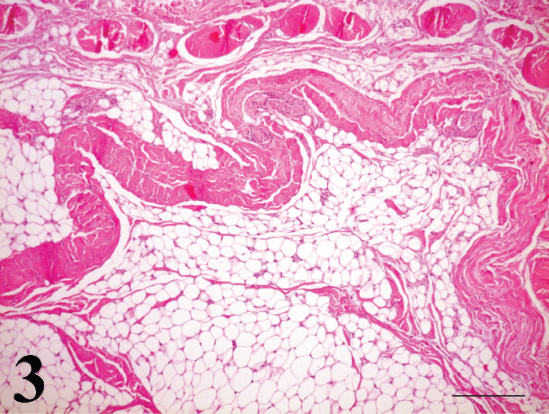
Infiltrative lipoma in a calf. Sheets of well-differentiated adipocytes are invading the adjacent muscle and fascia. Hematoxylin and eosin. Bar = 50 μm.

Infiltrative lipoma in a calf. Skeletal muscle is infiltrated by well-differentiated adipocytes that vary slightly in size and contain small eccentrically located nuclei. Hematoxylin and eosin. Bar = 50 μm.
associated with dyspnea due to infiltration of the ventral cervical musculature and impingement on the trachea. The neoplasm in this calf could not be completely excised, and regrowth occurred rapidly during the postoperative period. In human patients, infiltrating lipomas also are difficult to excise completely and are associated with a recurrence rate of up to 62.5%. 13
Infiltrating lipomas must be differentiated histologically from routine lipomas that lack invasive characteristics and from liposarcomas. Liposarcomas are characterized by a pleomorphic cell population, occasional large nuclei with a prominent nucleolus, and variably sized cytoplasmic lipid vacuoles. 8 Based on cellular morphology, liposarcomas can be further classified as well-differentiated liposarcoma, pleomorphic liposarcoma, and myxoid liposarcoma. 8 They are rare in domestic animals but have been reported in many species, including cows. 15 The neoplasm of the present report lacked the morphologic characteristics of a liposarcoma.
Congenital cutaneous neoplasms occasionally occur in various animal species, including humans. 6,8 Lipoblastomas and lipoblastomatosis are benign mesenchymal tumors of fetal white fat tissue that are almost exclusively observed in young children. 3 Lipoblastomas differ from lipoma or lipomatosis by their cellular immaturity and close resemblance to low-grade liposarcoma, especially its myxoid variant. 10 Two different forms of lipoblastomas occur in humans; the well-circumscribed type is more common than the diffuse and infiltrative variety. Histologically, lipoblastomas contain adipocytes in different stages of maturation. 2 A case report of congenital lipoblastoma in a calf has been published. 14 The neoplasm in the calf of the present report contained mature adipocytes and exhibited infiltration but lacked immature cells and mitotic figures of liposarcoma and lipoblastoma. In addition, the congenital infiltrating lipoma in this calf originated in 2 different regions of the body.
In human neoplasms of adipocyte origin, various chromosomal alterations have been described, and some of these chromosomal changes have been investigated for their diagnostic value. 1 In veterinary medicine, chromosomal and genetic analyses of adipose neoplasms have not been routinely performed and chromosomal abnormalities have not yet been described in adipose neoplasms of animals. However, a genetic predisposition for tumor development cannot be excluded.