
Abstract
An alpaca (Llama pacos), born and raised in Australia, was presented with multiple subcutaneous abscesses. Histological findings indicated a severe necrotizing and histiocytic myositis and cellulitis associated with central caseation and multiple sarcocysts. Ultrastructural examination supported the diagnosis; however, cyst wall characteristics were not consistent with the 2 known species found in alpacas. While seroconversion in camelids is reported to be near ubiquitous, myositis is rare, and this is the first case reported outside of the Americas.
Sarcocystis spp. are apicomplexan protozoa affecting a broad range of vertebrates. Typically, Sarcocystis spp. rely on a 2-stage life cycle involving an intermediate and definitive host. The intermediate host (typically a herbivore or omnivore) becomes infected by ingesting sporocysts excreted in definitive host feces (typically carnivores). 1,2
The presence of microscopic cysts (sarcocysts) is commonly noted in most production species as incidental findings, particularly sheep and cattle. Within Australia, 93% of sheep have demonstrable microscopic cysts within striated muscle, and 97% have serological evidence of exposure. 11 In South America, camelids are similarly prone to infection with Sarcocystis spp., with the incidence of infection in alpacas considered to be ubiquitous by 2 years of age (AC Hung, personal communication, 2010). 8 Similarly, a recent study demonstrated serological evidence for infection in 96% of llamas from Argentina. 10 No such studies have been carried out in Australia. The 2 species that have been identified and associated in the veterinary literature with alpacas are Sarcocystis aucheniae and Sarcocystis lamacanis (AC Hung, pers. comm., 2010). 4,6
Sarcocystis spp. vary greatly in their pathogenicity. In most species, an acute syndrome (Dalmeny disease) is associated with 2 phases of schizogony within endothelial cells of small blood vessels. 1 Experimental studies have characterized the initial hemorrhagic phase, followed by invasion of parenchymatous tissues by first-generation merozoites, and this can be followed by an early, severe inflammatory response. 3 Following the acute phase, tissue cyst formation occurs. In cattle and sheep, degenerating cysts are associated with a rare condition, eosinophilic myositis, 13 which is presumed to be a delayed hypersensitivity reaction. It is this chronic condition that is most visible to producers, since muscular necrosis typically leads to carcass downgrading.
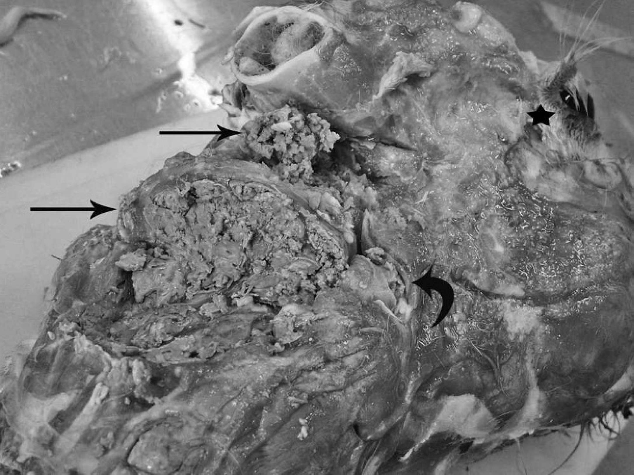
Alpaca; head and neck, skin removed. Multifocal to coalescing extensive zones of caseous necrosis within the musculature (arrows), extending into the dermis. Smaller adjacent focal lesion (curved arrow). Asterisk indicates anterior aspect.
In camelids, disease due to Sarcocystis spp. is considered rare, and aside from the economic impact of carcass downgrading (which is significant in South America), the presence of cysts is considered incidental (Stambaugh A: 2009, Incidental parasites in alpacas in Peru: Lamanema chavezi and Sarcocystis spp. Senior Seminar Paper, College of Veterinary Medicine, Cornell University, Ithaca, NY). 4 A single report of acute sarcocystosis has been published outside of South America, a case with disease manifestation noted in an imported animal transported to the United States. 7 There have been no reports of eosinophilic myositis in alpacas outside of the Americas.
The present case report describes a severe case of necrotizing and histiocytic myositis in an alpaca from southwest New South Wales (NSW), Australia. To the authors' knowledge, this is the first case of myositis associated with sarcocysts in an Australian camelid and was not associated with direct animal importation.
An alpaca from southwestern NSW presented with progressively developing, raised, subcutaneous abscess-like structures located bilaterally along the head and cervical region over a 1-month period. Fine-needle aspirates of one of the masses contained large numbers of eosinophils admixed with lesser neutrophils and mononuclear cells. The findings were suggestive of eosinophilic inflammation.

Alpaca; skin and dermis. A, focally expansile, well-circumscribed abscess filled with abundant necrotic debris (arrow). Bar = 300 μm. B, focal, concentric accumulation of histiocytes (curved arrow) with a central core composed of cellular debris (star). Remnant myofibers are at the center of the abscess (arrowhead). There is a distinct rim of lymphocytes, plasma cells, and lesser eosinophils (straight arrow). Bar = 200 μm. C, intact sarcocyst, in situ. Bar = 300 μm. D, sarcocyst wall and fine detail. Packeting of bradyzoites (curved arrow) by cyst wall projections (straight arrow). Bar = 10 μm.
Concurrently, hematology and biochemistry indicated numerous significant findings. Of these findings, peripheral eosinophilia (6.3 × 10⁹ cells/l, reference [ref.] interval: <2.3 × 10⁹/l), left shift (0.8 × 10⁹/l, ref. interval: <0.1 × 10⁹ cells/l), hyperproteinemia (81 g/l, ref. interval: 51-67 g/l), and hyperglobulinemia (54 g/l, ref. interval: 13-31 g/l) were most significant. The findings were supportive of inflammation but were not suggestive of any specific etiology. Over a period of 1 month, the lesions failed to resolve, and the animal was euthanized. A field necropsy was performed by the referring veterinarian. Innumerable multiple foci of caseating, pale, white, streaking lesions associated with the cranial and cervical musculature were noted (Fig. 1) extending to the intercostal musculature. There were no gross lesions noted elsewhere. The head and multiple fixed tissues were submitted to the laboratory for further investigation. Culture of the caseating lesions for bacterial and fungal pathogens was unrewarding. Tissue for histology was immersed in 10% formalin and processed in a standard manner. Histologically, there was a severe, extensive, and multifocal caseous necrosis, associated with myriad histiocytes and palisading rows of multinucleated giant cells effacing and replacing normal tissue architecture (Fig. 2A, 2B). Moderate numbers of lymphocytes and plasma cells surrounded histiocytic infiltrates, and lesser eosinophils were noted. The inflammatory foci included deep dermal foci extending into and beyond the panniculus muscle. Dense cords of fibrous tissue were noted dissecting the muscle bundles. The large focal necrotic cores were frequently mineralized, and the supporting connective tissue was heavily infiltrated with lymphocytes, plasma cells, and neutrophils. Occasional eosinophils were noted.
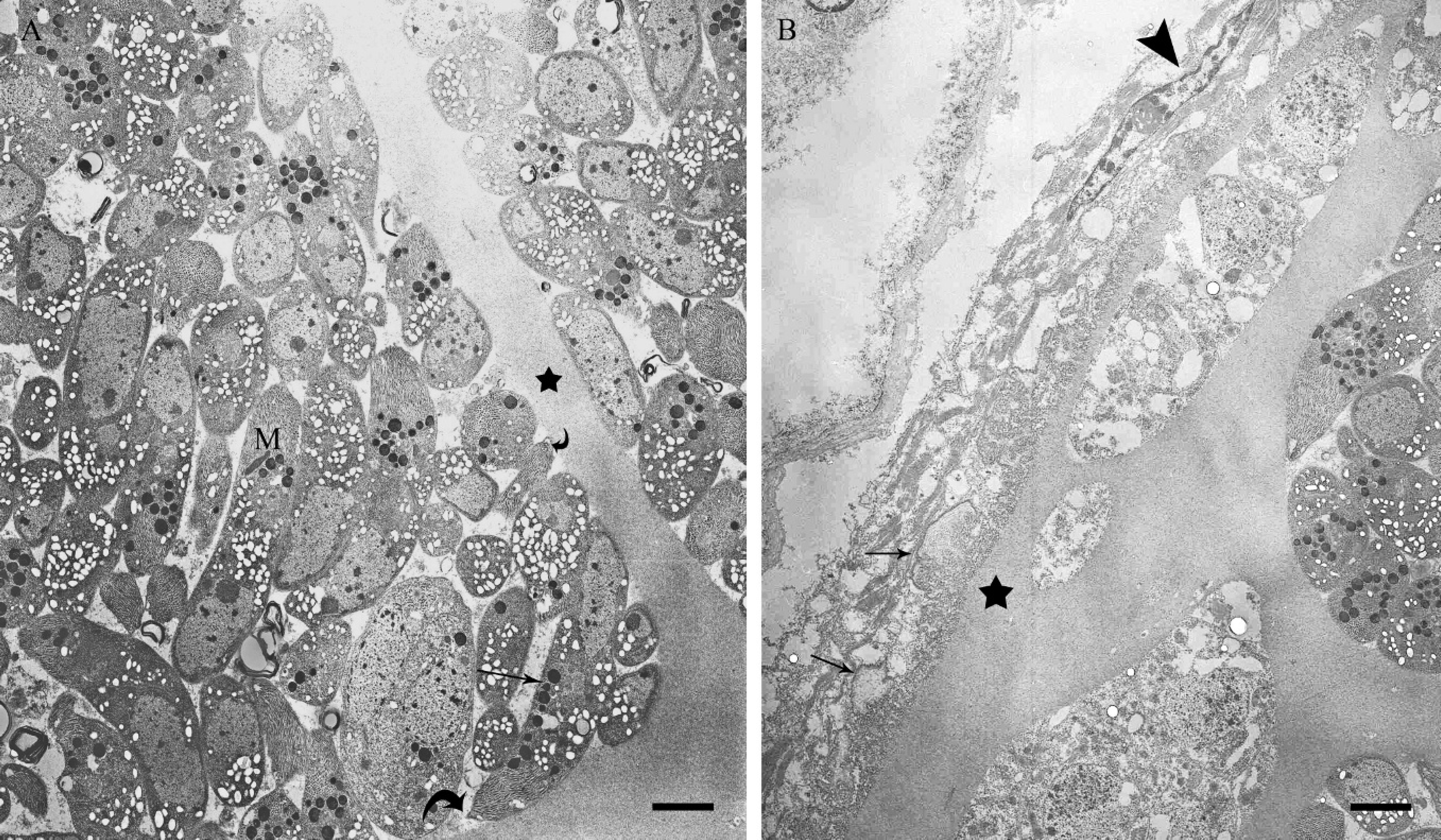
Alpaca; ultrastructure of sarcocyst. A, packeting of bradyzoites by cyst wall projections (star). Individual bradyzoites contain micronemes (M) and amylopectin granules (straight arrow). Cross sections of individual conoids (curved arrows). Bar = 1 μm. B, details of sarcocyst wall. Compressed and elongated host cell nucleus (arrowhead). The numerous irregular cyst wall projections are noted (arrows). The underlying cyst wall ground substance is marked (star). Bar = 1 μm.
Scattered through the tissue were numerous ovoid sarcocysts (Fig. 2C), ranging from 400 to 800 μm along the longest axis. The cysts were surrounded by a 2- to 4-μm, PAS-positive wall and filled with myriad, 1-μm, elongated bradyzoites (Fig. 2D). There was frequent compartmentalization of bradyzoites by cyst wall projections.
Select tissue samples were selected from extant paraffin blocks and processed for ultrastructural examination. Ultrathin sections were mounted on copper grids, stained with uranyl acetate and lead citrate, and examined and photographed using a transmission electron microscope. The cyst was filled with abundant bradyzoites, which were elongate, with distinct micronemes present. A distinct zone of ground substance was noted (Fig. 3A). The tissue cyst was surrounded by an irregular cyst wall with irregular, short, exophytic projections (Fig. 3B).
The term eosinophilic myositis in sheep and cattle is a well-described yet uncommon entity, induced (it is believed) by degeneration of mature sarcocysts. Upon review of the veterinary literature, the degree of eosinophilic infiltration is highly variable, dependent on host species, parasite species, and, perhaps most importantly, the temporal progression of the disease. In a recent publication, sarcocyst-associated myositis was considered eosinophilic to granulomatous and was termed chronic polymyositis, 5 which is arguably a more fitting term for a condition with such wide variations in cellular response.
The incidence of sarcocyst-induced myositis and the associated downgrading of carcasses is surprisingly low considering the near ubiquity of seroconversion in ruminants and camelids and the presence of incidental microscopic cysts in necropsy material. In the current case, there was a relative lack of eosinophils compared with a more severe histiocytic to granulomatous response. Considering the initial tissue and peripheral eosinophilia noted on cytology and hematology, it is likely that the histiocytic predominance reflected the temporal progression of the present case between initial cytology and necropsy.
The potential significance of sarcocyst-induced myositis in camelids is high. In an early survey of alpacas from Peru, 100% of necropsied animals were found to carry microscopic cysts. 6 There are published reports in most camelid species of near ubiquitous seroconversion. 9,10,12 However, even in these instances, actual acute disease (Dalmeny-like disease) or myositis is rare. A single case report describes the condition. In that case, the affected animal was imported from South America. 7
Two species of sarcocysts (S. aucheniae and S. lamacanis) have been reported in the alpaca. In the present case, the histological findings, the prominent septae, and tightly packed bradyzoites of the cyst microscopically are similar to the previous published report of Dalmeny disease in an alpaca. 7 However, the ultrastructural findings in the current case varied considerably from the description of either S. aucheniae or S. lamacanis. In the present case, the cyst wall was regularly undulating with exophytic, short projections. In contrast, S. aucheniae displays an undulating, anastomosing cyst wall. Further molecular evaluation is required to identify this particular agent.
The current case is of significance to the camelid industry in Australia. The animal was born and housed at a single alpaca stud farm, with no history of movement. The implication is that this parasite, which is dependent on an intermediate and then final host interaction, is present in Australia. Importantly, the ultrastructural findings in the current case are significantly different from the 2 known sarcocysts that infect alpacas, emphasizing the need to determine the identity of this particular parasite. Considering the regional prevalence of both wild dogs and working dog populations and the widespread presence of poorly identified sarcocyts in native marsupial hosts, it is entirely conceivable that this interaction could become more widespread. Despite the significance of high-quality fiber to the Australian alpaca industry, the lack of data relating to sarcocystosis is absolute, and to date, no data exist examining the significance of either acute or chronic disease. The significance for veterinary diagnosticians is important, as camelid submissions are increasing in frequency.