
Abstract
The objectives of our study were to describe the pathology of ear hematomas in swine and to add to the comprehension of the pathogenesis of this condition. The pathogenesis of aural hematomas has been studied mainly in dogs; however, disagreements exist about the precise anatomic location of the hemorrhage. Sixteen pigs with ear hematoma at various stages of development were included in this study. The pigs were submitted for routine autopsy for various and unrelated reasons over a period of several years. Based on gross examination, the 16 cases of aural hematomas were subjectively classified as acute (n = 6), subacute (n = 3), and chronic (n = 7). The age of the animals at the time of autopsy ranged from 2 weeks to adulthood, with all acute cases being <7 weeks of age. Morphologic examination of all acute cases revealed that the hematoma developed predominantly in a subperichondral location on both sides of the cartilaginous plate simultaneously. Within these same cases, there were also some areas in which blood-filled clefts had formed within the cartilage itself. Besides fibroplasia, neoformation of cartilage was found to represent a significant part of the repair process. All chronic cases were characterized on cross-section of the ear by the presence of at least 2 distinct, wavy, focally folded, and roughly parallel plates of cartilage separated from each other by fibrous tissue.
Introduction
Ear hematomas in pigs are a common problem in modern swine herds. The lesion develops on the pinna and is often referred to as auricular hematoma, aural hematoma, or othematoma. In pigs, the external ear is composed of the auricle (pinna) and the external ear canal (external auditory meatus). The auricle consists of a flat plate of elastic cartilage covered on each side by thin skin. The skin of the convex or outer surface of the auricle is hairier, thicker, and less tightly attached to the auricular cartilage than the skin of the concave or inner surface. Two to 3 cutaneous ridges are also present on the inner surface. Vascularization of the pinna is provided by the caudal auricular artery and vein.3,4,8
Pigs and dogs with pendulous ears are reported to be particularly prone to hematoma formation.2,13 In these 2 species, excessive head shaking or scratching from aural discomfort (e.g., otitis externa and media, scabies, feed particles in the ears) are often suggested as initiating traumatic events (Sunstrum J, et al. Treatment of ear haematomas in swine. Proc Am Assoc Swine Pract 2005;36:27–28, http://tinyurl.com/jr8w9mg). 1 In swine, othematomas also commonly result from injuries caused by fighting and biting between pigs in a stressful environment (Sunstrum J, et al., 2005) 2 ; although the condition occurs at various ages, the maximal frequency is observed in 4–8-week-old pigs.2,12
In animals, the pathogenesis and pathology of auricular hematomas have been studied mainly in dogs, in which the lesion is considered to develop mainly on the concave surface of the external ear.1,13 There are, however, some disagreements about the precise anatomic location of the hematoma in relation to the cartilage of the pinna.1,13 Subcutaneous, subperichondral, and intrachondral sites have all been suggested, respectively, as probable locations for the initial damage and thus origin of the hematoma. 13 The objectives of our study were to describe the gross and microscopic pathology of ear hematomas in swine, a species in which little has been reported on this particular aspect of the condition, and to add to the comprehension of the pathogenesis of this condition.
Materials and methods
A total of 16 pigs with ear hematomas at various stages of development were included in our study. These pigs were submitted for routine autopsy for various and unrelated reasons over a period of 11 years. The external ears of 2 unaffected nursery pigs also submitted for autopsy were used as controls. Based on gross examination, the 16 cases of aural hematoma in which the age of the lesion was unknown were subjectively classified as follows. 1) Acute cases (n = 6): the external ear was swollen and fluctuant. On cut section, there was partially coagulated blood (recent hematoma). 2) Subacute cases (n = 3): the external ear was swollen but not significantly distorted. On cut section, the hematoma may have been partially dehemoglobinized, and there was some evidence of early tissue repair such as fibroplasia (organizing hematoma). 3) Chronic cases (n = 7): the external ear was variably thickened, firm, distorted, and folded (cauliflower-shaped). On cut section, the pinnal cartilage was very irregular, and there was obvious fibroplasia.
Detailed gross and microscopic examination of the affected ears was conducted to try to localize the initial site of hemorrhage and to characterize the repair process that took place in this condition. For light microscopic examination, up to 5 cross-sections of the ear, at the level of the hematoma, were obtained for each animal depending on the size of the lesion. Samples were fixed in 10% neutral buffered formalin, embedded in paraffin, cut at 4 µm, and stained with hematoxylin–eosin–phloxine–saffron (HEPS). Selected sections were also stained with toluidine blue to better highlight the position of cartilage.
Results
The age of the affected animals ranged from 2 weeks up to adulthood (breeding animals), with all acute cases being <7 weeks of age, with a mean of 4.6 weeks of age. All cases but 1 were unilateral, with a nearly equal distribution between the right (n = 7) and left (n = 8) ear. The animal affected bilaterally had chronic lesions of both ears.
Control pigs
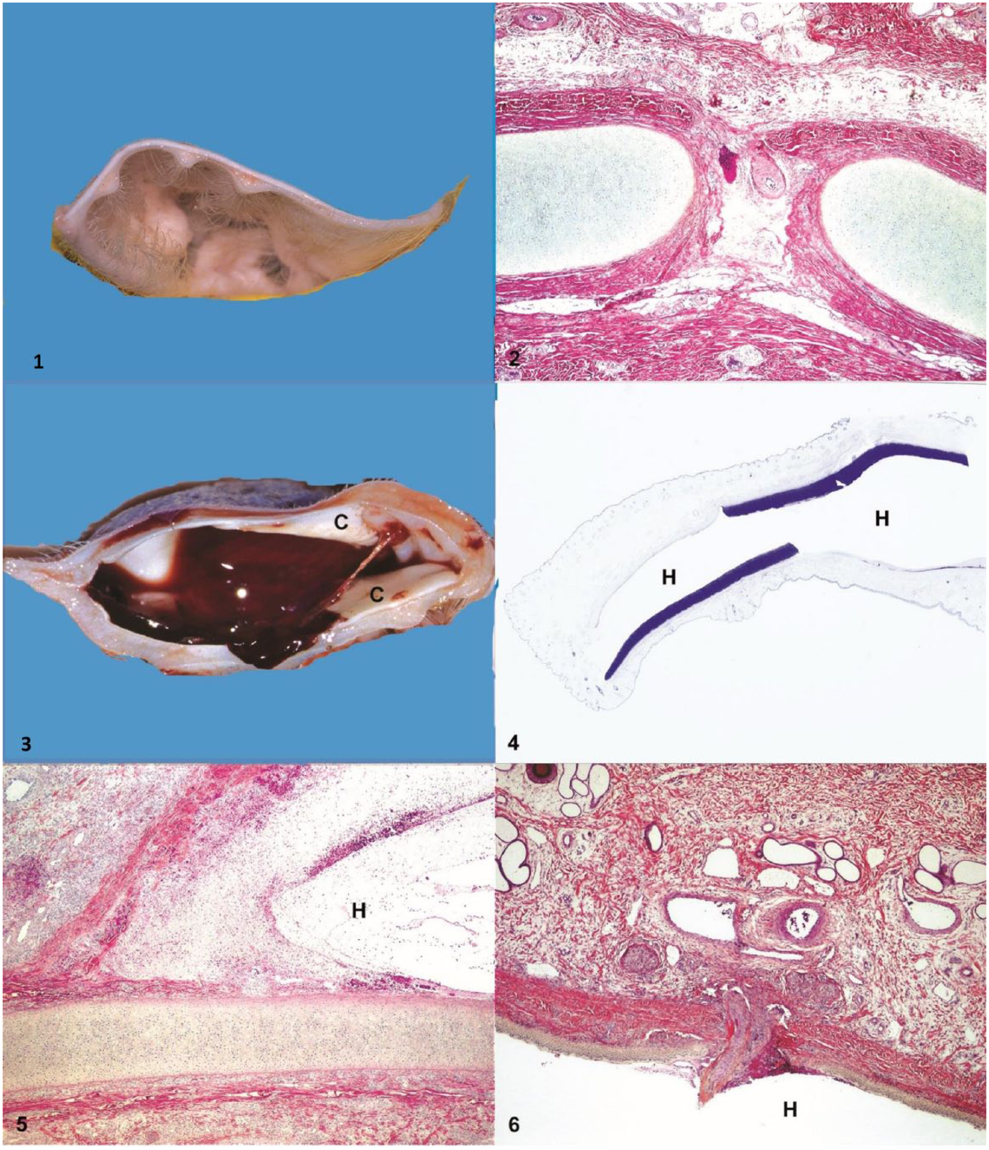
Control pigs were selected based on the absence of gross and microscopic lesions in the pinna. The skin was normal on both sides of the auricle, although hairier and thicker on the convex or outer surface of the auricle. Cutaneous ridges were more prominent near the base of the pinna and disappeared at the extremity of the ear (Fig. 1). Throughout the auricular cartilage, there were foramina that could be seen with the naked eye as small circular depressions once the skin was removed. Branches of the caudal auricular artery and vein, which ran on the convex surface of the ear, traversed these cartilaginous perforations to vascularize the inner surface of the pinna (Fig. 2).
Pigs with aural hematomas
Macroscopically, in all acute cases examined, the cleavage line produced by the hematoma was located in an irregular manner on both sides of the cartilaginous plate, convex or concave, within the same ear (Figs. 3, 4). Although the hemorrhages occurred on both sides of the aural cartilage, overall a larger volume of blood was observed on the concave side of the pinna. Microscopic examination of these cases showed that an important part of the tissue detachment produced by the hemorrhage took place between the perichondrium and the cartilage on both sides of the pinna simultaneously. In these cases, there was also some evidence of intrachondral cleavage that appeared either frankly intracartilaginous or more commonly very close to the perichondrium, leaving only a few rows of chondrocytes attached to the perichondrium (Figs. 5, 6). Dermal and subcutaneous tissues surrounding the aural cartilage were often edematous. Another significant change observed in 2 cases with acute lesions was the presence of thrombosed blood vessels crossing the cartilaginous foramina in the area of the hematoma.
Microscopically, one of the earliest repair processes observed in aural hematomas was the proliferation of fibrovascular (granulation) tissue in variable amounts at the interface of blood and cleaved connective tissues. In some cases, this tissue was focally and mildly infiltrated by a mixture of neutrophils and mononuclear leukocytes. Another relatively early change was the neoformation of cartilage that occurred either from the detached perichondrium or adjacent to the ruptured edge of the cartilaginous plate (Fig. 7). The newly formed cartilaginous tissue was often irregular and of variable thickness and maturity. The repair processes described above were observed in all subacute cases as well as in most acute cases, although more focally and at an earlier stage of development.
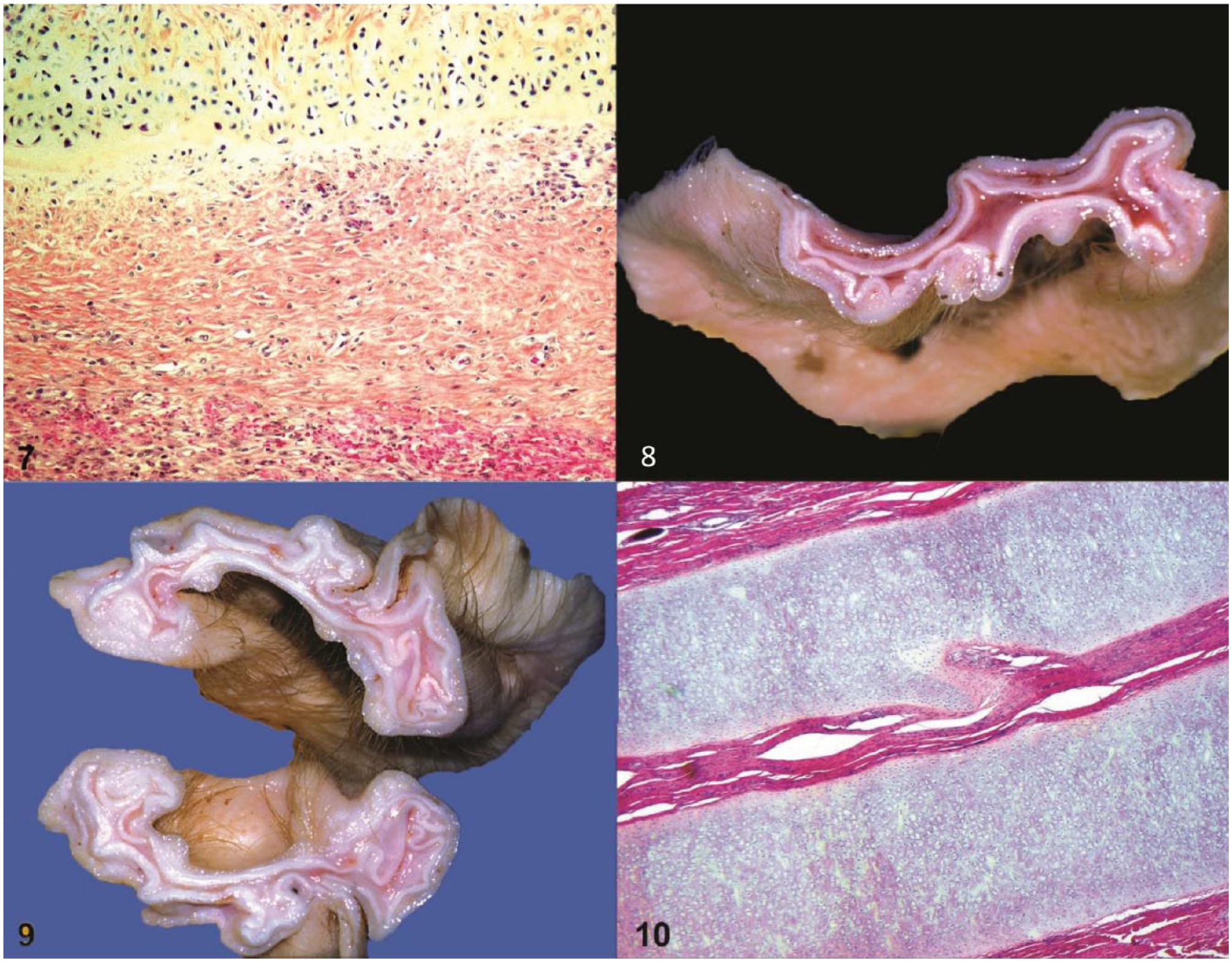
All chronic cases were characterized grossly, on cross-section of the ear, by the presence of at least 2 distinct, wavy, focally folded, and roughly parallel plates of cartilage separated by fibrous connective tissue. Both of these cartilaginous plates were often partially interrupted and appeared either similar to or thicker than the normal aural cartilage (Fig. 8). Some lesions appeared more complex focally with several sheets of randomly oriented cartilaginous tissue (Fig. 9). Some islands of cartilage were also occasionally observed distant from the main cartilaginous plates. Histologically, the appearance of the cartilaginous tissue varied greatly, ranging from well-organized and differentiated (Fig. 10) to irregular and more immature cytologically. In most chronic cases, both patterns of cartilaginous differentiation were present multifocally.
Discussion
The gross and histologic examination of these 16 cases of porcine ear hematoma at different stages of development was helpful in determining the likely pathogenesis of the condition, namely the initial location of the hemorrhage and the repair processes that eventually take place. To our knowledge, there are no other similar published studies in swine. In dogs, othematomas are reported to develop mainly on the concave side of the pinna.1,13 It has been suggested that this seemingly higher frequency on the concave side of the ear flap may result from the fact that the skin and subcutis are thinner in this area, making the hematoma more visible. 6 Observations from our study showed that although hemorrhages appeared globally more extensive on the concave side, the hemorrhages, in fact, occurred on both sides of the pinna simultaneously.
Opinions concerning the precise anatomic location of hematoma in dogs in relation to auricular structures have differed. The hemorrhage has been reported to occur subcutaneously,5,8 subperichondrally, 7 or within the cartilage itself.1,6,11 A subperichondral location appears to be the rule for hematoma auris in humans but this issue has been challenged and has not been resolved. 9 Examination of the acute cases in our study indicates that the formation of the hematoma in pigs takes place in both subperichondral and intrachondral locations. Considering these 2 locations, it is likely that the hemorrhages resulted from torn vascular branches at the points where they enter, pass through, or emerge from the foramina of the cartilage rather than from the larger blood vessels from which they derive and that run over the convex surface of the ear. The intrachondral cleavage, when present in a given section, often occurred near the perichondrium, leaving only a few rows of cartilaginous cells attached to it. Histologically, it was sometimes difficult to determine whether this represented remnants of the torn aural cartilaginous plate or a reactive perichondral regeneration of cartilage because the precise age of the hematomas was unknown. Chondrogenesis has been detected as early as day 4 following experimentally induced subperichondral ear hematoma in rabbits. 10 Neoformation of cartilage, as part of the reparative process, was found to be a constant and significant feature among all chronic cases examined in this series. This is consistent with the location of the tissue detachment produced by the hemorrhage. Subcutaneous aural hematomas, as opposed to those originating from the perichondrium or cartilage, would not result in neoformation of cartilage as demonstrated experimentally in rabbits. 10
Chronic cases in our study were all characterized grossly, on cross-section of the pinna, by apparent “duplication” of the aural cartilage. When examined at this stage of development, an observer could be misled to conclude that it is inevitably the sequel of intrachondral hemorrhage; however, similar changes could also be the end result of a subperichondral hematoma. Islands of cartilage occasionally observed at some distance from the main cartilaginous plates could have been the result of chondroprogenitor cells displaced at the time of hematoma formation. Distortion of the pinna, which becomes hard and cauliflower-shaped, as observed in all chronic cases of our study, is likely caused by a combination of several factors including myofibroblastic contraction of maturing granulation tissue and the presence of excess cartilaginous tissue (preexisting plus neoformed) often multifocally folded on itself. It has also been suggested that the separated perichondrium may retract and act as a “bow string,” which gradually folds back the original cartilage causing a buckle over the hematoma. 10
Under farm conditions, treatment of othematomas in pigs is not usually attempted or likely to be successful because treated ears will most often become infected and will heal more slowly than untreated ears (Sunstrum J, et al., 2005). There are, however, some exceptions in which treatment should be instituted, namely when a large hematoma impedes food intake and alters the general condition of the animal. There are some limitations to our study associated with the relatively small sample size, especially for subacute cases, because this lesion is not routinely investigated or sampled at autopsy. Furthermore, its retrospective nature somewhat limits the scope of lesions that can be examined. A prospective, on-farm study would likely yield othematomas at earlier and different stages, and thus may provide additional information, especially with regards to pathogenesis.
Footnotes
Acknowledgements
We thank Marco Langlois and Jacques Lagacé for their photographic assistance.
Authors’ contributions
R Drolet and S D’Allaire contributed to conception and design of the study, and contributed to acquisition, analysis, and interpretation of data. P Hélie contributed to analysis and interpretation of data. All authors drafted the manuscript; critically revised the manuscript; gave final approval; and agreed to be accountable for all aspects of the work in ensuring that questions relating to the accuracy or integrity of any part of the work are appropriately investigated and resolved.
Declaration of conflicting interests
The author(s) declared no potential conflicts of interest with respect to the research, authorship, and/or publication of this article.
Funding
The author(s) received no financial support for the research, authorship, and/or publication of this article.