
Abstract
In South America, capybaras (Hydrochoerus hydrochaeris) as well as coatis (Nasua nasua) are the reservoir hosts of Trypanosoma evansi. Capybaras from a T. evansi nonendemic area in the State of São Paulo, southeastern Brazil, were culled because of an ongoing outbreak of Brazilian spotted fever; serum samples from these capybaras were tested for antibodies to T. evansi. Of the 172 sera tested, 17 (9.9%) were seropositive by card agglutination test, with antibody titers of 1:8–1:128; 14 (8.1%) of these 17 seropositive sera were also seropositive by indirect fluorescent antibody test, with antibody titers of 1:16–1:256. Both serologic techniques proved to be efficient, with similar results for detection of antibodies to T. evansi in capybaras from a nonendemic area in Brazil.
Trypanosoma evansi is a flagellate protozoan parasite that can infect many domestic and wild animals, including horses, dogs, cats, rabbits, capybaras, ring-tailed coatis, cattle, buffalo, armadillos, and occasionally humans.4,6,8,11 This protozoan is the agent of trypanosomosis, a disease with broad distribution in Africa, Asia, and Latin America.4,6,11 Trypanosoma evansi trypomastigotes circulate in the blood of hosts and can be transmitted mechanically by blood-sucking insects such as tabanid flies of the genera Tabanus, Chrysops, and Haematopota.4,6 Transmission has also been reported by bites of vampire bats, oral transmission to dogs fed T. evansi–infected tissues, and iatrogenic transmission through contaminated needles.4,6,11 Two forms of the disease have been reported in domestic animals in Brazil: the acute, usually fatal form of the disease characterized by intermittent fever, widespread subcutaneous edema, progressive anemia, blindness, lethargy, and hemostatic alterations; and the chronic form characterized by weight loss, incoordination, and neurologic signs. 4 The chronic form also affects wild animals such as capybaras (Hydrochoerus hydrochaeris) and South American (ring-tailed) coatis (Nasua nasua), which can develop clinical signs similar to domestic animals4,11; however, infected capybaras are usually asymptomatic. Capybaras and coatis are considered to be reservoirs of T. evansi9,11,14,17 and are regarded as sources of infection for domestic animals.12,18
Detection of T. evansi infection can be challenging by light microscopy given the chronicity of infection, fluctuating levels of parasitemia, and parasite sequestration in tissues.2,5 Serologic tests that have been used to detect T. evansi infection include the indirect fluorescent antibody test (IFAT), enzyme-linked immunosorbent assay (ELISA), card agglutination test (CATT/T. evansi), an immunochromatographic test (Surra Sero K-SeT), and polymerase chain reaction (PCR).2 –5 The commercial CATT/T. evansi test kit, with a sensitivity of 80.2% and specificity of 98.5%, 6 has been used in studies for the detection of the flagellate in horses and camels.2,10
In Brazil, trypanosomosis has been restricted to the state of Mato Grosso (Pantanal wetland), in the Central-West region of the country. Outbreaks or isolated cases of T. evansi infection have also been reported in other areas of the country including north, south, and southeast Brazil.4,16,20 Infection by T. evansi in capybaras in Brazil has been reported only in the Pantanal, a unique biome characterized by a large wildlife biomass and abundance of tabanids.8,19 However, in other Brazilian regions, authors describe the presence of capybaras on farms with T. evansi outbreaks in horses, and suggest that capybaras may be the parasite reservoir.4,16,18,20
A large number of capybaras were culled in an area (State of São Paulo, southeastern Brazil) endemic for Brazilian spotted fever, a zoonotic tick-borne disease caused by Rickettsia rickettsii. 13 We had the opportunity to test serum samples from these animals for antibodies against T. evansi. This is a trypanosomosis nonendemic area, as cases of T. evansi infection have not been reported in this Brazilian state to date. Before being culled, blood samples were collected, and the sera was separated by centrifugation, and then stored frozen at −20°C until tested. Two serological tests were used to detect antibodies against T. evansi.
The commercial card agglutination test CATT/T. evansi a was used to determine the levels of serum antibodies reactive against T. evansi antigen according to the manufacturer’s instructions. 2 IFAT was run with minor changes to the methodology. 11 Slides were prepared with trypomastigotes of T. evansi (±30/well) obtained in blood from infected rats, and purified with the aid of a diethylaminoethyl cellulose column. Sera were previously diluted in phosphate buffered saline (pH 7.2) up to 1:16. Sera were incubated for 30 min at 37°C in a humidified chamber. A secondary antibody, an anti–guinea pig immunoglobulin G conjugated to fluorescein, b was added, and incubated for 30 min at 37°C in a humidified chamber. Positive and negative controls used were those provided by the manufacturer. Horse, sheep, and rat serum samples that tested positive for T. evansi in other studies were used as positive controls in the CATT/T. evansi test and IFAT used in our study. Additionally, serum samples from horses positive for Trypanosoma vivax and rats positive for Trypanosoma cruzi were used to check cross-reactivity.
No clinical signs were observed in any of the capybaras tested; 17 (9.9%) of these animals had antibodies reactive against T. evansi by CATT/T. evansi; 14 (8.1%) of these 17 had antibodies reactive against T. evansi by IFAT. The T. evansi antibody titers ranged from 1:8 to 1:128 by CATT/T. evansi, and 1:16 to 1:256 by IFAT (Table 1). Equine, ovine, and rat serum samples that in other studies tested positive for T. evansi by PCR and on blood smears (unpublished data) were used as positive controls in the CATT/T. evansi test and IFAT used in our study, confirming the sensitivity and specificity of the techniques for different vertebrate species. Equine serum samples positive for T. vivax by PCR and rat serum samples positive for T. cruzi by blood smear on other occasions were both negative by CATT/T. evansi. These results demonstrate the lack of cross-reactivity in this test.
Capybara (Hydrochoerus hydrochaeris) serum samples reactive against Trypanosoma evansi in a nonendemic area of the state of São Paulo, southeastern Brazil by card agglutination test (CATT/T. evansi) and indirect fluorescent antibody test (IFAT).
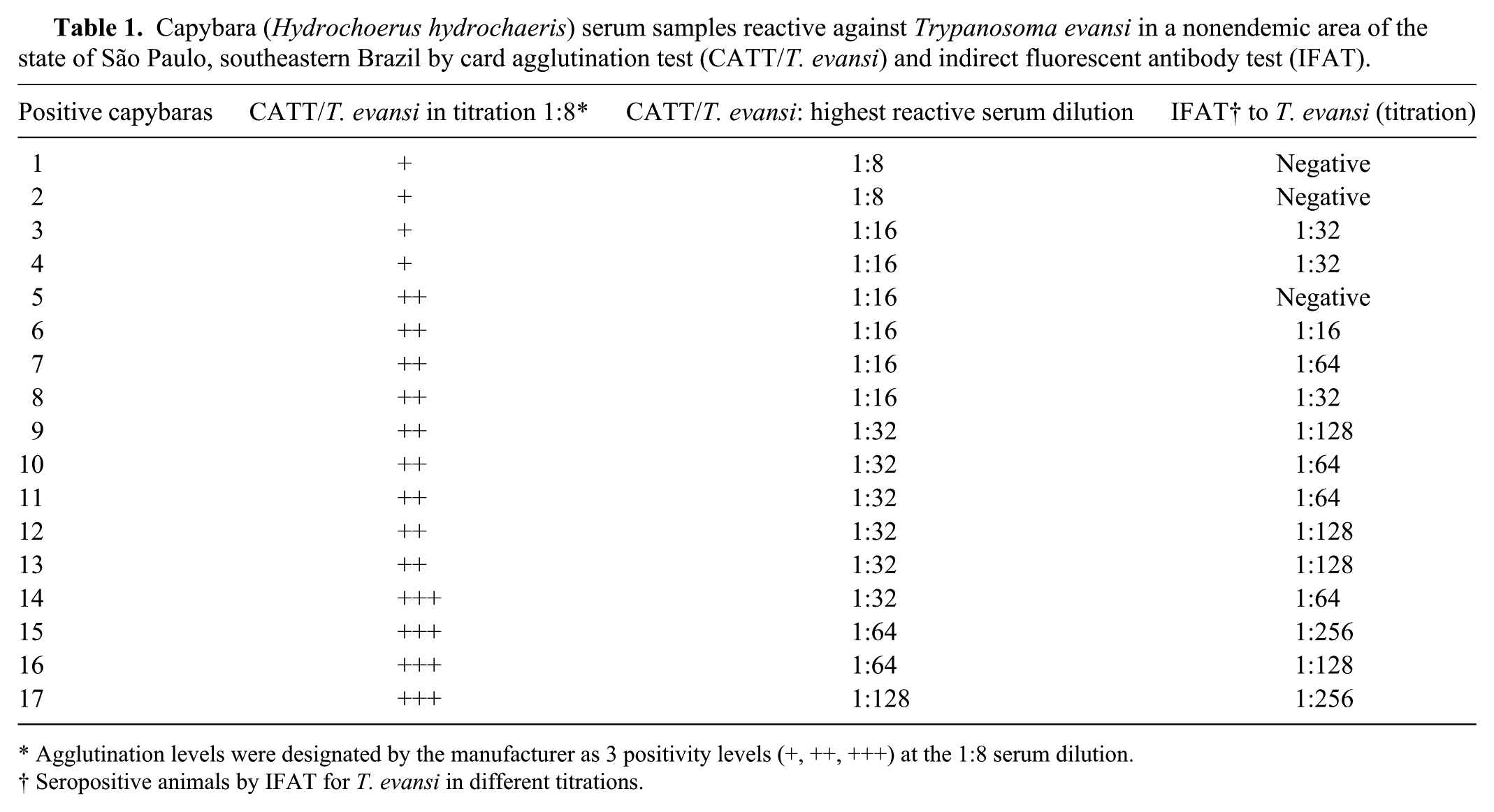
Agglutination levels were designated by the manufacturer as 3 positivity levels (+, ++, +++) at the 1:8 serum dilution.
Seropositive animals by IFAT for T. evansi in different titrations.
The CATT/T. evansi technique was chosen for our study because of its high specificity and ease of use. Our study revealed that 9.9% of the capybaras were seropositive for T. evansi by CATT/T. evansi. A similar result (8.1%) was obtained when a different technique (IFAT) was used. These results show that capybaras from this area in southeast Brazil (nonendemic for trypanosomosis seroreact against T. evansi. In Poconé County (Pantanal of Mato Grosso, Brazil), a region where the disease is endemic in Brazil, researchers using CATT/T. evansi found that 22% of capybaras were positive. 8 Capybara infection with T. evansi has been reported in the state of Mato Grosso, Pantanal complex, using IFAT (positivity of 33.3% 11 and 27.0% 19 ). T. evansi infection has been reported not only in capybaras from Brazil but also in capybaras from other South American countries, including Argentina, Colombia, Peru, and Venezuela.1,7,14,15 Capybaras are considered reservoir hosts of T. evansi in South America,1,7,8 and if parasitemia were only at low levels, they may be asymptomatic carriers.9,14 There are a few reports of clinical disease and death of capybaras resulting from T. evansi infection, but those are anecdotal and undocumented. 18
Based on the findings of this study, asymptomatic capybaras in southeastern Brazil have antibodies against T. evansi. This finding could not be supported by other techniques, such as blood smear and PCR, in our study, because we only had access to sera from an unrelated study. 13 To our knowledge, there is only 1 previous report of T. evansi infection in southeastern Brazil. The diagnosis was based on PCR results and response to therapy, and the few horses affected possibly became infected after a stallion from southern Brazil was brought to the farm. 16 Tropical regions such as southeastern Brazil have a large number of blood-sucking insects (tabanids, mainly) that can act as vectors of T. evansi. Capybaras could serve as reservoirs of this infection in southeast Brazil.
Footnotes
Authors’ note
This project was authorized by the Environment State Secretary of the state of São Paulo, Brazil (authorization no. 96/2012) and was approved by the Ethical Committee of Animal Use of the Faculty of Veterinary Medicine of the University of São Paulo, Brazil (protocol no. 3104/2013).
Authors’ contributions
AS Da Silva contributed to conception and design of the study; contributed to acquisition, analysis, and interpretation of data; drafted the manuscript; gave final approval; and agreed to be accountable for all aspects of the work in ensuring that questions relating to the accuracy or integrity of any part of the work are appropriately investigated and resolved. FS Krawczak contributed to conception and design of the study; contributed to acquisition, analysis, and interpretation of data; drafted the manuscript; and gave final approval. JF Soares contributed to acquisition, analysis, and interpretation of data; critically revised the manuscript; and gave final approval. V Klauck and R Pazinato contributed to analysis of data; drafted the manuscript; and gave final approval. A Marcili contributed to analysis and interpretation of data; critically revised the manuscript; and gave final approval. MB Labruna contributed to conception and design of the study; contributed to acquisition, analysis, and interpretation of data; critically revised the manuscript; and gave final approval.
a.
Prince Leopold Institute of Tropical Medicine, Antwerp, Belgium.
b.
Sigma-Aldrich, St. Louis, MO.
Declaration of conflicting interests
The author(s) declared no potential conflicts of interest with respect to the research, authorship, and/or publication of this article.
Funding
The author(s) declared that this work received financial support from FAPESP (Fundação de Amparo à Pesquisa do Estado de São Paulo) and CAPES (Coordenação de Aperfeiçoamento de Pessoal de Nível Superior).