
Abstract
An outbreak of goiter with high morbidity and mortality in a flock of budgerigars (Melopsittacus undulatus) in California is described. Forty-five out of 400 adult birds exhibited signs of illness, weight loss, and enlargement in the crop area; 15 of the 45 birds died over a 2–3-month period. Diet consisted of a commercial mixture with the addition of broccoli, whole oats, and carrots, but no minerals or supplements. Six budgerigars were subjected to necropsy; all 6 birds had severely enlarged thyroid glands. Thyroid follicular hyperplasia was histologically observed in all birds examined, while granulomatous thyroiditis and microfollicular adenoma were observed in 2 birds, respectively. Virological, bacteriological, parasitological, and heavy metal analyses were negative or within normal limits. The total iodine in the thyroid glands of affected birds was measured by inductively coupled plasma–mass spectrometry. Following iodine supplementation and removal of broccoli from the diet, the owner reported weight gain and a reduced death rate among clinically affected birds; no additional birds became sick. The presence of broccoli with its iodine-binding ability and the complete lack of added minerals in the diet of these animals were thought to be the predisposing factors for the outbreak in the present study. Outbreaks of goiter accompanied by high mortality are rare in any species and, to the best of the authors’ knowledge, have not been described previously in any avian species. Recognition of this condition may help improve medical, welfare, and trade standards concerning this species.
Introduction
Goiter (follicular thyroid hyperplasia) has been reported in its congenital and acquired forms, in human beings and in most domestic 6 and some wild mammalian and avian species, 17 with widely varying interspecies frequency. Goiter is often the result of dietary iodine deficiency 7 but has also been attributed to consumption of goitrogenic substances, 13 toxicity by iodine and other substances, 11 and to hereditary factors, including autosomal recessive inheritance in goats. 20 It remains endemic in some areas because of a lack of, or despite, iodine supplementation. 21
Goiter outbreaks have been reported in human beings, 10 albeit with decreasing frequency, but are infrequently reported in other species. Such reports include outbreaks in cattle in Japan 32 and in sheep in Slovakia 16 and Australia. 8 Outbreaks accompanied by high mortality are rare in any species and, to the best of the authors’ knowledge, outbreaks of goiter have not been described previously in any avian species. Goiter has been reported in psittacines (budgerigars and cockatiels) in the form of individual cases,25,28,30 and anecdotal evidence suggests that psittacines are more susceptible to goiter22,23,27 than other avian species, with a few reports suggesting that the prevalence of goiter in these birds is higher than in other avian species.3,4,12 However, these reports lack epidemiologic analysis and/or interspecies prevalence comparisons. For example, a 1963 study on 129 budgerigar submissions reported that 23.8% of the birds died as a result of thyroid dysplasia attributed to iodine deficiency in the seed mixtures; however, these birds were exclusively individual pet submissions examined in one center over time and no data on other avian species was provided. 4 In another analysis of 257 individual budgerigar postmortem examinations, the most common disease reported was neoplasia of the gonads, kidneys, and fat, while hepatitis and focal hepatic necrosis, thyroid dysplasia, and septicemia were also common 2 but less frequent. A Berlin Zoo study of 3,314 postmortem examinations of zoo birds from 18 avian orders allowed interspecies prevalence comparisons, and the average incidence of goiter was 1.2%; the highest rates were found in orders Falconiformes (6.6%), Phoenicopteriformes (3.9%), and Ciconiiformes (2.8%). 31 Furthermore, in a California survey of nearly 12,500 avian cases, 26 only 30 individuals from various species had a diagnosis of thyroid follicular hyperplasia, 29 of which also had other diagnoses, and only 4 psittacines were recorded, including 2 budgerigars, 1 lovebird, and 1 cockatoo. Among other avian species, goiter has been reported in chickens, 15 pigeons, 17 geese, 18 penguins, 24 great cormorants, 9 white backed vultures, 14 southern caracara, 14 and black stilts. 1 The current study describes an outbreak of severe thyroid hyperplasia with high morbidity and mortality in a large flock of young adult budgerigars (Melopsittacus undulatus).
Materials and methods
Clinical history
Ten live (4 male, 6 female; birds 1–10) and 2 dead (female; birds 11 and 12), approximately 1-year-old budgerigars were submitted for necropsy and diagnostic workup to the California Animal Health and Food Safety Laboratory (CAHFS; San Bernardino, California) in January 2013. Ten additional birds (5 live, 5 dead; birds 13–17 and 18–22, respectively) were submitted on a 4-month post–initial diagnosis follow-up. The flock had a 2–3-month history of illness, weight loss, and high mortality with 30 out of approximately 400 adult birds (7.5%) having died in the previous 2–3 months. The owner noticed subcutaneous enlargement in the crop area in the birds that died and in at least 15 other birds showing signs of illness. The birds were kept in an open but covered aviary in Orange County, California. Diet consisted of a commercial ration (approximately 70% of total feed) mixed with broccoli and occasionally other greens, soaked whole oats, blended carrot, apple cider, lemons, and yam. The commercial mixture had been recently introduced (approximately 1 month prior to the start of the signs). No added minerals and no mineral supplements were provided. The owner had been administering a water-soluble formulation of amphotericin B a for the prevention of Macrorhabdus sp. (megabacteria) infections since first noticing signs of illness. Most birds in the aviary were genetically related to each other, although breeding among first degree relatives was avoided. According to the owner, overall inbreeding was relatively low when compared to the usual levels of the industry. The aviary was thoroughly and regularly cleaned and disinfected.
Clinical procedures and necropsy
On physical examination, the initial 10 birds submitted live (birds 1–10) appeared moderately depressed and in variable nutritional state (from poor to good), while birds 13–17 appeared mildly depressed. Whole ethylenediamine tetra-acetic acid blood and serum samples were collected from birds 1–10 via brachial vein venipuncture, and stored at 4°C and −70°C, respectively. A total of 15 birds were necropsied. Birds 1–3 and 13–17, submitted live, were subjected to necropsy immediately following euthanasia by carbon dioxide inhalation. Necropsy was also performed on the 2 budgerigars submitted dead initially (birds 11 and12) and all 5 birds submitted dead 4 months post–initial diagnosis (birds 18–22).
Histology and histochemistry
The following ancillary tests were performed on all budgerigars subjected to postmortem examination, following CAHFS standard operating procedures, unless otherwise specified. Samples of thyroid glands, brain, paranasal sinuses, beak, trachea, lung, air sacs, heart, liver, kidney, spleen, crop, esophagus, proventriculus, ventriculus, small and large intestine, sciatic nerves, and skeletal muscle were fixed by immersion in 10% buffered formalin (pH 7.2) for 24–72 hr. All tissues were processed by standard histological techniques for the production of 4-µm-thick hematoxylin and eosin–stained sections. Selected sections of the thyroid glands and liver were stained with Perl blue (to detect iron), modified Ziehl–Neelsen (to detect acid-fast bacteria), Steiner (to detect bacteria), Congo red (to detect amyloid), periodic acid–Schiff, and Gram histochemical stains.
Bacteriology, virology, and parasitology
Samples of small intestine (bird 1), kidney (bird 1), and liver and lung (birds 1 and 3) were aseptically collected, inoculated onto 5% sheep blood Columbia agar plates, b and incubated in 5–10% CO2 at 37°C for 48 hr. DNA extracts of liver samples were subjected to real-time polymerase chain reaction (PCR) with primers designed to detect a fragment of the Salmonella-specific invA gene, and culture was performed as previously described. 8 DNA extracts from pharyngeal swab and cloacal swab pool samples (birds 1–3) were subjected to quantitative reverse transcription (qRT)-PCR to detect the avian Influenza A virus matrix gene according to the national testing standard operating procedure published by the National Animal Health Laboratory Network (NAHLN). Tissue pool samples (bird 1) containing thyroid, spleen, kidney, liver, and lungs were aseptically collected in virus transport medium and subjected to virus isolation in embryonated specific pathogen–free chicken eggs via the chorioallantoic membrane and the chorioallantoic sac route. A pooled fecal sample from birds 1–3 was analyzed by fecal flotation exam.
Toxicology
Liver from bird 1 was analyzed for heavy metals (lead, manganese, iron, mercury, arsenic, molybdenum, zinc, copper, and cadmium) by inductively coupled argon plasma–atomic emission spectrometry. Selenium concentration in the liver of bird 1 was determined by inductively coupled plasma–mass spectrometry (ICP-MS) using hydride generation. Total iodine concentration was measured in the thyroid glands of birds 1 and 3, and from 2 unrelated budgerigar submissions (birds 23 and 24), 4 chickens (birds 25–28), and a peahen (bird 29) without thyroid pathology, submitted for routine diagnostic workup to CAHFS and included in the study as controls (Table 1). Thyroid gland tissue (0.1 g per sample) was microwave digested using 3 ml of a 5:1 chilled mixture of nitric acid and perchloric acid. A rapid digestion was performed c and consisted of a ramp to 200°C in 10 min, to convert all available forms of iodine to stable iodate quickly so as to minimize any losses from the formation of volatile iodine. After digestion, an additional 1:10 dilution was performed, and samples were analyzed d by ICP-MS.
Signalment, pathology, and total iodine concentrations (mg/kg wet weight) in the thyroid gland of budgerigars (Melopsittacus undulatus) with goiter and in control birds.*
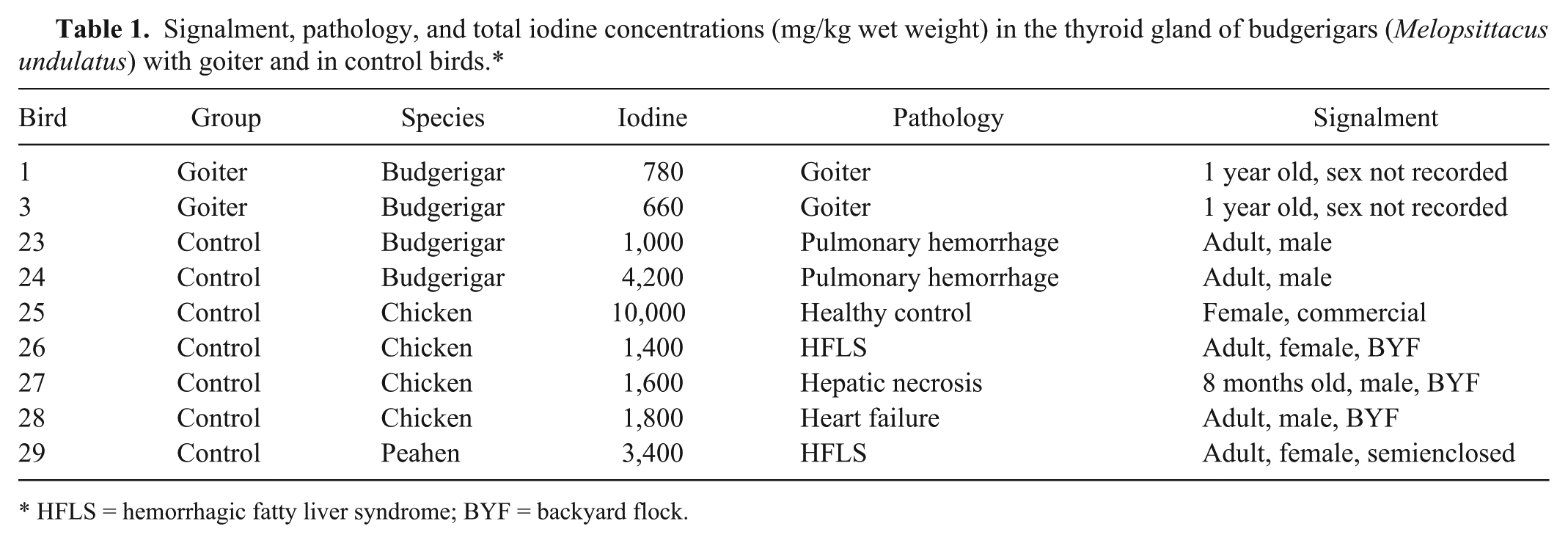
HFLS = hemorrhagic fatty liver syndrome; BYF = backyard flock.
Morphometric analysis and archival case retrieval
Thyroid gland sections of birds from the outbreak with (birds 1–3, 11) or without (birds 13–17) goiter, of 2 additional unrelated budgerigars without thyroid pathology (birds 30 and 31), a peahen (bird 29), and 6 additional chicken submissions (birds 32–37) were subjected to morphometric analysis. e The maximum follicular length, the individual follicle area, and the proportion of follicles with intraluminal papillary projections or blood were assessed in 107.5 ± 64.6 (34–228) follicles in each of the birds from the outbreak, and in 78.3 ± 77.4 (22–251) follicles in each of the control birds (Table 2). Intergroup comparisons of the parameters analyzed were performed using the χ2 and analysis of variance on ranks tests; p value indicating statistical significance was set at 0.05. The CAHFS laboratory system-wide electronic case archives f were searched for budgerigar submissions with thyroid pathology for the 5-year period from January 1, 2008 to March 1, 2013.
Morphometric parameters of the thyroid gland follicles of budgerigars (Melopsittacus undulatus) from the outbreak in the current study at initial presentation (group A), and following intervention (group B) and budgerigar (group C) and chicken (group D) controls.*
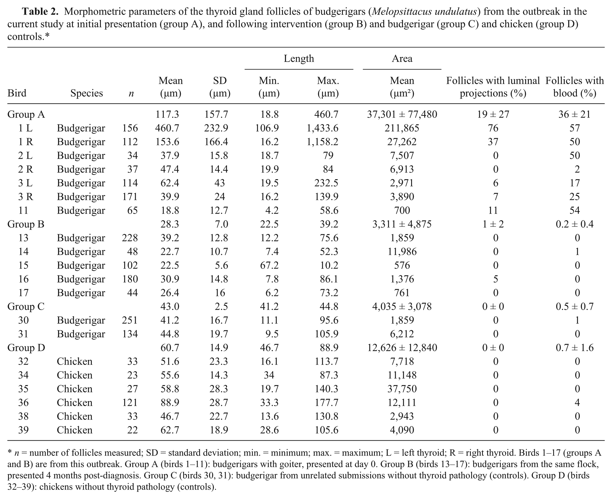
n = number of follicles measured; SD = standard deviation; min. = minimum; max. = maximum; L = left thyroid; R = right thyroid. Birds 1–17 (groups A and B) are from this outbreak. Group A (birds 1–11): budgerigars with goiter, presented at day 0. Group B (birds 13–17): budgerigars from the same flock, presented 4 months post-diagnosis. Group C (birds 30, 31): budgerigar from unrelated submissions without thyroid pathology (controls). Group D (birds 32–39): chickens without thyroid pathology (controls).
Results
Gross pathology
At necropsy, bird 1 was in poor nutritional condition, with minimal fat reserves and moderate to severe pectoral muscle atrophy, while birds 2, 3, 11, and 12 were in fair to good nutritional condition, and were well fleshed with adequate fat reserves. All birds necropsied initially (birds 1–3, 11, 12) had severely enlarged thyroid glands bilaterally, more pronounced on the left side. The largest thyroid gland was observed in bird 1 (left), measuring 2.6 cm × 1.2 cm × 1.5 cm, and weighing 2.8 g, or 6.3% of the total body weight of 44.4 g. This represents a 31.5-fold increase compared to normal thyroid gland weight, which is approximately 0.2% of the total body weight. 19 The weight of the largest thyroid gland in each of the other birds (birds 2, 3, 11, 12) ranged from 0.4 g (0.78% of total body weight of 50.7 g; bird 2) to 0.7 g (1.7% of total body weight of 40.2 g; bird 3) and the dimensions ranged from 1 cm × 0.9 cm × 0.5 cm (bird 2) to 1.1 cm × 1 cm × 1 cm (bird 3), respectively. In addition, the left thyroid gland of bird 2 was diffusely red, and bore an approximately 1 mm semicircular raised nodule on its surface. No significant gross abnormalities were observed in the rest of the carcass in birds 1–3, 11, and 12. No significant gross changes were noted in birds 13–22.
Histology, histochemistry, and morphometric analysis
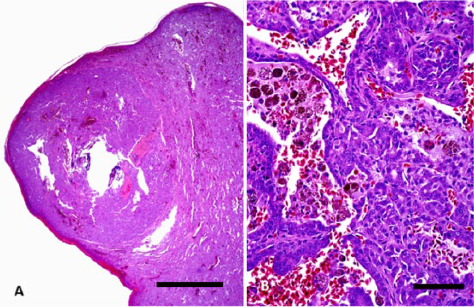
Histologically, the thyroid lesions of all birds necropsied during the initial presentation (birds 1–3, 11, 12) were similar. Follicles were enlarged to a variable degree (Table 2), and contained pale-staining colloid, while 20–30% of the follicular luminae of the thyroid glands were collapsed and lacked colloid. The follicular epithelium was moderately hyperplastic. Many follicles contained large numbers of red blood cells (Table 2), foamy hemosiderin-laden macrophages, and sloughed epithelial cells. In addition, most thyroid follicles of the left thyroid gland in bird 1 (Fig. 1) were severely dilated, lined by hyperplastic and hypertrophic cells that multifocally formed intraluminal papillary projections (Table 2) and were filled with blood, numerous Perl blue–positive, hemosiderin-laden macrophages and sloughed epithelial cells, fibrin, and moderate to abundant pale-staining colloid-like material (Fig. 2A). Many follicles had luminal granulomatous inflammation. The interstitium was multifocally mildly fibrotic.
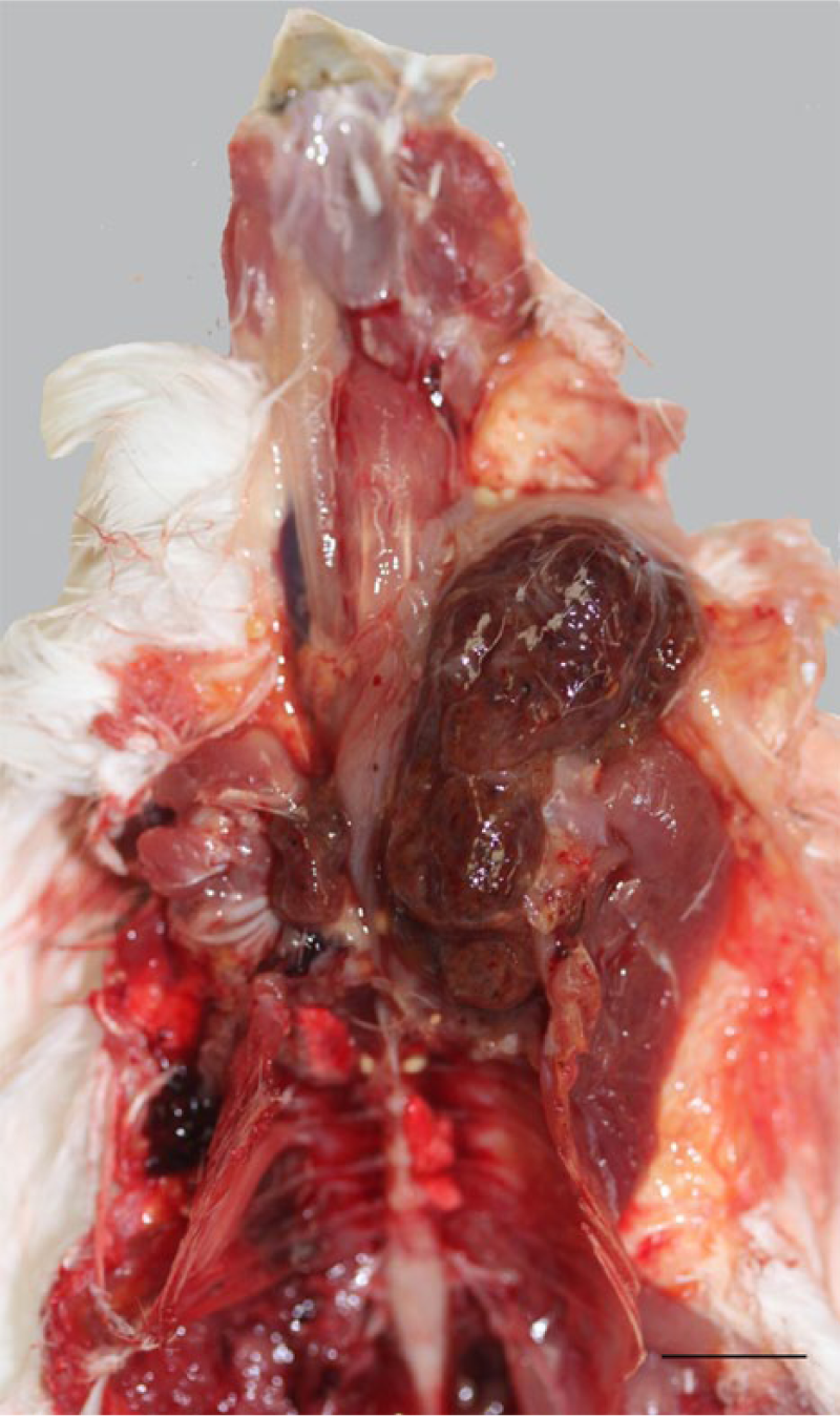
Budgerigar (Melopsittacus undulatus). Severe bilateral thyroid gland hyperplasia (goiter), more pronounced on the left thyroid gland in bird 1. The weight of the left thyroid gland of this bird was equivalent to 6.3% of total body weight (reference value: 0.2%). Bar = 1 cm.
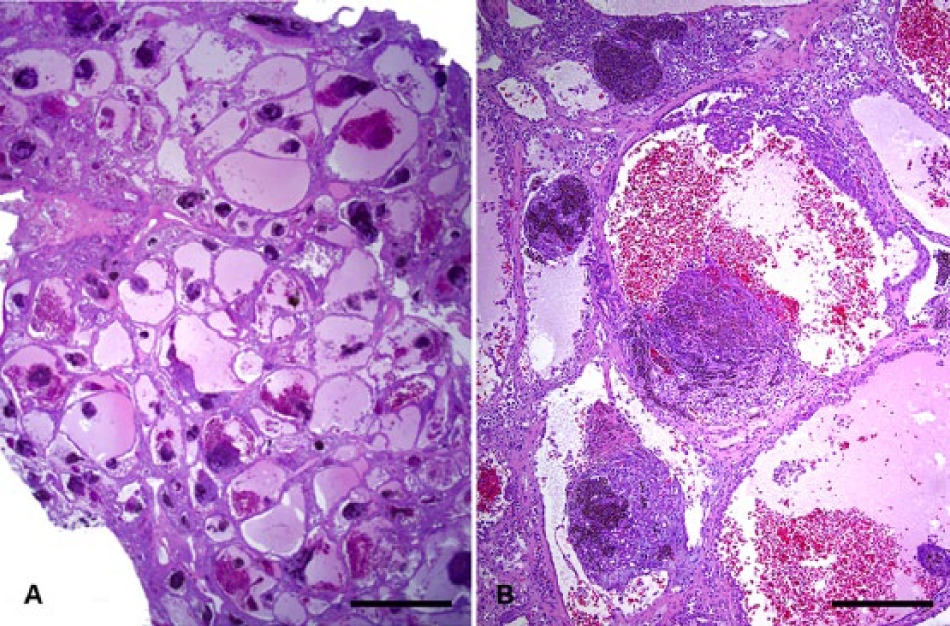
Budgerigar (Melopsittacus undulatus). Left thyroid gland, bird 1.
Furthermore, in bird 2, there was a solitary, round, expansile, microfollicular adenoma, partly encapsulated by a thin layer of fibrous tissue that moderately compressed the adjacent parenchyma of the left thyroid (Fig. 3). Most follicles in the non-neoplastic parenchyma were hyperplastic and collapsed, or were of small size and contained little discernible colloid. A focal region of lymphocytes forming a sheet, morphologically reminiscent of thymic remnant, was noted in birds 2 (with goiter) and 14–17 (current outbreak, without goiter). No histologic changes were observed in the right thyroid gland of bird 2.
Budgerigar (Melopsittacus undulatus). Thyroid adenoma, bird 2.
The lungs of birds 1–3 and 11–12 (with goiter) were mildly diffusely congested. Mild periportal lymphocytic infiltrates were present in the liver; no bacteria were identified on Gram-stained sections. A large number of epithelial cells with intracytoplasmic hemosiderin granules was noted in the renal parenchyma. Multifocally, there were multiple small fissures of the keratinized layer of the beak of bird 1 that were severely infiltrated by bacteria. No significant gross or histologic abnormalities were observed in birds 13–22 (submitted 4 months post-diagnosis).
The proportion of follicles containing blood was significantly higher in the birds examined at the initial presentation (with goiter) compared with the birds presented 4 months post-diagnosis (P = 0.003; Table 2). The maximum follicular length, individual follicle area, and proportion of follicles with intraluminal papillary projections was markedly higher in the birds examined at initial presentation (with goiter) compared with the birds presented 4 months post-diagnosis; however, the differences did not reach statistical significance (P = 0.098, 0.078, and 0.078, respectively; Table 2).
A diagnosis of thyroid follicular hyperplasia (goiter) was established in all affected birds examined histologically. Mild granulomatous thyroiditis and fibroplasia, and thyroid microfollicular adenoma arising in thyroid hyperplasia were also diagnosed in 2 birds, respectively.
Bacteriology, virology, and parasitology
Gallibacterium anatis biovar anatis and mixed bacterial flora were isolated from the liver and lungs of bird 1. Mixed flora was also isolated from the kidney of bird 1, and the small intestine, lung, and liver of bird 3. Liver samples were negative for Salmonella sp. by real-time PCR and confirmatory culture. Pharyngeal swab and cloacal swab pool samples were negative for avian Influenza A virus matrix gene by qRT-PCR. Virus isolation techniques were negative for the presence of viruses. No parasite eggs were detected on examination of a fecal pool sample by flotation.
Toxicology
Heavy metal concentrations in livers were within the normal range for all birds. Selenium levels (1.7 mg/kg wet weight) were slightly above what is typically seen in budgerigars (1.0 ± 0.4, range: 0.4–1.7 mg/kg wet weight). Iodine levels in the thyroid glands of the 2 birds analyzed were consistently low, based on the comparison with controls (Table 1), albeit statistical comparison was not performed.
Twenty-day and 4-month post-diagnosis follow-up
On a 20-day post-diagnosis follow-up, the owner reported that he had stopped adding broccoli to the feed mixture immediately and supplemented the water with iodine drops, and white vinegar, after the diagnosis of goiter was established, and that 5 more birds (all of which were sick at the time of submission) had died, while the other birds that were sick at the time of submission were putting on weight and appeared to be in overall better health. Furthermore, he did not observe any additional birds exhibiting clinical signs. On a 4-month post-diagnosis follow-up, the owner reported that the problem was resolved, with the possible exception of 4 out of sixty 3-year-old birds that despite normal appetite had lost weight and were weak and depressed, and 3 out of 60 neonates that appeared weak. Iodine mineral blocks designed for rabbits were provided ad libitum to the birds.
Archival case retrieval results
No other goiter cases were identified in a total of 30 budgerigar necropsy cases (38 birds) retrieved from the CAHFS laboratory system-wide electronic case archives d for the period from January 1, 2008 to March 1, 2013. Thyroid gland lesions were observed in only 1 other budgerigar, a 39-g adult male with atherosclerosis of major and thyroid gland vessels, and thyroid gland dysplasia associated with severe vitamin A deficiency.
Discussion
Similar to most goiter cases, animals in the current outbreak responded to iodine supplementation, indicating that iodine deficiency was most likely responsible for the development of goiter. Inappropriate iodine testing methodology has in the past led to false reporting of nonexistent iodine deficiency. 11 A method for the accurate measurement of total iodine levels in fresh or frozen thyroid tissue was developed for the current study based on the principle of oxidation and subsequent detection by ICP-MS. Although the small size of the groups did not allow meaningful statistical comparisons to be performed, iodine levels in the thyroid of affected birds were lower compared to those of controls (Table 1), a finding consistent with iodine deficiency being the primary etiologic agent for the development of goiter in the current outbreak.
The 31.5-fold increase of the thyroid gland weight reported in the current outbreak is much higher than that observed in earlier reports. The normal thyroid gland weight is approximately 0.02% of the total bodyweight, 19 and in a budgerigar weighing 35 g, each thyroid gland measures approximately 2 mm × 1 mm × 1 mm, 5 while previous studies reported thyroid glands exceeding 1 cm in size and 1 g in weight in budgerigars, 4 and a 3-fold increase in the relative thyroid gland weight in geese. 18 Furthermore, adenomas are rarely described 29 and may be difficult to differentiate from hyperplasia, although the adenoma described in the present report was grossly nodular and clearly delineated from the adjacent hyperplastic thyroid gland. It is possible that, similar to other organs, thyroid hyperplasia, dysplasia, and adenoma are part of a lesion continuum and that hyperplasia predisposes to the development of adenoma.
Budgerigars are anecdotally considered to be particularly susceptible to developing goiter; however, the published literature and a search of the laboratory archives do not seem to support this assumption. Epidemiologic studies may be warranted to determine whether the propensity of budgerigars to thyroid gland hyperplasia development, if present, represents true species susceptibility or is due to poor management, higher inbreeding rates, or nutritional factors that may be more prevalent in the management of this species, and to define the mechanisms that infer such species susceptibility.
Broccoli, as well as other members of the Brassicaceae family, contains goitrogenic compounds, which can interfere with the uptake and organification of iodine, and subsequently lead to the inability to form active thyroid hormones. Budgerigars and other species may therefore develop goiter when consuming large quantities of goitrogenic agents including cabbage, broccoli, kale, turnips, rapeseed, and soybean or when fed an iodine-deficient diet. 13 Goiter can also develop in budgerigars as a result of iodine-deficient drinking water and provisions of a seed mixture based on millet. 27 The presence of large quantities of broccoli and the lack of mineral supplementation in the diet of these birds were considered major contributors to the development of goiter, as the onset of disease coincided with the change of diet. Iodine supplementation of the feed and water and removal of broccoli from the feed resulted in rapid clinical improvement and stopped mortality. Reexamination of the breeding scheme in such cases may also help prevent the recurrence of goiter in the flock. Recognition of this condition may help improve medical, welfare, and trade standards concerning this species in practice.
Footnotes
Acknowledgements
The authors thank Drs. M. Hawkins and R. Poppenga for insightful discussions on the case, and colleagues at CAHFS for providing avian thyroid gland tissues.
a.
MegaBac-S, Vetafarm Pty Ltd, Wagga Wagga, New South Wales, Australia.
b.
Hardy Diagnostics, Santa Maria, CA.
c.
MARS Xpress, CEM Corp., Matthews, NC.
d.
Agilent 7500ce, Agilent Technologies Inc., Santa Clara, CA.
e.
MicroSuite Basic Edition 5.0, Olympus Corp., Shinjuku, Tokyo, Japan.
f.
StarLims version 10, 2013, StarLims Corp., Hollywood, FL.
Declaration of conflicting interests
The author(s) declared no potential conflicts of interest with respect to the research, authorship, and/or publication of this article.
Funding
Funding for the current report was provided by the California Animal Health and Food Safety Laboratory, School of Veterinary Medicine, University of California, Davis.