
Abstract
Non-neoplastic thyroid hyperplasia is common in terrestrial animals, secondary to nutritional imbalances or other goitrogenic compounds. Thyroid hyperplasia is relatively common in teleost fish; however, malignant thyroid neoplasia is rarely reported. We diagnosed cases of thyroid neoplasia in a population of jade perch (Scortum barcoo). The 3,000 affected fish had grossly apparent, bilateral pharyngeal swellings. Histologic examination confirmed proliferative thyroid lesions ranging from hyperplasia to well-differentiated follicular cell carcinoma. In addition, the younger population of animals on the farm also had bacterial septicemia and mild Dactylogyrus sp. gill infections. Feed analysis revealed a severe deficiency of iodine and vitamin C in the homemade fish diet used on the farm. The concentrations of other minerals, such as zinc, were also on the lower end of the recommended requirements for freshwater fish. The farm was using surface water in its recirculating aquaculture system. We recommended a switch to a commercial aquaculture diet, as well as to use well water rather than surface water to avoid any contaminants, and to treat the younger fish with an antibiotic for bacterial septicemia. Our case provides evidence of progression from nutritional-associated thyroid hyperplasia to neoplasia in farmed teleost fish.
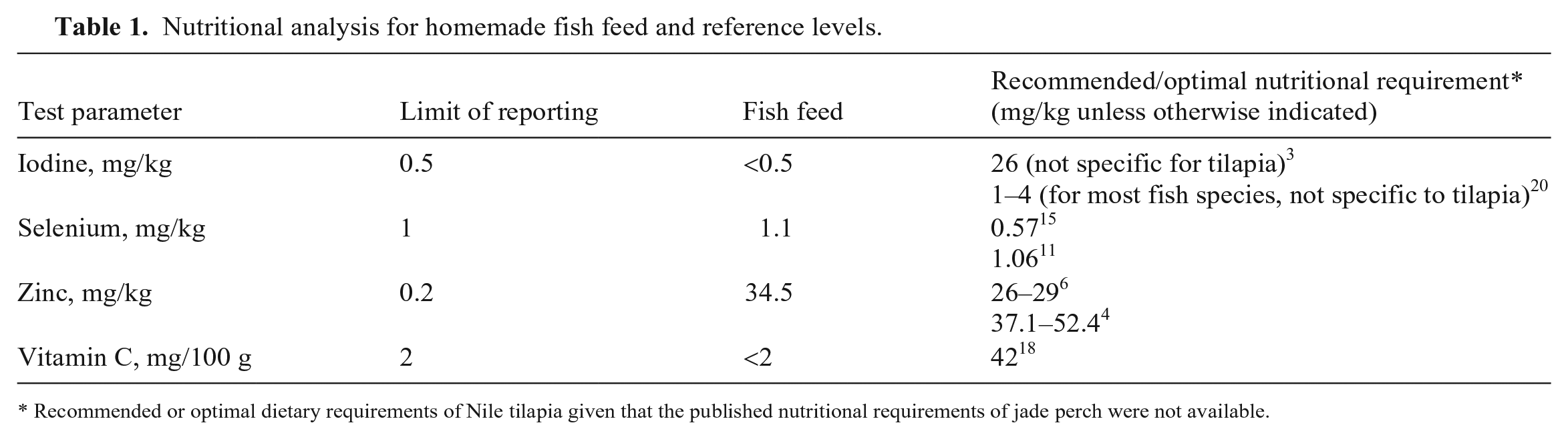
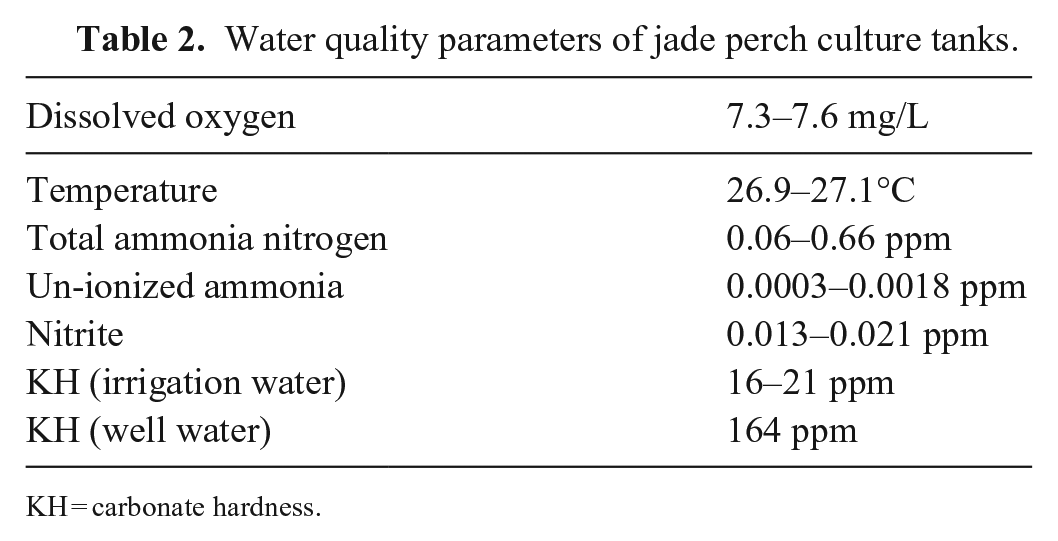
Approximately 3,000 jade perch (syn. barcoo grunter; Scortum barcoo), weighing 300–400 g and stocked in ten 7-tonne tanks on a small farm in Hong Kong, were reported to have high mortality (estimated by the farmer at 4%/d for several weeks). During a visit to the farm site, veterinarians from City University observed several moribund fish with large, 2–3-cm superficial skin lesions on the flank, tail peduncle, and ventral abdomen, along with patchy scale loss and occasional subcutaneous abscesses. Additionally, many fish in the tanks appeared anorexic, lethargic, and darker than normal. Some moribund fish also had necrotic gills and eroded fins. Internally, moribund fish had splenomegaly and bilateral, 1–1.5-cm swellings in the pharyngeal region (Fig. 1). These tissue swellings were noted in all fish examined, even those without other clinical abnormalities.
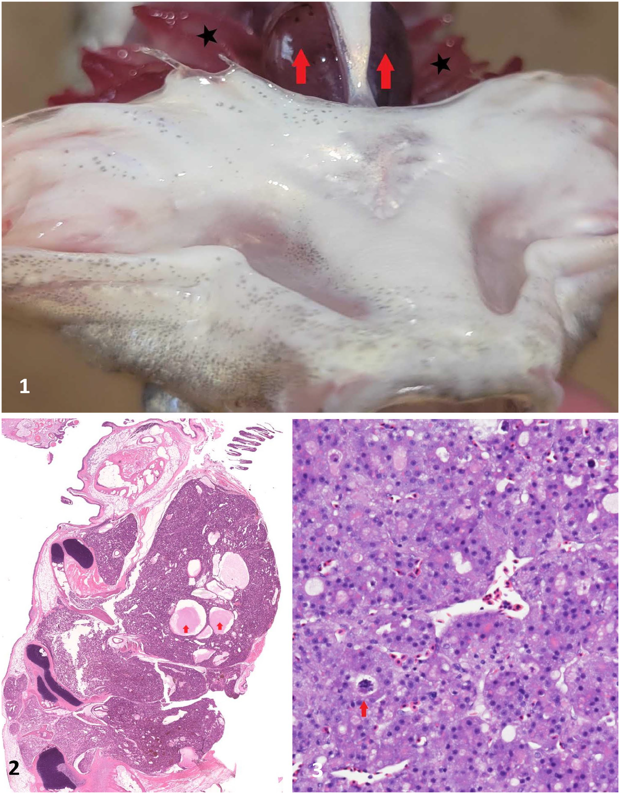
Jade perch gross and histologic lesions.
All of the culture tanks on the farm were freshwater and were equipped with their own mechanical and biological filter units and air supply. Surface water was used to top up the tanks and/or as replacement water when the quality of the water appeared poor (turbid or murky). The farm manager fed the fish a homemade diet, which was created using ingredients such as fish meal, soybean meal, peanut meal, bone meal, and bread. Ingredients were mixed using a small on-site feed extrusion unit. Because of supply-chain issues, the farmer disclosed that he had been unable to purchase mineral or vitamin premixes for over 6 mo before the onset of clinical signs.
The site also had 2 other groups of fish: 524 larger grow-out fish weighing 800–1,000 g, and 21 broodstock fish weighing >3 kg. Despite being on the same diet and utilizing the same water source as the younger grow-out fish, neither of these groups experienced mortality, and they did not exhibit the skin lesions described above. Close inspection of these 2 groups of fish suggested that the larger grow-out fish also had enlarged bilateral masses in the pharyngeal region. These lesions were not grossly visible in the broodstock fish.
Six of the early grow-out moribund fish were collected for gill and skin wet mount assessment for ectoparasites under light microscopy at 400× magnification. Skin wet mounts were unremarkable; however, gill assessments revealed the presence of a light infection with a Dactylogyrus sp. Given the level of infection in these fish and the common occurrence of these parasites in subclinical warm-water fish populations in Hong Kong, this finding was considered incidental.
Samples were taken from the posterior kidneys of 3 moribund fish for bacterial culture, conducted at the City University–Veterinary Diagnostic Laboratory (CityU-VDL; City University of Hong Kong, Kowloon Tong, Hong Kong, SAR China). Culture swabs were inoculated onto sheep blood agar, MacConkey agar, chocolate agar, and thiosulfate–citrate–bile salts–sucrose agar, and incubated for 48 h. The swab was also incubated in trypticase soy broth and then transferred to chocolate medium plates after 24 h; all media were incubated aerobically at 25°C. Specific bacterial colonies were identified to the species level with a matrix-assisted laser desorption/ionization time-of-flight mass spectrometer (MALDI-TOF MS; Bruker). Aeromonas hydrophila and Streptococcus iniae were isolated from the kidneys of all 3 fish.
The spleen, liver, and pharyngeal bilateral masses from 3 moribund fish were collected and submitted to the CityU-VDL for histologic evaluation. Increased numbers of peri-ellipsoid macrophages were observed in the spleen and reported as indicative of nonspecific systemic inflammation. The liver from 1 of the 3 fish had hepatocellular degeneration of unknown origin. The pharyngeal mass in one fish was diagnosed as thyroid hyperplasia. The pharyngeal masses in the other 2 fish had histologic features consistent with thyroid follicular cell carcinoma (Figs. 2, 3) as described in previous case reports. 2 Specifically, histologic features included solid sheets of small dense nests of cells or single scattered cells, sometimes within haphazard scirrhous stroma; areas of necrosis were also present. There was moderate anisocytosis and anisokaryosis, nuclei had densely clumped chromatin and prominent nucleoli, and one mitotic figure was noted in 10 hpf (400×, 2.37 mm2). Further, in some cases, tumor cells had invaded the adjacent tissues, including the fibrocartilage and the superficial dermis.
Given the histopathologic changes in the thyroid tissue, ~500 g of the homemade fish feed was submitted to the Australian Laboratory Services (ALS; Hong Kong) for analysis of iodine, vitamin C, zinc, and selenium levels. We chose these ingredients for analysis because their deficiencies have been known to affect thyroid function.8,10,13 ALS used inductively coupled plasma–mass spectrometry (ICP-MS; 7900, Agilent) to measure iodine, selenium, and zinc, and high-performance liquid chromatography–fluorescence detector (HPLC-FLD; Dionex UltiMate 3000; ThermoFisher Scientific) to measure vitamin C (Table 1). Because there were no published nutritional requirements for jade perch at the time of our report, we provide the nutritional requirements for Nile tilapia (Oreochromis niloticus) and other nonspecified freshwater species for reference (Table 1). The homemade diet was severely deficient in iodine and vitamin C. Zinc and selenium were borderline adequate, depending on which standard reference is used (Table 1).
Nutritional analysis for homemade fish feed and reference levels.
Recommended or optimal dietary requirements of Nile tilapia given that the published nutritional requirements of jade perch were not available.
We also conducted routine water quality testing at the farm site. Water samples were collected from all affected fish tanks and tested (SL1000 portable parallel analyzer; Hach) for total ammonia nitrogen (TAN), nitrite (NO2), pH, and alkalinity (KH). Dissolved oxygen and water temperature were assessed directly (ProSolo digital water quality meter; YSI). Un-ionized ammonia levels (UIA) were calculated based on TAN, pH, salinity, and temperature levels (Hamza’s Reef ammonia cycling calculator, https://www.hamzasreef.com/Contents/Calculators/AmmoniaCycling.php). All water quality parameters were within normal ranges except for KH, which was low (Table 2). Once the farmer switched to well water and stopped using surface water as his source of replacement water, the average KH level increased to 164 ppm (Table 2).
Water quality parameters of jade perch culture tanks.
KH = carbonate hardness.
Given our test results, we concluded that mixed bacterial septicemia associated with A. hydrophila and S. iniae likely caused the acute mortality event in the smaller grow-out fish. We suggested treatment with in-feed antibiotics (oxytetracycline) to address the immediate cause of death. We also suggested monitoring these fish for Dactylogyrus sp. infestation and following up with a formalin bath treatment if parasite numbers increased. However, chronic mortalities persisted for nearly 2 mo.
Given the poor response to our antibiotic treatment, we suspected there may have been underlying immunosuppression associated with the observed thyroid lesions. The thyroid gland is a key modulator of homeostasis and the metabolic rate of fish, which affects somatic growth, metamorphosis, reproductive events, and the ability to tolerate environmental changes, 1 as well as immune development and function.7,17 Correlation between histologic pattern, neoplastic differentiation, and thyroid function has been described previously. 16 Given the severity of the changes noted on gross and histologic examination, especially in the small fish that were in a rapid growth phase, lesions in the thyroid may have caused systemic metabolic or immune aberrations that predisposed them to infectious diseases.
Although the incidence of thyroid neoplasia is rare in fish compared to thyroid hyperplasia, chronic exposure to suboptimal conditions, natural carcinogens, and/or tumor promoters in water systems or diets has been associated with thyroid neoplasia. 19 Many extrinsic and intrinsic factors can cause stimulation of the thyroid follicles. These include exposure to goitrogenic substances (endosulfan, malathion, carbamate pesticides, cyanide compounds, trihalomethanes, ammonia, perchlorate) and deficiencies of iodine within the diet and/or the environment.5,9 As a result, serum thyrotropin increases to abnormal levels, causing stimulation of the thyroid follicles and resulting in proliferation and hyperplasia. Although teleost endocrinology is poorly understood, elevation in the risk of developing thyroid carcinoma as a result of higher serum thyrotropin is well documented in human clinical endocrinology and oncology. 12 Our case may represent a population-level example of this phenomenon.
We identified 2 primary issues that could have resulted in thyroid hyperplasia and/or neoplasia on this farm: endocrine-disrupting chemical–contaminated surface water, and dietary deficiencies in trace minerals such as iodine. The former could not be fully assessed because of restricted resources and the difficulty in assessing large volumes of water for intermittent low levels of chemicals. Therefore, we suggested that the farmer change his source of replacement water to well water to remove the water source as the issue.
The other possible cause of thyroid disease on this farm was the diet, which was low in iodine. Even though fish are known to absorb iodine from the water through their gills, in freshwater fish, a percentage of iodine is absorbed from the diet through the gut. 6 It is therefore biologically plausible that the chronic nature of the dietary deficiencies on this farm, especially in the fast-growing animals, could have triggered prolonged elevation in serum thyrotropin, which resulted in thyroid hyperplasia, and in some fish, eventually progressed to thyroid neoplasia. To address the low iodine and vitamin C levels in the diet, we recommended that the farmer change his homemade fish diet to a commercial feed and improve his feed storage (dry, cool, dark area) to reduce vitamin C degradation.
Thyroid proliferative lesions can resolve with iodine supplementation in zebrafish. 14 The new commercial feed used by the farmer in our case was analyzed to have iodine and zinc concentrations of 7.7 mg/kg and 149 mg/kg, respectively, using the method described above. We revisited the farm 10 wk after the diet change and found no reported mortalities and that only 30% of the fish examined had grossly visible swellings in the pharyngeal region compared to the first visit at which all animals exhibited these signs.
The etiology of the thyroid issues in our case was never confirmed and may have been multifactorial. Because we recommended several mitigative strategies at the same time to improve the welfare of the fish as quickly as possible, we are uncertain whether the anecdotal improvements in the condition of the fish were from the new diet provided and/or the change in the water source. Regardless, our case illustrates a possible progression from thyroid hyperplasia to thyroid neoplasia in a population of aquaculture fish and may provide a model for further work to understand the pathogenesis and prevention of environmentally (or dietary) induced neoplasia of the thyroid gland.
Footnotes
Acknowledgements
We thank Jeffrey Wolf and David Groman for manuscript support through review of the histologic images.
Declaration of conflicting interests
The authors declared no potential conflicts of interest with respect to the research, authorship, and/or publication of this article.
Funding
Our project was funded under the Sustainable Fisheries Development Fund at the Agriculture, Fisheries and Conservation Department in Hong Kong Special Administrative Region, PR China (grant SFDF_0042).