
Abstract
An ~10-y-old male sheep had anorexia and progressive weight loss for ~1 mo. The sheep was emaciated, and 20 d later, became recumbent and lethargic, and was hypoglycemic (0.33 mmol/L; RI: 2.6–4.4 mmol/L). The sheep was euthanized because of poor prognosis, and submitted for autopsy. We found no gross lesions in the pancreas; however, histologically, focal proliferations of round-to-polygonal cells were separated by connective tissue into small nests. These proliferating cells, which had abundant eosinophilic-to-amphophilic cytoplasm and hyperchromatic nuclei, were immunopositive for insulin and negative for glucagon and somatostatin; the lesion was diagnosed as an insulinoma. Insulinoma has not been reported previously in sheep, to our knowledge. In addition, autopsy and histologic examination revealed the presence of an adrenocortical carcinoma with myxoid differentiation and a thyroid C-cell carcinoma. Our case indicates that multiple endocrine neoplasms can occur in sheep, as in other animal species.
In human medicine, concurrent occurrence of hyperplasia, adenoma, or carcinoma in ≥ 2 endocrine organs has been termed multiple endocrine neoplasia (MEN) syndrome, which results from the mutation of a specific gene. 11 In veterinary medicine, although the association of genetic mutations has not been elucidated, concurrent endocrine tumors have been reported in dogs,2,3,23 cats,15,16 cattle,21,24 and a horse. 5
Insulinoma is the most common pancreatic endocrine tumor encountered in veterinary medicine. Given that the neoplasm is often functional, affected animals exhibit depression, weakness, and periodic convulsive seizures associated with hypoglycemia.7,18 Insulinoma is most prevalent in dogs, and has also been reported in ferrets, cats, and horses. 7 Although the incidence of insulinoma in ruminants is rarer than in companion animals, insulinoma has been described in slaughtered adult cows 8 and an aged goat. 25 We retrieved no ovine cases of MEN or insulinoma in a search of Google, PubMed, and Scopus, suggesting that those conditions have not been reported in sheep.
An ~10-y-old castrated male sheep was referred to the Veterinary Medical Center of Obihiro University of Agriculture and Veterinary Medicine (Obihiro, Hokkaido, Japan) with an ~1-mo history of anorexia and progressive weight loss. Blood examinations revealed malnutrition that was evident from moderate hypoproteinemia (59 g/L; RI: 65–90 g/L), hypoalbuminemia (19 g/L; RI: 29–47 g/L), and anemia (PCV 0.15 L/L; RI: 0.26–0.41 L/L). The sheep was emaciated; however, the cause of the emaciation could not be determined. The sheep was treated with fluid therapy and intensive care to improve its nutritional condition. Twenty d later, the sheep became recumbent and lethargic, and developed arrhythmia and deep and slow breathing. On that day, the sheep was hypoproteinemic (57 g/L), hypoalbuminemic (17 g/L), hypolipidemic (triglyceride 0.03 mmol/L, RI: 0.28–0.51 mmol/L; total cholesterol 0.57 mmol/L, RI: 1.13–2.57 mmol/L), and hypocalcemic (total calcium 1.8 mmol/L; RI: 2.4–2.9 mmol/L). Blood glucose levels were measured several times on that day, and the sheep was persistently hypoglycemic (0.33 mmol/L; RI: 2.6–4.4 mmol/L). Ultimately, the sheep was euthanized given poor prognosis, and submitted for autopsy, with the owner’s consent.
At autopsy, serous atrophy of adipose tissue was observed systemically; ~500 mL of yellow, translucent, serous ascitic fluid was noted. The pancreatic-duodenal lymph node attached to the right lobe of the pancreas was enlarged (1.5 × 1.0 × 1.0 cm); however, no gross lesions were evident in the pancreatic parenchyma. Gray-white masses were observed in the left adrenal and right thyroid glands (1.0 cm and 0.2 cm diameter, respectively). Samples of systemic organs were collected and fixed in 15% neutral-buffered formalin for histologic examination. The right lobe of the pancreas was sliced at 1-cm intervals, and all of the sections were examined histologically.
In addition to routine histologic examination, immunohistochemistry (IHC) was performed on sections of the pancreas, adrenal gland, and thyroid gland (Table 1). Sections were pretreated with citrate buffer pH 6.0 at 98°C for 20 min for antigen retrieval, then endogenous peroxidase was blocked with 3% H2O2 at room temperature for 10 min. Sections were incubated with each primary antibody at 4°C overnight, followed by incubation with polymer reagent (MAX-PO; Nichirei Bioscience) for 30 min at room temperature. Immunoreactivity was detected with 3,3′-diaminobenzidine, and sections were counterstained with Mayer hematoxylin. As negative controls, each primary antibody was replaced by mouse or rabbit immunoglobulin (Dako) based on the host of the primary antibody.
Primary antibodies used for immunohistochemistry of tumors in a sheep.
— = polyclonal antibody.
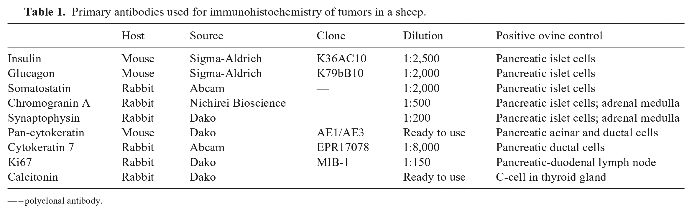
In the H&E-stained section of the pancreas, we observed ~3-mm proliferations of round-to-polygonal cells that were separated by connective tissue into small nests (Fig. 1A). There was no fibrous capsule around the lesion, and the proliferating cells invaded the pancreatic parenchyma. The proliferating cells had abundant eosinophilic-to-amphophilic cytoplasm and uniform, round-to-oval, hyperchromatic nuclei (Fig. 1B). Mitotic figures were not observed in the section. With IHC, the proliferating cells were strongly immunopositive for insulin (Fig. 1C, 1D), synaptophysin, and chromogranin A, and negative for glucagon, somatostatin, pan-cytokeratin, and cytokeratin 7. Ki67-positive cells were observed rarely (0–2 cells/hpf). Although the pancreatic-duodenal lymph node was enlarged grossly, no metastatic lesion was detected histologically. Based on these findings, we diagnosed the pancreatic tumor as an insulinoma.
Pancreatic insulinoma in a sheep.
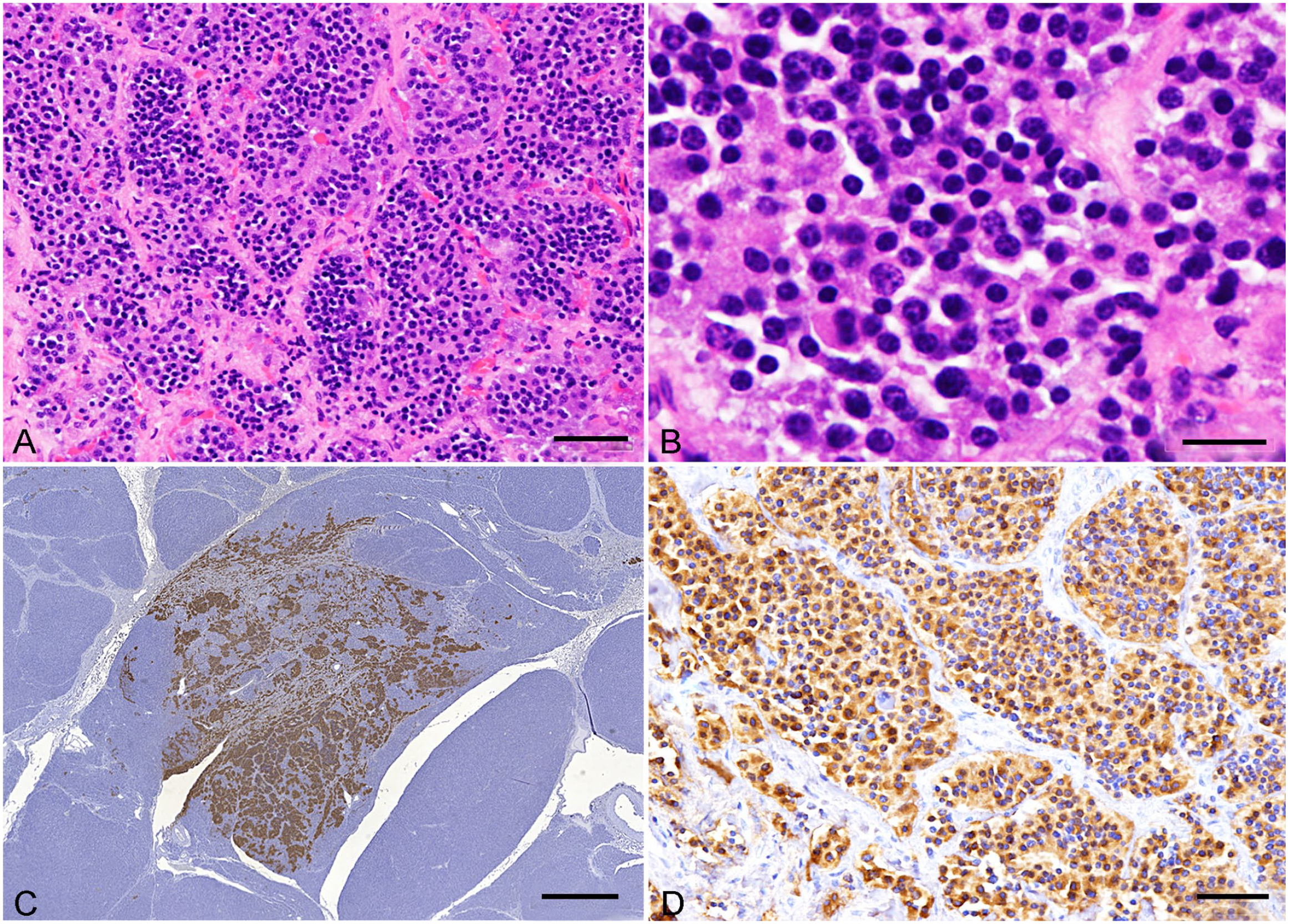
The mass in the left adrenal gland was characterized histologically by a sheet-like proliferation of polygonal cells with abundant eosinophilic cytoplasm. Focally, neoplastic cells formed lumen-like structures surrounding mucinous material (Fig. 2A). Abundant mucinous materials, which stained bright blue with an Alcian blue pH 2.5 stain, were also observed between the neoplastic cells (Fig. 2B). Nuclear atypia of neoplastic cells was moderate to high, and mitotic figures were rare. Binucleate neoplastic cells were also observed. Some neoplastic cells had intranuclear cytoplasmic invaginations, characterized as round eosinophilic-or-amphophilic inclusions surrounded by a thin basophilic membrane (Fig. 2C). The size of these inclusions varied from the size of a nucleolus to ones that filled most of the nucleus. The neoplastic cells were weakly immunopositive for chromogranin A and synaptophysin, compared to the adrenal medulla internal control, and immunonegative for pan-cytokeratin and insulin. We considered that the myxoid differentiation indicated that the neoplastic cells were poorly differentiated and that the tumor may be malignant. Although there was no invasion or metastasis, nuclear atypia was moderate to high. Therefore, we diagnosed this tumor as an adrenocortical carcinoma with myxoid differentiation.
Adrenocortical carcinoma with myxoid differentiation in a sheep.
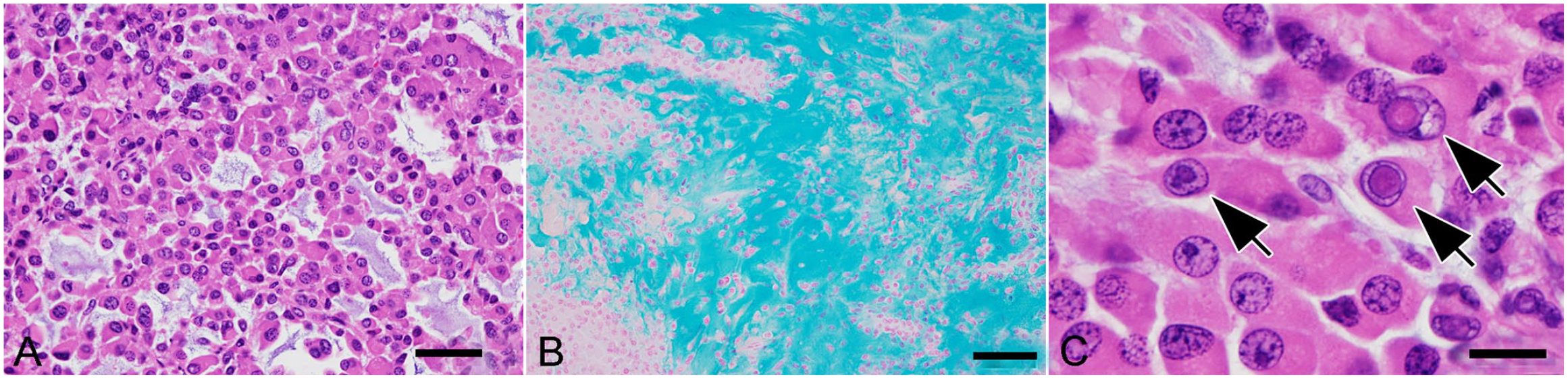
The mass in the right thyroid gland was characterized by bundle-like proliferations of spindle cells subdivided by thin fibrovascular connective tissue (Fig. 3A). This mass was partially surrounded by a fibrous capsule; however, neoplastic cells invaded focally into the capsule and adjacent normal tissue. Nuclear atypia was moderate, and the mitotic count was 3 per 10 hpf (2.37 mm2; 10 fields with FN 22/40× objective). Given that the spindle cells were immunolabeled with calcitonin (Fig. 3B), we diagnosed the lesion as a thyroid C-cell carcinoma. In addition, there was multifocal-to-diffuse C-cell hyperplasia between follicles in both thyroid glands (Fig. 3C, 3D).
Thyroid C-cell carcinoma (A, B) and C-cell hyperplasia (C, D) in a sheep.
Insulinoma has been reported in various animal species including dogs, cats, ferrets, horses, cows, and a goat.7,8,25 We found no reports of insulinoma in sheep in a search of Google, PubMed, and Scopus. Insulinomas are classified as benign or malignant depending on the behavior of the tumor. In veterinary medicine, insulinomas are classified as malignant in most cases in small and large animals. 18 It is difficult to differentiate benign insulinomas from malignant counterparts histologically; however, local invasion of neoplastic cells may indicate malignancy, whereas metastasis is confirmatory. 12 In our case, the neoplastic cells invaded the pancreatic parenchyma, and this finding suggested a diagnosis of malignant insulinoma. Furthermore, given the persistent hypoglycemia, the insulinoma in our case was considered to be functional.
Because the insulinoma in our case did not form a mass lesion, nesidioblastosis was also included as a differential diagnosis. Nesidioblastosis is characterized histologically by an increase both in size and number of islets.7,20 The enlarged islets consist of a large number of β-cells with a few α-cells and δ-cells (immunolabeled with insulin, glucagon, and somatostatin, respectively).7,22 These histologic changes in islets are observed throughout the entire pancreas in most cases. 4 Nesidioblastosis causes congenital hypoglycemia in human neonates and infants; however, it is usually an incidental finding in animals.7,11 The lesion observed in our case was focal, and suspected as functional. In addition, glucagon- or somatostatin-immunolabeled cells were not detected in the lesion in our case by IHC. Based on these findings, we diagnosed our case as insulinoma, not as nesidioblastosis.
Adrenocortical carcinoma with myxoid differentiation is a common subtype in ferrets,9,18 and has also been reported in a cat 1 and cows. 6 We did not find any reports of this subtype of adrenocortical carcinoma in sheep in a search of Google, PubMed, and Scopus. A retrospective study in ferrets indicated that this subtype of adrenocortical tumor is associated with high malignancy. 14 Given that metastatic lesions were not detected in our autopsy and histologic examination, the malignancy of this subtype in sheep is unclear. Cytoplasmic invaginations into the nuclei were observed frequently in our case. The significance and pathogenesis of these invaginations are unclear; however, the presence of invagination has been described sporadically in specific tumors, such as Leydig cell tumor in dogs 19 and hepatocellular tumor in mice. 10 Interestingly, intranuclear cytoplasmic invaginations were described in a case of feline adrenocortical carcinoma with myxoid differentiation 1 ; thus, such invaginations may be a common histologic finding in adrenocortical carcinoma with myxoid differentiation. In our case, the neoplastic cells were weakly immunopositive for chromogranin A and synaptophysin, which are markers for adrenal medullary tumors. A few adrenocortical tumors immunopositive for those neuroendocrine markers have been reported in various animal species. 18
Thyroid C-cell hyperplasia and tumor are common findings in aged bulls fed diets with a high-calcium content intended for dairy cows. 17 C-cell hyperplasia has been reported in sheep with hypercalcemia experimentally induced by the administration of vitamin D3. 13 Our case was fed coarse feed; therefore, the C-cell hyperplasia that we observed was probably not related to hypercalcemia.
Our case had an insulinoma, adrenocortical carcinoma, and thyroid C-cell carcinoma and C-cell hyperplasia. In humans, MEN syndrome is a hereditary disease resulting in the simultaneous development of hyperplasia, adenoma, or carcinoma in various endocrine organs. 11 In veterinary medicine, the simultaneous development of endocrine tumors and/or hyperplasia in multiple organs have been reported, especially in dogs and cats.2,3,15,16,23 In ruminants, concurrent multiple endocrine tumors and/or hyperplasia have been reported in a family of Guernsey bulls, 17 a Holstein bull, 24 and a Japanese black bull. 21 Given that the genetic backgrounds of subjects have not been elucidated in veterinary medicine, unlike in humans, it is unclear whether the MEN and hyperplasia in our case are incidental findings or an aspect of the syndrome similar to that identified in humans. Nevertheless, our case indicates that MEN can occur simultaneously in sheep, as in other animal species.
Footnotes
Acknowledgements
We thank Ms. Akiko Tomikawa for her technical support with histological processing.
Declaration of conflicting interests
The authors declared no potential conflicts of interest with respect to the research, authorship, and/or publication of this article.
Funding
The authors received no financial support for the research, authorship, and/or publication of this article.