
Abstract
Biliverdin is an intermediate of heme degradation with an established role in veterinary clinical diagnostics of liver-related diseases. The need for chromatographic assays has so far prevented its wider use in diagnostic laboratories. The current report describes a simple, fast, high-throughput, and inexpensive assay, based on the interaction of biliverdin with infrared fluorescent protein (iRFP) that yields functional protein exhibiting infrared fluorescence. The assay is linear in the range of 0–10 µmol/l of biliverdin, has a limit of detection of 0.02 μmol/l, and has a limit of quantification of 0.03 µmol/l. The assay is accurate with relative error less than 0.15, and precise, with coefficient of variation less than 5% in the concentration range of 2–9 µmol/l of biliverdin. More than 95% of biliverdin was recovered from biological samples by simple dimethyl sulfoxide extraction. There was almost no interference by hemin, although bilirubin caused an increase in the biliverdin concentration, probably due to spontaneous oxidation of bilirubin to biliverdin. The newly developed biliverdin assay is appropriate for reliable quantification of large numbers of samples in veterinary medicine.
Introduction
Biliverdin is a green pigment that is produced during heme metabolism. Heme is converted by heme oxygenase to biliverdin, which is then reduced to bilirubin by biliverdin reductase. 17 In mammals, the transformation of biliverdin by biliverdin reductase is rapid. Biliverdin is thus a short-lived species and, compared to bilirubin, is present in serum in relatively low concentrations. Increased serum concentrations of biliverdin (hyperbiliverdinaemia or green jaundice) in human beings are rare, and are mostly caused by mutations in genes for metabolic enzymes.3,14 Biliverdin has not been included in standard diagnostic testing; however, several studies have suggested the administration of biliverdin in therapy due to its antioxidant and anti-inflammatory activity. 16 Such administration could increase the demand for a biliverdin assay in the future.
On the other hand, a need for a biliverdin diagnostic assay already exists in veterinary medicine. Birds and reptiles have little biliverdin reductase activity, and produce very low amounts of bilirubin. 5 Increased concentrations of biliverdin in serum, bile, or excreta can point to anemia due to intoxication10,11 or infection (e.g., with Plasmodium gallinaceum 18 or Chlamydophila psittaci 15 ) or to liver problems or biliary obstruction. 5 Biliverdinuria is almost always the sign of hepatic disease and occurs due to a serum accumulation of biliverdin that exceeds the renal threshold.7,15 Biliverdin is therefore an alternative analyte in bird and reptile disease diagnostics 5 for which the reference values remain to be established. Additionally, biliverdin is also an important pigment of eggshells in several species. 1 Egg coloration studies also require a biliverdin assay to determine biliverdin content in the eggshells.1,13
A clear need for an efficient, simple, and cost-effective method for biliverdin determination thus exists. Reported assays involve low-throughput, high-performance liquid chromatography (HPLC),6,10,12 or enzyme assays involving measuring the rate of change of absorbance. 4 The current study describes a fluorescence biliverdin assay, based on selective binding of biliverdin to infrared fluorescent protein (iRFP), which utilizes biliverdin as a cofactor to gain functionality. 2 A basic method validation was also performed according to published guidelines (Ederveen J: 2010, A practical approach to biological assay validation. Progress report number 08090. Dutch Ministry of Housing, Spatial Planning and the Environment. Available at: http://www.edraservices.nl/documenten/Assay-validation-P08090.pdf ). A similar fluorescence assay has been reported for assaying bilirubin, using fluorescent protein UnaG from eel. 9
Materials and methods
Molecular cloning
Infrared fluorescent protein
2
was back-translated into irfp gene, which was synthesized.
a
Promoter CP25
8
was amplified by polymerase chain reaction (PCR) with 2 overlapping primers (CP25F: 5′-TAAT
Expression of iRFP and preparation of biliverdin determination reagent
Escherichia coli DH5α, which contains plasmid pGEM::CP25-IRFP, was grown overnight in lysogeny broth medium supplemented with 100 µg/ml of ampicillin (LBA) at 37°C with vigorous shaking. The culture was diluted (1:100) with fresh LBA to a final volume of 1 liter and grown to an optical density at 600 nm of 2.50. Following centrifugation at 5,000 × g and resuspension in 100 ml of phosphate buffered saline (PBS; pH 7.2), the bacteria were frozen, thawed on ice, and sonicated for 15 min (UP200S d ). Bacterial lysate was centrifuged at 11,000 × g for 20 min. The supernatant (biliverdin determination reagent) was removed, aliquoted, and stored at −20°C for the biliverdin assay.
Preparation of biliverdin solutions
Biliverdin HCl e stock solution (25 mmol/l) was prepared in dimethyl sulfoxide (DMSO). The stock solution was diluted in DMSO to prepare other biliverdin solutions, which were aliquoted and stored at −20°C. Biological biliverdin samples were prepared by spiking chicken serum (1-ml aliquots) with 2.5 mmol/l of biliverdin to give various concentrations. Hemin e was prepared at 10 mg/ml in DMSO and bilirubin e at 1 mg/ml in chloroform. Appropriate dilutions of each were made in DMSO.
Dimethyl sulfoxide extraction
Aliquots of biliverdin-spiked chicken serum (20 µl) were mixed with 80 µl of DMSO. The samples were vortexed vigorously for 30 sec and centrifuged at 13,000 × g for 10 min. Ten-microliter aliquots of the cleared supernatant were used for the biliverdin assay.
Measurement of fluorescence
Ten microliters of biliverdin solution were pipetted onto a black, flat-bottom, 96-well plate, f followed by the addition of 90 µl of biliverdin determination reagent. After 15-min incubation at room temperature, fluorescence was measured with a microplate reader. g Excitation was set at 690 nm and emission at 713 nm. Gain was set at optimal, Z-position at 20,903 µm and number of flashes at 200. Fluorescence was recorded in multiple 9-reads-per-well mode and the values for a particular well averaged. All samples were measured in triplicate, except for the assessment of precision, where 6 replicates were measured.
Determination of biliverdin concentration, and limits of detection and quantification
Calibration standards in the range of 0.25–10.0 µmol/l were added to the plate with each measurement. Linear regression was used to calculate the equation for the determination of sample concentration. The limit of detection (LOD) was determined by calculating the concentration of biliverdin from the yield of fluorescence of the negative control (containing no biliverdin), increased by 3× standard deviation (SD). The limit of quantification (LOQ) was determined by calculating the concentration of biliverdin from the yield of fluorescence of the negative control (containing no biliverdin), increased by 10× SD.
Results
Determination of linear range
The fluorescence of biliverdin solutions was determined over a concentration range of 0–25 µmol/l. Linear response was observed in the range of 0–10 µmol/l (Fig. 1).
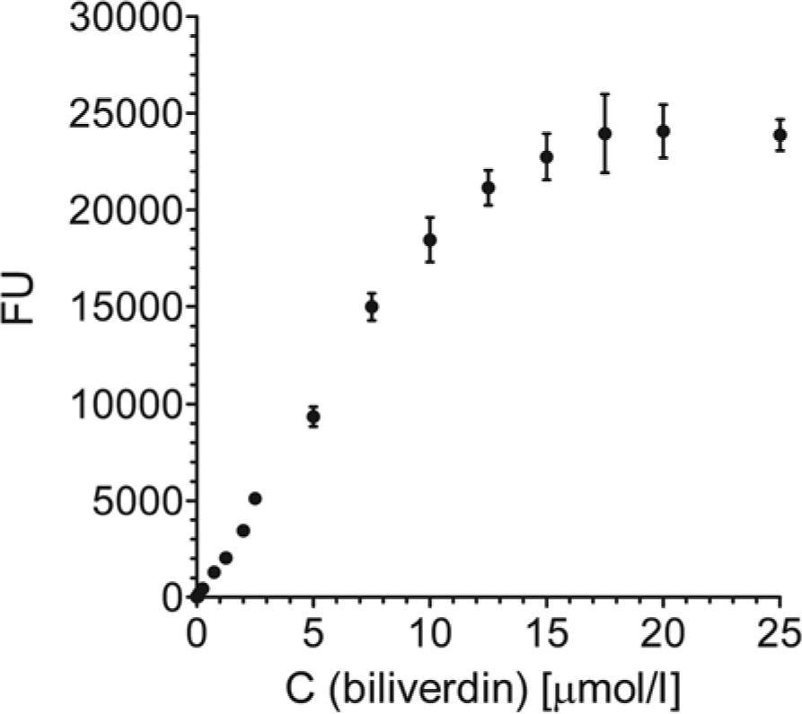
Determination of the linear range of the biliverdin assay. FU = fluorescence units.
Selection of calibration standards
The following concentrations of biliverdin were selected for preparing the calibration curve: 0.25, 1.25, 2.5, 5.0, 7.5, and 10.0 µmol/l. All calibration curves used for determining sample concentrations had R2 values greater than 0.9900.
Determination of optimal incubation time, and limits of detection and quantification
The optimal time of incubation of sample with biliverdin determination reagent was determined by measuring the fluorescence of calibration standards at 5-min time intervals. Temporal change of the calibration curve parameters (average coefficient of variation [CV] and coefficient of determination [R2]) were monitored (Fig. 2). Fifteen-minute incubation was selected as optimal, with the lowest average CV and high R2. The LOD and LOQ of the assay were determined to be 0.02 and 0.03 µmol/l of biliverdin, respectively.
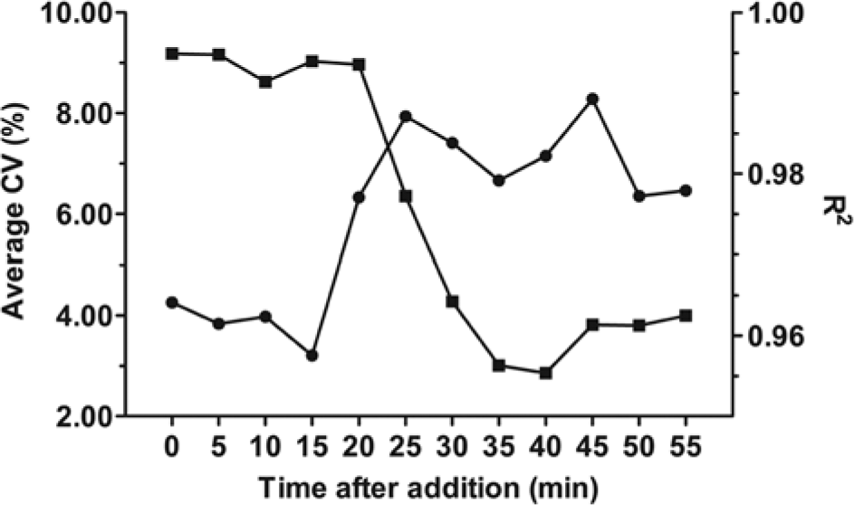
Average coefficient of variation (CV; circles) and coefficient of determination (R2; squares) of the calibration curve as a function of time following the addition of biliverdin determination reagent.
Assessment of accuracy
Eight biliverdin solutions with 4 different concentrations were prepared, and biliverdin concentrations determined with the proposed assay on 2 consecutive days. There was little day-to-day variation with any of the biliverdin solutions. The deviations of determined concentrations from the actual values were between 0.67% and 15.17% (Table 1).
Accuracy of determination of biliverdin concentration.*
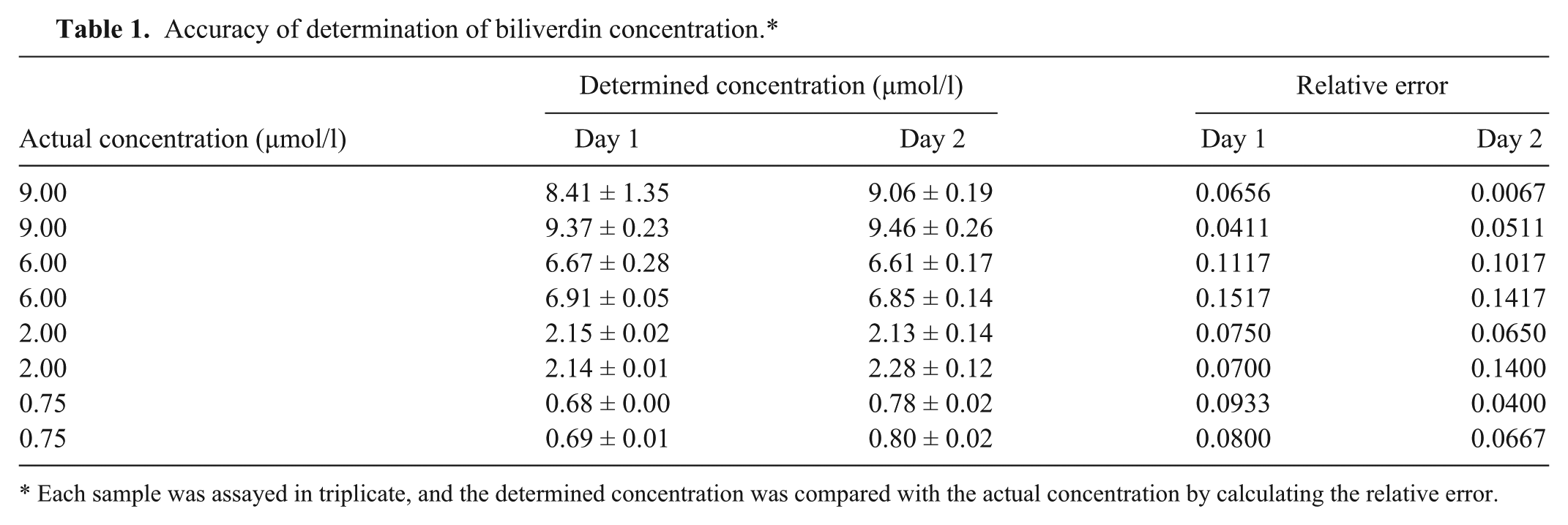
Each sample was assayed in triplicate, and the determined concentration was compared with the actual concentration by calculating the relative error.
Assessment of precision
Four biliverdin solutions were assayed for biliverdin concentration in 6 replicates on 2 consecutive days. The CVs lay between 0.88% and 3.96%, except for determination of the sample with the lowest concentration, for which CV was 14.30% (Table 2).
Precision of determination of biliverdin concentration.*
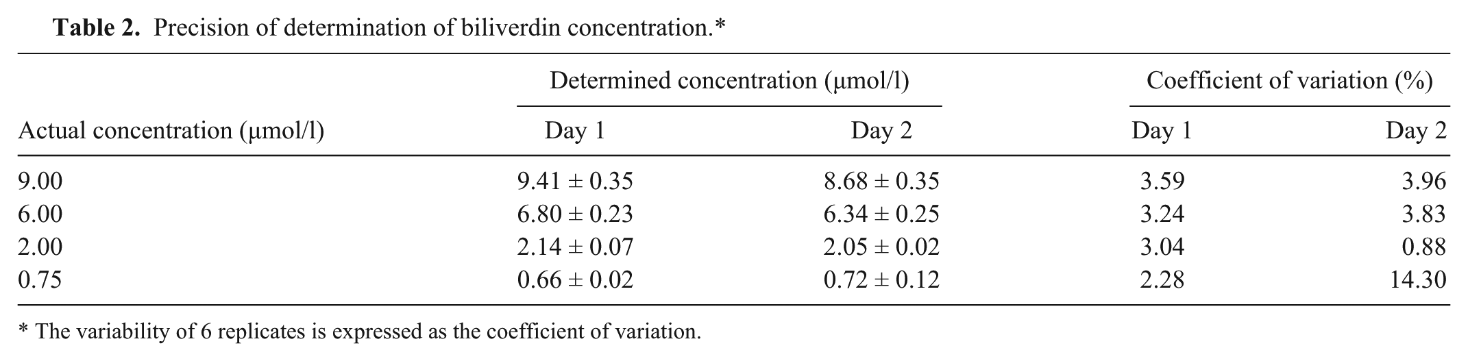
The variability of 6 replicates is expressed as the coefficient of variation.
Interference by hemin and bilirubin
Aliquots of a biliverdin solution were spiked with different concentrations of hemin and bilirubin. The accuracy of biliverdin determination, with and without the addition of hemin or bilirubin, was monitored. Hemin did not influence the determination of biliverdin regardless of its concentration (no significant difference in the assay accuracy was observed). Bilirubin, however, interfered with biliverdin determination, resulting in apparently higher biliverdin concentrations. This interference was concentration dependent in a nonlinear fashion, and resulted in a maximum 1.5-fold increase in apparent biliverdin concentration, when the molar ratio of bilirubin to biliverdin was 19:1 (Table 3).
Accuracy of determination of biliverdin concentration in the presence of various concentrations of hemin and bilirubin.*
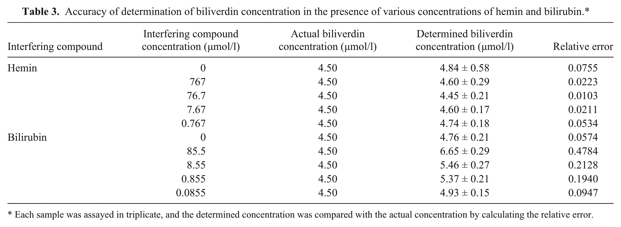
Each sample was assayed in triplicate, and the determined concentration was compared with the actual concentration by calculating the relative error.
Determination of biliverdin in chicken serum
The concentrations of biliverdin spiked in 4 samples of chicken serum were determined with high accuracy (relative error <0.0987; Table 4). The recovery of biliverdin from the serum, achieved with DMSO extraction, was very high (95–100%).
Accuracy of determination of biliverdin concentration in chicken serum.*
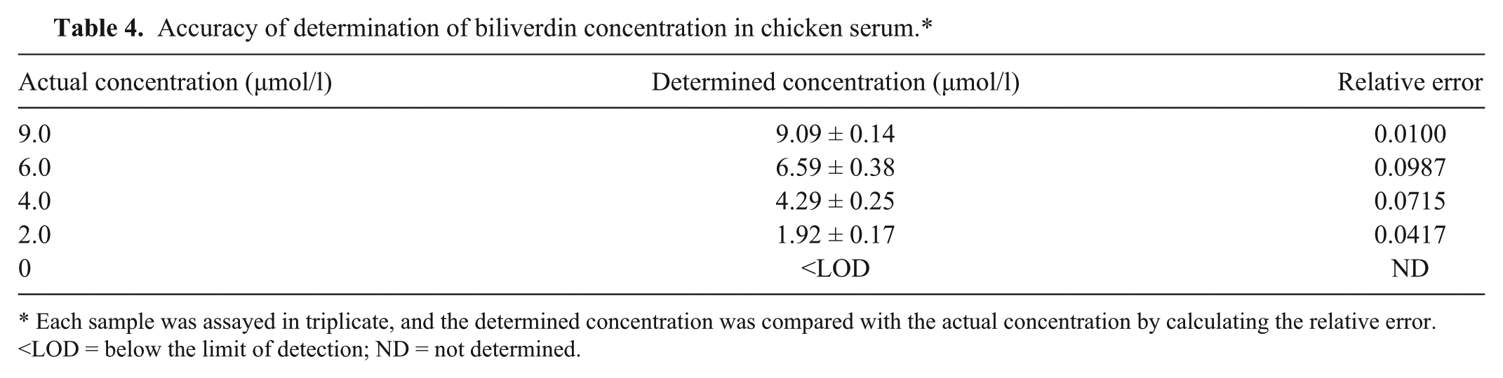
Each sample was assayed in triplicate, and the determined concentration was compared with the actual concentration by calculating the relative error. <LOD = below the limit of detection; ND = not determined.
Discussion
The current study describes a new, simple, inexpensive, and rapid biliverdin assay that is amenable for high-throughput. The flow chart of the assay is shown in Figure 3. The developed assay, contrary to those previously reported,4,6,10,12 involves neither chromatography nor enzyme assay. It relies on the specific interaction of biliverdin with iRFP, which results in strong fluorescence in the infrared part of the spectrum. The use of infrared light minimizes interference from background autofluorescence by biomolecules from living systems. 19 The specific interaction with iRFP removes the need for separation of the sample components. The reaction is set in a microtiter plate, which enables testing of multiple samples in parallel. With the exception of a microplate reader capable of measuring fluorescence in the infrared region, the assay requires no special instrumentation. The assay is fast and simple; samples are mixed with biliverdin detection reagent in the microplate and can be measured after 15 min of incubation.
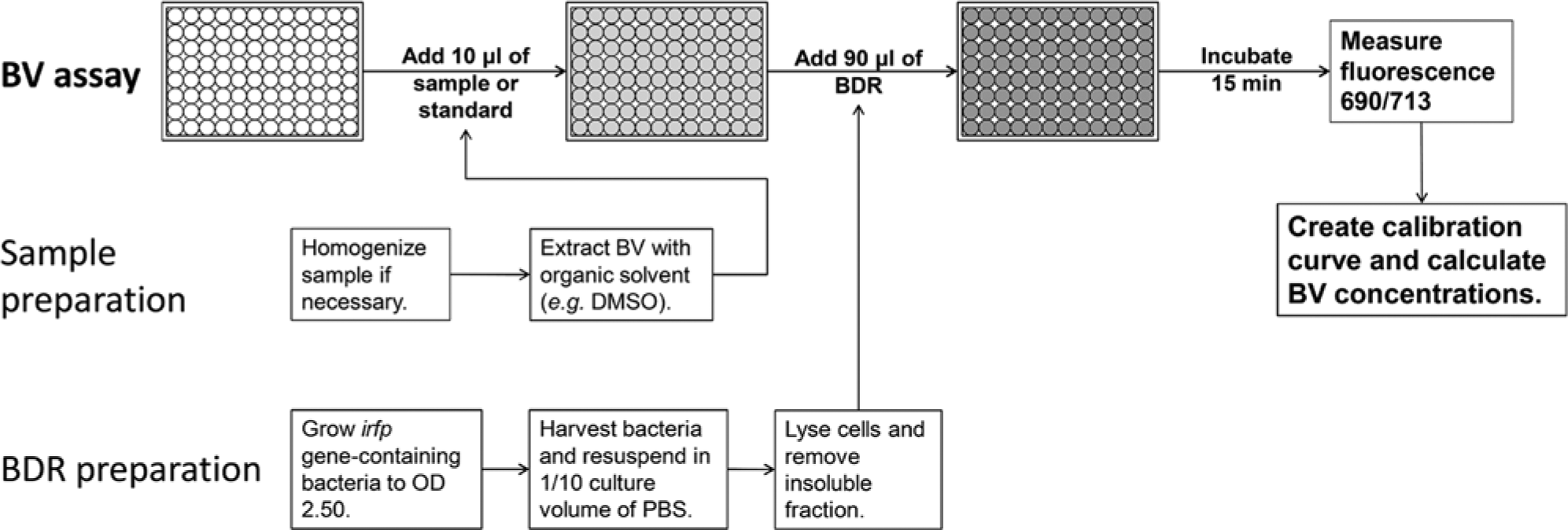
Scheme of the proposed biliverdin assay. Potential samples include serum, plasma, urine, tissue homogenate, and eggshells. BV = biliverdin; BDR = biliverdin detection reagent; DMSO = dimethyl sulfoxide; PBS = phosphate buffered saline; OD = optical density.
Biliverdin detection reagent is prepared by expressing iRFP in E. coli. The expression is driven by a constitutive promoter and requires no inducers. Infrared fluorescent protein for biliverdin assay requires no downstream processing; a simple concentrated soluble fraction of iRFP-expressing E. coli cell lysate can be used as the biliverdin detection reagent. The major requirement for its preparation is the growth of a sufficient amount of the bacteria. This can be done at low cost. At the price of LB growth medium of US$5 per liter, the costs of biliverdin detection reagent amount to approximately 1.9 cents per sample (measured in triplicate), making the assay easily affordable.
The assay is linear in the concentration range from 0 to 10 µmol/l, with the lowest calibration point set at 0.25 µmol/l. The calibration curves had correlation coefficients above 0.99, demonstrating robustness of the assay. The concentration range of the assay is similar to those of previously reported HPLC assays (i.e., 0.3–6.8 µmol/l12 and 0.138–2.2 µmol/l10), while the concentration range of the spectrophotometric assay is higher (8.5–257 µmol/l). 4
The LOD and LOQ were determined to be 0.02 and 0.03 µmol/l of biliverdin, respectively, demonstrating the ability of the assay to quantify even smaller amounts of biliverdin. The assay has good inter- and intra-day accuracy, with the determined concentrations deviating from the actual ones by no more than 15%. The precision was also good, with CV not exceeding 5%, except for 1 sample with 0.75 µmol/l concentration, where CV was below 15%. Precision and accuracy are also comparable to those of previous assays,4,10 and are in accordance with biological assay validation guidelines (Ederveen J: 2010, A practical approach to biological assay validation).
Biliverdin is a product of heme metabolism. Two compounds, hemin and bilirubin, that are upstream and downstream of biliverdin, respectively, in the heme degradation pathway, were tested for possible interference with the assay of biliverdin. Hemin had almost no influence on the accuracy of the assay. Bilirubin, on the other hand, caused an apparent increase in the biliverdin concentration by a maximum 1.5-fold, when bilirubin was in 19-fold molar excess. This could be attributed to the partial spontaneous oxidation of bilirubin samples. It is less important in avian samples, which lack bilirubin. Nevertheless, special care should be given to the storage of samples and prevention of oxidation.
Determination of biliverdin in a typical physiological sample was simulated by adding biliverdin to chicken serum. Excellent recovery (95–100%) was achieved with a simple DMSO extraction step. Accuracy and precision were good, similar to those for DMSO-prepared samples, with the determined concentration deviating by no more than 10% from the expected concentration.
The proposed biliverdin assay has a high potential for reliable quantification of the large numbers of samples generated in veterinary clinical diagnostics and research. However, it has to be kept in mind that further optimization of the assay is necessary before it can be introduced to clinical or research laboratories. First, the feasibility of the assay has to be demonstrated for clinical samples other than serum, including urine and tissue homogenates, for which appropriate extraction methods have to be developed. Second, the assay has to be validated on pathological clinical samples of avian or reptilian origin, in which an increase in biliverdin concentration is to be expected. Third, an effective method of extracting biliverdin from eggshells would enable the assay to be used in egg coloration studies.
Footnotes
Acknowledgements
The authors are grateful to Prof. Roger H. Pain for critical reading of the manuscript, Dr. Dušan Benčina for the donation of chicken serum, and Ana Nikolić for excellent technical assistance.
a.
GeneArt, Regensburg, Germany.
b.
Promega Corp., Madison, WI.
c.
Eurofins MWG Operon, Ebersberg, Germany.
d.
Hielscher Ultrasonics GmbH, Teltow, Germany.
e.
Sigma-Aldrich Chemie GmbH, Steinheim, Germany.
f.
Greiner Bio-One GmbH, Frickenhausen, Germany.
g.
Infinite M1000, Tecan Austria, Salzburg, Austria.
Declaration of conflicting interests
The author(s) declared no potential conflicts of interest with respect to the research, authorship, and/or publication of this article.
Funding
This study was supported by the Slovenian Research Agency Grant P4-0127.