
Abstract
The current report describes 3 rare cases of mammary diffuse fibroadenomatoid hyperplasia in water buffalo (Bubalus bubalis). All of the animals were between 10 and 12 months of age. Grossly, the lesions consisted of severe diffuse swelling with homogeneous large masses in the udder. Surgical removal of the masses was curative. Microscopically, there was severe hyperplasia of the mammary epithelium and numerous well-differentiated and mildly pleomorphic acini and their associated ducts. Moderate proliferation of the fibrous connective tissue and the myoepithelial cells near the proliferating acini was also evident. The hyperplastic epithelial cells exhibited positive immunostaining for cytokeratin, estrogen receptors, and progesterone receptors. In addition, the myoepithelial cells displayed moderate positivity for alpha smooth muscle actin. Based on the clinical, morphologic, and immunohistochemical findings, a diagnosis of mammary diffuse fibroadenomatoid hyperplasia with probable hormonal influence was made.
Proliferative changes in the mammary gland are rarely observed in farm animals. Sporadic cases of mammary neoplasia have been described in mares,1,6 cattle,11,14 goats, 16 and water buffalo, 10 whereas hyperplastic lesions have been diagnosed only in cattle. 7 Mammary epithelial hyperplasia is classified into 3 distinct types: ductal, lobular, and adenosis. 2 A fourth type of this disorder, called fibroadenomatoid hyperplasia, has been reported in women as a benign lesion that is characterized by prominent hyperplasia of the epithelial elements and multifocal proliferation of the interlobular fibrous stroma. Fibroadenomatoid hyperplasia may be a localized or diffuse process and is found in approximately 5–7% of all benign surgical biopsies from American and Japanese women. 5 In human beings, this type of lesion was formerly known as sclerosing lobular hyperplasia, fibroadenomatosis, and fibroadenomatoid mastopathy. 5 The objective of the current study was to describe the clinicopathologic and immunohistochemical findings of 3 rare cases of mammary diffuse fibroadenomatoid hyperplasia in young water buffalo.
Case 1, observed in 1985, was a 10-month-old intact female buffalo of the Mediterranean breed (Bubalus bubalis) that presented a swelling in the udder that had progressively evolved over 4 months. Two cases with similar gross lesions had been observed on this farm during the previous year. This animal was from a herd of 110 water buffalo heifers of similar age that were raised in the county of Camaquã, state of Rio Grande do Sul, Brazil. Treatment with chloramphenicol was unsuccessful. The lesion, which affected all 4 quarters, was surgically removed. The excised mammary gland was white, spongy, and swollen, presenting 2 poorly circumscribed masses that measured 44 cm × 24 cm × 24 cm and 35 cm × 20 cm × 14 cm. The cut surface of the mammary gland showed numerous coalescing lobules separated by fibrous septa. The second case, in a 12-month-old intact Mediterranean-breed female buffalo, was observed on the same farm in 1987 in a herd of 150 heifers of the same age. The farmer reported that the animal had exhibited the progressive enlargement of the mammary gland throughout at least 4 months. Surgery was also performed to remove the mammary gland. Grossly, the lesions were similar to and of approximately the same size as those observed in the previous case.
Case 3, observed in 2011, was a 10-month-old intact female mixed-breed buffalo with a 6-month history of progressive and diffuse enlargement of the udder (Fig. 1A), affecting all 4 quarters. This animal was from a herd of 40 water buffalo that were raised in the county of Cromínia, state of Goiás, Brazil. Treatment with dexamethasone and penicillin was administered, but no clinical improvement was observed. The gland was removed surgically. The entire excised mammary gland was a whitish, smooth, homogeneous round mass with spongy areas, measuring 45 cm × 44.5 cm × 43.5 cm. The lesion was not well-circumscribed but rather had infiltrated the entire udder. The cut surface was multilobulated, with the lobules separated by fibrous septa.
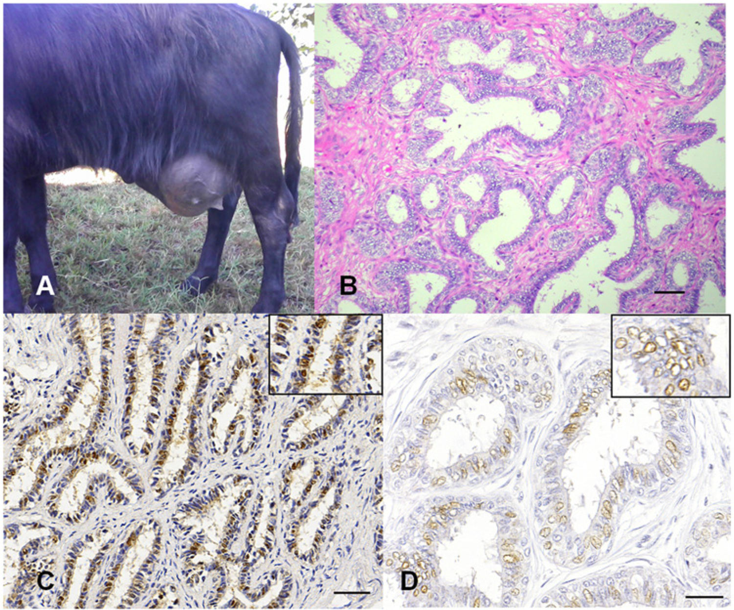
Mammary fibroadenomatoid hyperplasia; water buffalo (Bubalus bubalis).
No remaining normal mammary tissue was observed in the mammary region after surgery in all 3 cases. Furthermore, there was no indication of the recurrence of mammary lesions at 2 years after the surgical removal of the masses.
Samples of the mammary lesions from the 3 cases were collected, fixed in 10% formalin for 24 hr, and processed using routine methods for histological evaluation. Sections were stained with hematoxylin and eosin, and immunohistochemical staining (IHC) was performed using a biotin–peroxidase system, with labeling of the secondary antibody with a polymer a and diaminobenzidine as the chromogen. Antigen retrieval was performed by pressurized heating at 125°C in a citrate-buffered solution at pH 6.0 b (for cytokeratin and alpha smooth muscle actin) or in an ethylenediamine tetra-acetic acid buffer solution at pH 9.0 (for the estrogen receptor and progesterone receptor). b To block the endogenous peroxidase activity, the slides were incubated in a solution of H2O2 (3%) in methyl alcohol. The reagents were applied manually, with a 1-hr incubation for the monoclonal primary antibodies and a 30-min incubation for the other reagents, except for the diaminobenzidine chromogen, which was applied for 5 min. The IHC antibody panel is described in Table 1. The IHC sections were counterstained using Harris hematoxylin. The positive controls for IHC consisted of normal mammary glands. For the negative controls, the primary antibodies were replaced with normal serum. c
Panel of antibodies used in the current study.
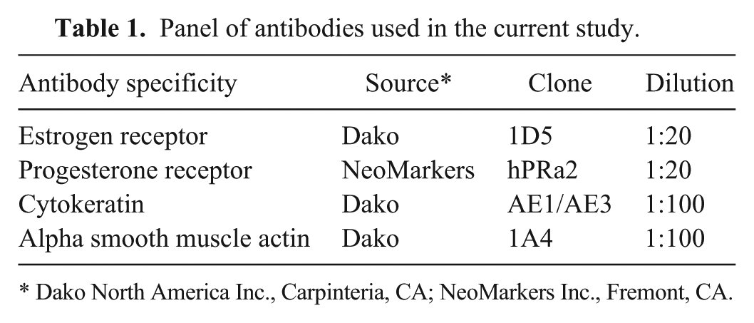
Dako North America Inc., Carpinteria, CA; NeoMarkers Inc., Fremont, CA.
Microscopically, all the cases displayed evidence of severe proliferation of the mammary epithelium with the formation of numerous, well-differentiated, and mildly pleomorphic acini and their associated ducts (Fig. 1B). The acini had 1–4 layers of columnar or cuboidal epithelial cells and were moderately dilated. The cell borders were distinct. The cytoplasm was slightly eosinophilic, and the nucleus was round and hypochromatic. Moderate proliferation of the fibrous connective tissue and the myoepithelial cells among the proliferated acini was evident (Fig. 1B). In the 3 cases, many of the affected epithelial cells showed strongly positive cytoplasmic labeling for cytokeratin and mild to moderate nuclear staining for estrogen receptors (Fig. 1C) and progesterone receptors (Fig. 1D). In addition, the myoepithelial cells exhibited moderately positive reactions for alpha smooth muscle actin. The location, intensity, and distribution of the IHC labeling were very similar in all 3 cases. Based on the morphologic and immunohistochemical findings, a diagnosis of mammary diffuse fibroadenomatoid hyperplasia with probable hormonal influence was made.
Hyperplastic changes in the mammary gland are diagnosed frequently in dogs and cats but very rarely in herbivores.7,12 The pathogenesis of this proliferative disorder is poorly understood, but hormonal changes appear to be involved in female dogs and cats. 12 A case of mammary hyperplasia in a 13-year-old cow was attributed to chronic irritation in association with chronic mastitis. 7 The authors considered hyperplasia to be a regenerative response to compensate for the damaged mammary tissues. In the buffalo in the current study, there were no inflammatory lesions in association with the fibroadenomatoid hyperplasia. Endocrine influences on the hyperplasia were suspected, given the IHC-labeling patterns (positive reactions for estrogen and progesterone receptors). In contrast, a mammary diffuse fibroadenomatous change (fibroepithelial hyperplasia) in cats usually presents with high concentrations of only progesterone receptors. 12 In cattle, estrogen and progesterone receptors have been observed in mammary tissues during many physiological stages, such as in prepubertal heifers, primigravid cows, lactating pregnant cows, and lactating nonpregnant cows. 3 Compared with the condition in normal mammary epithelium, 3 the affected animals in the present study showed increased progesterone receptor expression.
The 3 affected buffalo in the current investigation were young animals, between 10 and 12 months of age. Many cases of proliferative mammary changes (including neoplasms) in dogs and cats have been observed in adult animals, mainly after previous therapeutic or spontaneous exposure to ovarian hormones.12,15 In human beings, cases of mammary fibroadenomatoid hyperplasia have been diagnosed in adult women with mean ages of 32–33 years5,13 and 58 years. 9
The possible differential diagnoses in these cases included mammary neoplasia and mastitis. Initially, because of the large size of the lesions, a presumptive diagnosis of mammary neoplasia was indicated, but the histology combined with the clinical and gross features confirmed a diagnosis of mammary diffuse fibroadenomatoid hyperplasia. The histological findings observed in the present cases are similar to those of a mammary fibroadenoma, which is a common neoplasm in young rats and women and has been described occasionally in a heifer, 17 a young cow, 11 and a lamb. 4 Mammary fibroadenomas have been reported in some rat strains, such as Sprague–Dawley, and mainly in animals used in carcinogenicity studies. 8 The morphologic aspects of this tumor are very similar in all of the affected species. This benign neoplasm generally forms well-delimited nodules, whereas fibroadenomatoid hyperplasia is characterized by extensive and diffuse lesions throughout the mammary glands, 5 as in the present cases. The results of the present investigation suggest that the prognosis for this lesion is good because there was no recurrence of the mammary masses in any of these cases for at least 2 years after their surgical excision.
Footnotes
a.
Advance HRP enzyme, Dako North America Inc., Carpinteria, CA.
b.
Dako North America Inc., Carpinteria, CA.
c.
Ultra V Block, NeoMarkers Inc., Fremont, CA.
Declaration of conflicting interests
The author(s) declared that they have no conflicts of interest with respect to their authorship or the publication of this article.
Funding
The author(s) received no financial support for the research, authorship, and/or publication of this article.