
Abstract
Squamous cell carcinoma (SCC) is the most common tumor arising in the periocular and penile areas of horses. Both ultraviolet radiation and papillomaviruses have been implicated in the pathogenesis of SCC in various species, including the horse. This retrospective study used polymerase chain reaction (PCR) to detect papillomavirus DNA in archival biopsy samples from equine periocular and penile SCC from 3 different geographic areas (northeast, southeast, and central United States). Forty-two periocular SCCs were tested; none contained papillomavirus DNA. Twenty-two penile SCCs were tested, and papillomavirus DNA was identified in 10 (43%) cases. Sequencing of the PCR products revealed homology with Equus caballus papillomavirus 2 (EcPV-2). No geographic distribution in the detection of papillomavirus was identified. Penile SCCs were significantly more likely to be papillomavirus positive than the periocular SCCs (P < 0.001). The role of papillomavirus in the development of penile SCC requires further investigation. The differing pathogeneses of periocular and penile SCC suggest that the tumors may respond differently to treatment.
Squamous cell carcinomas (SCCs) are the most common tumors of the eye and ocular adnexa, and the penis and prepuce in the horse.7,19 Periocular SCC commonly affects the eyelids, nictitans, conjunctiva, and limbus.3,7 Appaloosas and American Paint Horses are most commonly affected. Because such tumors result in part from prolonged exposure to ultraviolet radiation, this predisposition is explained by the frequent lack of periocular pigmentation in these breeds. 3 As with most neoplasms, the prevalence of periocular SCC increases with age. 3 Ocular SCC can invade local soft tissues, the bony orbit, sinuses, and brain. Metastases to regional lymph nodes, salivary glands, and the thorax also rarely occur. 21
Penile SCCs typically arise on the glans portion of the penis and occur in equal frequency in stallions and geldings. 6 Although some report no breed predispositions, 19 others report that Appaloosas and American Paint and Quarter Horses are overrepresented. 18 The tumors frequently infiltrate the corpus cavernosum and may metastasize to regional lymph nodes, but distant metastases are rare. 6 The average age of diagnosis is 19.5 years. 19 Penile papillomas are the second most common tumor arising on the penis and prepuce, and like SCCs, most occur on the glans penis. 19 Penile papillomas are thought to progress to in situ carcinomas and ultimately SCCs. 12
Since 2010, there have been several reports describing the variable presence of equine papillomavirus DNA in periocular and penile SCC. The purpose of the current study was to detect equine papillomavirus DNA, via polymerase chain reaction (PCR), in both periocular and penile SCC, from 3 regions of the United States, to further elucidate the etiopathogenesis of these tumors.
Pathology records from the University of Tennessee (Knoxville, Tennessee), Tufts University (Medford, Massachusetts), and Colorado State University (Fort Collins, Colorado) were searched retrospectively for cases of equine SCC that affected the periocular or penile region. Histologic slides from the cases were reviewed by a board-certified pathologist (KMN, JAL, EJE) to confirm the diagnosis. Cases were selected for further diagnostics based on availability of sufficient tumor tissue and to achieve a similar number of cases from each periocular location and submitting institution. A representative slide from each case was selected for PCR analysis. Samples dated from 1996 to 2012. Samples of DNA were extracted from 25-µm-thick scrolls of formalin-fixed, paraffin-embedded tissue with a commercial kit a according to the manufacturer’s protocol. Between each sample, the microtome blade was changed and forceps were cleaned with 95% alcohol to avoid contamination. The DNA was stored at −20°C prior to analysis.
To amplify papillomavirus DNA from the examined samples, multiple primer sets were tested using previously reported techniques. The primer sets were FAP59/FAP64 and the CP65/CP70 and CP66/CP69 nested PCR sets that have been shown to amplify various papillomavirus types from both human beings and animal species.1,5 These degenerate primers amplified a feline papillomavirus DNA template, which was used as a positive control. Additional primer sets used were EPV-F/EPV-R and EPV1F/ EPV1B that were designed to amplify EcPV19,13; EcPV 2-NB forward and reverse primers that were designed to amplify EcPV2 2 ; and SPVF/SPV that were designed to amplify bovine papillomavirus from equine sarcoid. 20 The primer sets did not amplify a feline papillomavirus control DNA template. All 6 primer sets were applied to each DNA sample.
Additionally, to ensure that the extracted DNA contained amplifiable DNA, primers that amplify a 267-bp region of an exon in the equine immunoglobulin gamma 7 heavy chain constant region gene (GenBank accession no. AJ302058) were used in a PCR assay. The amplification was performed as follow: 94°C for 10 min, 35 cycles of 94°C for 1 min, 50°C for 1 min, 72°C for 1 min, and finally 72°C for 10 min. Samples that did not amplify this reference gene were excluded from the study.
Each PCR mixture contained 1 µl each of a forward and reverse primer (concentration: 50 µM), 8 µl of nuclease-free water, 12.5 µl of Taq premix, and 2.5 µl of DNA template. All PCR reactions included a negative control that did not contain template DNA. The PCR reactions were conducted, according to published criteria, in an automated thermocycler, and the PCR products were analyzed by electrophoresis in a 1.4% agarose gel containing ethidium bromide. Negative controls were included for each primer set, and none amplified DNA from nontemplate samples.
Although positive controls were not available for all primer sets, all PCR products were sequenced to confirm the fidelity of the primers and the amplified product. To sequence PCR products, primers were digested using ExoSAP-IT, b according to the manufacturer’s instructions. Samples were sequenced at the University of Tennessee Molecular Biology Resource Facility using a commercial kit c and a capillary electrophoresis instrument. c The sequences were compared with known sequences from GenBank using the basic local alignment search tool (BLAST; http://www.ncbi.nlm.nih.gov/blast/Blast.cgi).
All statistical tests were carried out using a commercial software program. d Continuous data for age was normally distributed and expressed as mean and range. Comparison of source (University of Tennessee, Colorado State University, and Tufts University) with samples positive or negative for papillomavirus was done using the Fisher exact test. The association of age with location (penile vs. periocular), periocular location (third eyelid, conjunctiva, or lid), and papillomavirus (positive or negative) was conducted using the t-test procedure. A P value of 0.05 was used for statistical significance in all tests.
The 42 periocular SCC samples included 17 tumors from the University of Tennessee in the southeastern United States, 13 tumors from Tufts University in the northeastern United States, and 12 from Colorado State University in the central United States. Locations of the periocular tumors included third eyelid (nictitans; 20), cornea or limbus (10), eyelid (8), and conjunctiva (4). The average age of horses with periocular SCC was 12.8 years (range 3.5–24 years). No papillomavirus DNA was amplified from any periocular SCC with any of the primer sets (Table 1). There was no association between age of the horse and the periocular location of the SCC (P = 0.76).
Squamous cell carcinomas: location, source, signalment, year sample was collected, and papillomavirus polymerase chain reaction (PCR) results.*
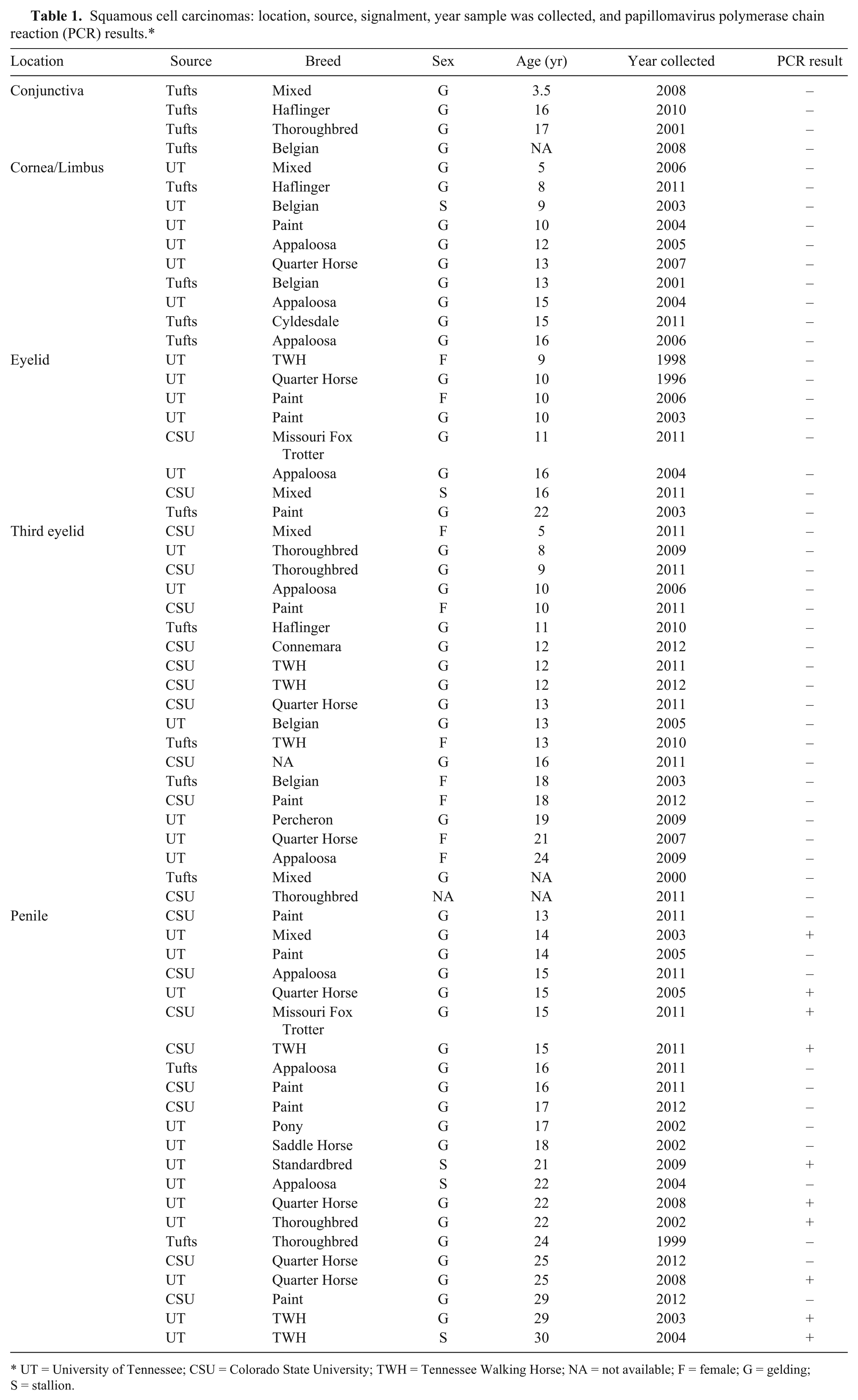
UT = University of Tennessee; CSU = Colorado State University; TWH = Tennessee Walking Horse; NA = not available; F = female; G = gelding; S = stallion.
The 22 penile SCC samples included 12 tumors from the University of Tennessee, 2 tumors from Tufts University, and 8 from Colorado State University. The average age of horses with penile SCC was 19.7 years (range 13–30 years; Table 1). The horses with penile SCC were significantly older than the horses with periocular SCC (P < 0.0001).
Papillomavirus DNA was amplified from 10 out of 23 (43%) penile SCCs using primers designed to amplify EcPV-2. All PCR products were sequenced; sequencing demonstrated 99% homology to EcPV-2 E1 gene, over 670 base pairs. Papillomavirus DNA was not detected with the primers designed to amplify EcPV-1 or Bovine papillomavirus. Penile SCCs were significantly more likely to be papillomavirus positive than the periocular SCCs (P < 0.001). Eight of the 10 (80%) papillomavirus-positive horses were from the University of Tennessee, which represented 66% of the horses from that location. Two (20%) of the papillomavirus-positive horses were from Colorado State University, which represented 25% of horses from that location. There was no association between the geographic origin of the sample and papillomavirus-positive test results (P = 0.17). The average age of the 10 papillomavirus-positive horses was 20.8 years (range 14–30). There was also no association between age and papillomavirus-positive test results (P = 0.41). Two of the papillomavirus-positive horses were stallions, and the remaining 8 were geldings. The breeds represented included American Quarter Horse (3), Tennessee Walking Horse (3), and 1 each of Standardbred, Thoroughbred, Missouri Fox Trotter, and mixed breed.
The exact pathogenesis of SCC is not known, but a combination of factors, including ultraviolet radiation, lack of pigmentation, and papillomavirus, have been implicated. The findings in the current study suggest that SCCs have differing pathogeneses and therefore, may respond differently to treatment.
Papillomaviruses are nonenveloped, double-stranded DNA viruses that are associated with papillomas and SCCs in a variety of species. 13 Since 2010, papillomavirus DNA has been repeatedly identified from penile papillomas, in situ carcinomas, and SCCs in horses from Switzerland, Belgium, Australia, Austria, and the United Kingdom.2,12,15 A Belgian study found Equus caballus papillomavirus 2 (EcPV-2) DNA in 94% of equine penile SCC (n = 16). 2 In the United States, EcPV-2 DNA has been identified in 45% (n = 20) 10 and 80% (n = 20) 10 of penile SCC. In samples from non-SCC penile disease in horses, reports of EcPV-2 DNA identification vary from as low as 5% (n = 20) 11 and 16% (n = 19) 10 to up to 55%.11,12 In samples of normal penile mucosa from slaughtered horses in New Zealand, EcPV-2 DNA was found in only 2% (n = 32) of samples. 10 Similarly, EcPV-2 DNA has been found in 10% of penile swabs from horses without penile lesions (n = 39). 2 Papillomavirus DNA has not been identified in smegma (n = 27) or scrotal skin (n = 13). 15 The current report identified EcPV-2 DNA in 43% of penile SCC; this is similar to a previous report, 11 but much lower than other reports.2,10 The reason for the variation in detection rates is unclear and may reflect differing methodologies or geographic variation. The significantly higher rate of EcPV-2 DNA isolation in equine penile SCC, compared with non-SCC disease, suggests a causal association between the virus and the development of SCC. 10 However, the variable presence of papillomavirus DNA in penile SCC may suggest that the precursor lesions (papillomas) are initiated by the virus, but subsequent promotion and progression to malignancy is not directly associated with the virus. 16 Future studies will investigate the presence of EcPV-2 in non-SCC penile diseases.
There are fewer reports investigating the association of papillomaviruses with periocular SCC. One group failed to find papillomavirus in ocular SCC (n = 10) from horses in Australia, Austria, and the United Kingdom. 15 Another group found EcPV-2 DNA in 1 out of 30 (3.3%) ocular swabs from healthy horses and in SCC-affected retrobulbar tissue, but not from a SCC-affected nictitans. 17 In 2013, a group failed to identify EcPV-2 DNA from the normal nictitans from 75 slaughtered horses in New Zealand. 10 There is a report of a horse with ocular and periocular SCC and the copresence of bovine papillomavirus 1 (BPV-1) and EcPV-2 DNA in the initial tumor and metastatic lesions. 8 Bovine papillomaviruses have also been associated with equine sarcoids. Periocular SCCs are common in cattle, and reports vary regarding bovine papillomavirus being associated with these lesions.4,14 The current report failed to amplify EcPV-2 DNA from any of the periocular SCCs.
In conclusion, EcPV-2 DNA was amplified from 43% of penile SCCs, but was not detected in any of the ocular SCCs, regardless of the geographic origin of the sample. Thus, penile SCCs were significantly more likely than periocular SCCs to be associated with EcPV-2 DNA, suggesting the possibility for differing pathogeneses for these 2 entities. Further investigation on the role of papillomavirus in the pathogenesis of penile SCC is warranted.
Footnotes
Acknowledgements
The authors thank Misty Bailey for technical editing of the manuscript.
Declaration of conflicting interests
The author(s) declared no potential conflicts of interest with respect to the research, authorship, and/or publication of this article.
Funding
The author(s) received no financial support for the research, authorship, and/or publication of this article.
a.
DNeasy blood and tissue kit, Qiagen Inc., Valencia, CA.
b.
ExoSAP-IT, Affymetrix, Santa Clara, CA.
c.
ABI prism dGTP BigDye terminator cycle sequencing kit, ABI 3730 capillary electrophoresis instrument; Applied Biosystems, Foster City, CA.
d.
SAS version 9.3, SAS Institute, Cary, NC.