
Abstract
Submission of a raccoon (Procyon lotor) for necropsy following exhaustion at a California wildlife care center revealed minimal gross pathologic changes and only mild vacuolar changes in the white matter of the brain. Turquoise granular material was noted in the gastrointestinal tract and was submitted for toxicological testing along with portions of the brain, liver, kidney, and mesenteric and perirenal adipose tissues. Testing of the turquoise material for 7 anticoagulant rodenticides, strychnine, 4-aminopyridine, starlicide, and salts revealed none of these compounds; however, desmethylbromethalin was detected by high-performance liquid chromatography–tandem mass spectrometry. Other tissues were subsequently analyzed; the mesenteric and perirenal adipose tissues contained desmethylbromethalin. Desmethylbromethalin is the active metabolite of bromethalin, uncouples oxidative phosphorylation, and results in cerebral edema. Bromethalin is a rodenticide that is visually indistinguishable from many other rodenticides, making identification of poisonings by appearance alone nearly impossible. Based on the pathological and toxicological findings, a diagnosis of bromethalin toxicosis was established. In cases of wildlife species with unknown deaths or inconsistent clinical signs with normal or minimal histological findings, bromethalin toxicosis should be considered as a differential. Adipose tissue is the tissue of choice and can be easily harvested from a live or deceased animal to help confirm or rule out bromethalin exposure or intoxication.
Keywords
Bromethalin is a rodenticide that targets the nervous system and has been available since 1985 as 0.01% bait pellets, bars, and place packs (U.S. Environmental Protection Agency (EPA): 1998, Reregistration Eligibility Decision (RED): rodenticide cluster. EPA-738-F-98-004. Available at: http://www.epa.gov/oppsrrd1/REDs/factsheets/2100fact.pdf). Unfortunately, bromethalin cannot visually be distinguished from other rodenticides, making identification of poisonings by color or appearance alone nearly impossible. 1 After ingestion, bromethalin is metabolized in the liver to its active metabolite desmethylbromethalin, which uncouples mitochondrial oxidative phosphorylation leading to decreased cellular adenosine triphosphate (ATP) production and disruption of sodium–potassium ATPase pumps. 13 Bromethalin and desmethylbromethalin are lipid soluble based on their log P values of 6.70 and 4.26, respectively; thus, both compounds are capable of crossing the blood–brain barrier and are not known to be substrates of multidrug resistance transporters that would lead to removal from the central nervous system (CNS).12,15 Inability to maintain a normal sodium–potassium gradient leads to fluid buildup inside the CNS resulting in the development of cerebral edema and increased cerebrospinal fluid pressures. 13 Experimental bromethalin intoxication has been described in cats, dogs, monkeys, and rodents.4,5 Interestingly, despite the reporting of significant numbers of bromethalin poisonings at animal and human poison control centers 9 across the United States, there is only one case report documenting a suspected poisoning in a cat 8 and one report of a fatal intoxication in a human being. 10 Thus, the current report describes the salient histological and toxicological findings in a raccoon poisoned by bromethalin.
Clinical signs of intoxication, which have been described in cats, dogs, and rodents, generally develop within 24 hr after ingestion of bromethalin at or above the median lethal dose (LD50) and include muscle tremors, hyperthermia, hyperexcitability, and focal and generalized seizures.3,13 At concentrations below LD50, a paralytic syndrome has been described in which clinical signs develop over several days and include ataxia, CNS depression, and paralysis. These signs can worsen over several weeks, and animals may progress to a comatose or semicomatose state.1,2 Diffuse spongy degeneration and glial cell hypertrophy within the white matter along with vacuolization of the optic nerve are characteristic postmortem microscopic findings.4,5
In October 2012, a male juvenile raccoon (Procyon lotor) was submitted to the California Animal Health and Food Safety Laboratory (CAHFS; Davis, California) for postmortem examination. The raccoon received subcutaneous fluids, oral corn syrup, and external warming after arriving hypothermic, semiconscious, and nonresponsive at a wildlife care center in San Rafael, California. The raccoon died shortly thereafter. Turquoise, granular fecal material was noted in the cage by the care center staff upon death.
At necropsy, the raccoon was in good flesh, previously frozen, and in fair postmortem condition upon thawing. There was light froth in the trachea and bronchi, and lungs were mottled dark pink-purple. Liver, kidneys, bladder, heart, spleen, thymus, brain, nasal passage, esophagus, and pancreas were grossly unremarkable. The stomach was filled with turquoise granular ingesta, and the colon was impacted with very firm, dry turquoise-colored feces (Fig. 1). No hemorrhage was evident in the carcass. No significant microscopic changes were seen on examination of select 5-µm tissue sections stained with hematoxylin and eosin, including brain, heart, trachea, lymph node, lungs, tonsils, kidneys, adrenal glands, thymus, spleen, liver, testes, stomach, intestines, pancreas, skeletal muscle, and peripheral nerves. Miscellaneous findings on microscopic examination were dense eosinophils in bronchial adventitia with few luminal neutrophils. There was mild vacuolar change in the white matter of the brain.
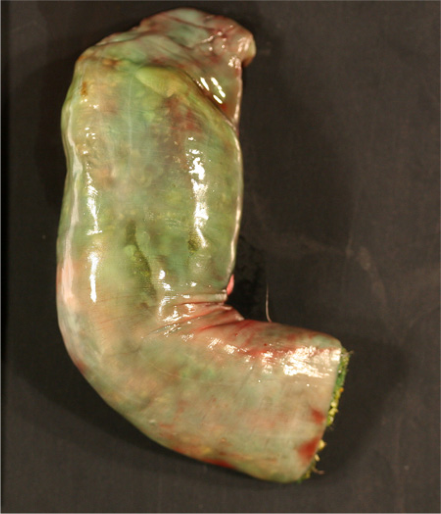
Raccoon (Procyon lotor). Turquoise-colored feces within the colon.
Selected pieces of formalin-fixed cerebellum were postfixed in 2.0% glutaraldehyde and then routinely processed and embedded in epoxy resin a ; selected thick sections were stained with toluidine blue. Ultrathin sections from selected white matter regions were examined using a transmission electron microscope. b Multifocal myelin splitting was seen in myelin sheaths in thin tissue section electron microscopy.
Portions of brain, liver, kidney, mesenteric fat, perirenal fat, and stomach content were submitted for toxicological testing. Although hemorrhages were not seen on gross or microscopic examinations, liver and stomach content were screened for 7 anticoagulants by high-performance liquid chromatography–tandem mass spectrometry (HPLC-MS/MS). 11 The compounds tested included first-generation anticoagulant rodenticides, warfarin, diphacinone, chlorophacinone, and coumachlor, as well as second-generation anticoagulant rodenticides, brodifacoum, bromadiolone, and difethialone. None of the listed anticoagulant rodenticides were detected in the liver or stomach content.
The unique turquoise coloring of the stomach contents was suggestive of the dyes used in other non-anticoagulant rodenticides, commercial fertilizers, and pesticides. Therefore, the stomach content was tested for bromethalin, strychnine, 4-aminopyridine, starlicide, and minerals that may suggest ingestion of fertilizer. Strychnine, 4-aminopyridine, and starlicide were not detected in the stomach content, and the concentrations of potassium and sodium (2,200 mg/kg wet weight [ww] and 1,400 mg/kg ww, respectively) were not consistent with those found in most fertilizers. Ocular fluid was analyzed for sodium, potassium, phosphorus, calcium, and magnesium, and all minerals were found within safe limits. Desmethylbromethalin was detected in the stomach content. Subsequently, brain, liver, kidney, mesenteric adipose, and perirenal adipose were also tested for desmethylbromethalin.
Briefly, 1-g aliquots of each tissue were extracted by homogenization with 5% ethanol in ethyl acetate. The extracts were evaporated under nitrogen, exchanged into 0.5 ml of methanol, and then analyzed by HPLC-MS/MSc,d using electrospray ionization. A 2.1 × 100 mm, 1.8-µm HPLC column e was used with a binary gradient consisting of 0.1% formic acid in water and 0.1% formic acid in methanol. Full-scan MS/MS of precursor ions at m/z 562 and 564 was used to identify desmethylbromethalin. Spectra and retention times of tissue analysis were compared with those obtained via analysis of a certified desmethylbromethalin standard f and with analysis of extracts of control samples (bovine adipose tissue) fortified with this standard. The limit of detection for this analysis was 0.050 µg/g (ww) for each of the tissue types. Desmethylbromethalin was detected in the mesenteric and perirenal adipose tissue but not in brain, liver, or kidney. Extracts of mesenteric and perirenal adipose tissue samples were also later analyzed using selective reaction monitoring MS/MS.g,h The HPLC column used was a 2.1 × 100 mm, 1.8-µm column. i Mobile phases consisted of 0.1% formic acid in water and 0.1% formic acid in acetonitrile, and gradient elution was utilized. Precursor/product ion transitions of m/z 562/278 and 562/254 with collision energies of 25 and 35 V, respectively, were used for desmethylbromethalin detection. The limit of detection was 0.001 µg/g (1 ng/g; ww), and both of the adipose tissue samples were again positive for desmethylbromethalin using this method. Full quantification of desmethylbromethalin in the positive specimens was not performed. Development of an HPLC-MS/MS analysis for bromethalin was also attempted on both instrument types. Analysis of certified bromethalin standard material j gave no signal consistent with the molecular structure of this compound on either instrument, likely due to poor ionization by the electrospray process. This information indicates that bromethalin is not amenable to analysis by electrospray HPLC-MS/MS and that a different analytical technique would be required for its detection.
Based on the postmortem finding, histology, and toxicological findings, a diagnosis of bromethalin toxicosis was confirmed in this raccoon. In acute, single-feeding studies of dogs, detection of bromethalin was documented in kidney, liver, adipose, and brain 15–63 hr after ingestion. 4 In the present case, desmethylbromethalin, not bromethalin, was detected in the adipose and stomach contents. The inability to detect bromethalin in the various tissues may be due to the rapid and efficient metabolism of bromethalin to desmethylbromethalin by cytochrome P450 N-demethylation in the liver, 1 the fact that the applied HPLC-MS/MS analysis for bromethalin did not allow for its detection, or because of lack of sensitivity of the method. In fact, in a plasma radiocarbon study in rats, 100% of the circulating radioactivity after injection of a single dose of bromethalin was due to desmethylbromethalin, confirming that bromethalin is rapidly metabolized. 13 Desmethylbromethalin is a lipophilic substance, and thus, may have been preferentially concentrated in adipose tissue in this raccoon. In the past 10 years, it was shown that P450 isoforms, specifically CYP1A1, CYP1B1, and CYP2B1, are present and functional in mammalian adipose tissue.7,14 Thus, it is also possible that the highly lipophilic compound bromethalin is distributed to and accumulates in adipose tissue where it is subsequently metabolized by adipose P450 to desmethylbromethalin.
It must be emphasized, from a veterinary diagnostic perspective, that desmethylbromethalin was not detected in liver, kidney, and brain, but in adipose tissue. Other studies have also shown that, following intoxication, the highest concentrations of desmethylbromethalin are found in adipose tissue. 4 The utilized HPLC-MS/MS method provides detection of desmethylbromethalin down to 0.001 µg/g (1 ng/g), which is considered a limit of detection of excellent diagnostic use as most animals poisoned by bromethalin will likely have desmethylbromethalin concentrations higher than 0.001 µg/g (1 ng/g) present in adipose tissue. The finding of desmethylbromethalin in adipose tissue has significant implications for diagnosticians, veterinarians, and wildlife personnel. In cases in which the cause of death is suspected to be from bromethalin toxicosis or is unknown, adipose tissue should be collected as it represents the tissue sample of greatest diagnostic use for toxicological testing to identify the metabolite desmethylbromethalin.
Light microscopic and ultrastructural findings in the current case were very mild as compared to those described in previous studies.4,5 The mild vacuolization in the white matter of the brain, evident on light microscopic examination, could have been attributed to either bromethalin intoxication or to autolysis. Moreover, myelin splitting demonstrated by transmission electron microscopy could not distinguish ultrastructural changes associated with bromethalin intoxication in contrast to autolysis, indicating light and ultrastructural changes cannot be used to confirm or rule out poisoning with bromethalin.
It is currently more common to find reports of anticoagulant rodenticide exposure in wildlife associated with exposure to compounds such as brodifacoum, bromadiolone, or diphacinone (Erickson W, Urban D: 2004, Potential risks of nine rodenticides to birds and nontarget mammals: a comparative approach. EPA ref. no. P.2004.27 A. Available at: www.fluoridealert.org/pesticides/EPA-HQ-OPP-2006-0955-0005.pdf). This trend is likely to change because of the removal of second-generation anticoagulant rodenticides from local retailers in 2011 (EPA: 2008, Risk mitigation decision for ten rodenticides. ID no. EPA-HQ-OPP-2006-0955-0764. Available at: http://www.epa.gov/oalj/filings/Reckitt_HrgReq_Ex03.pdf). Henceforth, the incidence of domestic and wildlife intoxication with faster-acting rodenticides such as bromethalin, strychnine, and zinc phosphide is likely to increase. 6 In fact, the American Society for the Prevention of Cruelty to Animals Animal Poison Control Center has documented an increase in reports from bromethalin-based rodenticides. 9 In addition, primary nontarget intoxication of raccoons may be underreported because this species is typically seen as pests and subsequent necropsy of deceased individuals is not pursued by local residents or animal control officers. Although the risk of secondary poisoning is low, the potential still exists (Jackson WB, Spaulding SR, Van Lier RBL, Dreikorn BA: 1982, Bromethalin—a promising new rodenticide. In: Proceedings of the Tenth Vertebrate Pest Conference, ed. Marsh RE, pp. 10–16, Monterey, CA). To the authors’ knowledge, no studies have been published regarding the LD50 in specific wildlife species such as the raccoon. Working more closely with wildlife care centers will enable better assessment of primary and secondary nontarget exposure and/or intoxication of wildlife to non-anticoagulant rodenticides such as bromethalin.
In cases of wildlife species with unknown deaths or inconsistent clinical signs with normal histological findings, bromethalin toxicosis should be considered as a differential. In addition, adipose tissue (mesenteric or perirenal) is the tissue of choice and can be easily harvested from a live or deceased animal for toxicological bromethalin testing.
Footnotes
Acknowledgements
The authors wish to thank Samuel Stump and Marcia Booth for their valuable technical assistance and Dr. Melanie Piazza for providing clinical information.
Declaration of conflicting interests
The author(s) declared no potential conflicts of interest with respect to the research, authorship, and/or publication of this article.
Funding
The author(s) received no financial support for the research, authorship, and/or publication of this article.
a.
Eponate-12 epoxy resin, Ted Pella Inc., Redding, CA.
b.
906 E transmission electron microscope, Zeiss, Oberkochen, Germany.
c.
HPLC Model 1200 rapid resolution HPLC system, Agilent Corp., Santa Clara, CA.
d.
LTQ linear ion trap mass spectrometer, Thermo Corp., San Jose, CA.
e.
Zorbax SB-C18 reverse phase HPLC column, Agilent Corp., Santa Clara, CA.
f.
Desmethylbromethalin standard procured from Toronto Research Chemicals, Toronto, Ontario, Canada.
g.
HPLC Model 1290, Agilent Corp., Santa Clara, CA.
h.
Model 6460 triple stage quadrupole mass spectrometer with JetStream ion source, Agilent Corp., Santa Clara, CA.
i.
Eclipse Plus C18, Agilent Corp., Santa Clara, CA.
j.
Bromethalin standard procured from the U.S. EPA National Pesticide Standard Repository, Fort Meade, MD.