
Abstract
The objective of the present study was to validate a previously described competitive enzyme-linked immunosorbent assay (cELISA) to detect antibody to Equine arteritis virus (EAV) based on GP5-specific nonneutralizing monoclonal antibody (mAb) 17B79 using the World Organization for Animal Health (OIE)–recommended protocol, which includes the following 5 in-house analyses. 1) The assay was calibrated with the OIE-designated reference serum panel for EAV; 2) repeatability was evaluated within and between assay runs; 3) analytical specificity was evaluated using sera specific to related viruses; 4) analytical sensitivity was evaluated with sera from horses vaccinated with an EAV modified live virus (MLV) vaccine; and 5) the duration of cELISA antibody detection following EAV vaccination was determined. The positive cELISA cutoff of ≥35% inhibition (%I) was confirmed by receiver operating characteristic plot analysis. Analytical sensitivity of the cELISA was comparable to the serum neutralization (SN) assay in that it detected EAV-specific antibody as early as 8 days postvaccination. The duration of EAV-specific antibody detected by cELISA was over 5 years after the last vaccination. This cELISA could detect EAV-specific antibody in serum samples collected from horses infected with various EAV strains. In the field trial performed by American Association of Veterinary Laboratory Diagnosticians–accredited state laboratories and OIE laboratory, the diagnostic specificity of the cELISA was 99.5% and the diagnostic sensitivity was 98.2%. The data using various serum panels also had consistently significant positive correlation between SN titers and cELISA %I results. The results further confirm that the EAV antibody cELISA is a reliable, simple alternative to the SN assay for detecting EAV-specific antibodies in equine sera.
Introduction
Equine arteritis virus (EAV; order Nidovirales, family Arteriviridae, genus Arterivirus) may cause clinical disease with signs, including fever, anorexia, conjunctivitis, nasal discharge, dependent edema, abortion, and infrequently, death in young foals.3,43 The disease is most frequently transmitted through direct contact with respiratory secretions containing infective virus. A serious consequence of infection is that 30–70% of infected stallions become clinically inapparent carriers following recovery from the acute phase of the infection. 44 Equine arteritis virus can persist in the reproductive tract of carrier stallions for many years (perhaps for life) and be a significant source of infection for mares by shedding virus in semen. Carrier stallions can transmit EAV to mares by natural service or artificial insemination, sometimes causing abortion epizootics. Therefore, careful screening and early identification of carrier stallions using sensitive and specific diagnostic tests are critical to the effective control of EAV on breeding farms.
Equine arteritis virus has a positive-sense RNA genome and is closely related to Porcine reproductive and respiratory syndrome virus (PRRSV), Lactate dehydrogenase-elevating virus of mice, and Simian hemorrhagic fever virus.5,13,20,39 The EAV genome is approximately 12.7 kb with at least 10 open reading frames (ORFs). Replicase polyproteins 1a and 1ab (from ORF1a and ORF1b) are cleaved into at least 13 nonstructural proteins. The structural proteins include E (encoded by ORF2a), GP2 (ORF2b), GP3 (ORF3), GP4 (ORF4), GP5 (ORF5), ORF5a protein (ORF5a), M (ORF6), and N (ORF7).19,39 Glycoprotein GP5 carries the major neutralizing antibody epitopes and is the most reactive structural protein in binding with antibody in EAV-positive sera.1,2,7
Various assays have been developed to detect EAV antibody, and some have been used in the field. However, most of these assays do not reliably identify horses with a positive serum neutralization (SN) test. Indirect enzyme-linked immunosorbent assay (ELISA) methods lacked specificity due to false-positive reactions resulting from previous host exposure to non-EAV biologicals,6,10 and most other ELISAs lacked optimal sensitivity and/or specificity relative to the SN assay.6,8,10,15 The SN assay principally detects antibodies to immunodominant GP5 protein, 7 and is considered the most sensitive assay to detect EAV-specific antibodies in horse serum.7,8 The SN assay is, to date, the prescribed test for international trade by the World Organization for Animal Health (OIE), and an SN antibody titer of ≥1:4 is considered positive for EAV. However, the SN assay is time-consuming and requires specific laboratory facilities, equipment, and technical expertise to perform. Considerable interlaboratory variation in results is another problem frequently encountered using this assay.32,38
An improved competitive blocking ELISA (cELISA) based on a nonneutralizing epitope on the EAV GP5 protein was previously reported with an initial diagnostic sensitivity and specificity of 99.8% and 95.5%, respectively. 9 This improved cELISA is not adversely affected by previous exposure of horses to non-EAV biologicals.8,9 The current study describes the in-house and field validation of the improved cELISA following OIE-recommended protocols as part of ongoing efforts to develop a simpler and more reliable serological assay for EAV.
Materials and methods
Equine sera tested
An EAV reference serum panel (n = 4) prepared by the OIE reference laboratory was used for primary calibration of the assay. An interdependency panel including 24 EAV SN-positive sera and 26 EAV SN-negative sera was prepared to test the robustness of the cELISA in 3 different laboratories. Additionally, 3 field serum panels comprised of 50–65 SN-positive sera and 129–135 SN-negative sera were prepared, 1 by each of 3 participating laboratories. Two horses (H631 and H632) were vaccinated with a single dose of EAV modified live virus (MLV), a and serum samples were sequentially collected before and after vaccination for cELISA evaluation. Another horse, H537, was vaccinated 4 times with a single dose of EAV MLV over a 2-month period followed by 2 additional vaccinations at 1,964 days post first vaccination (DPV1) and 2,001 DPV1; serum samples were sequentially collected between 1,923 and 2,342 DPV1. One panel was collected sequentially from horse C7 infected with virulent EAV strain KY84. 46 Another panel was sequentially collected from horse F19 infected with an infectious complementary DNA clone of virulent EAV strain Bucyrus. 18
Mouse monoclonal antibody against EAV
Production and characterization of mouse monoclonal antibody (mAb) 17B7 (immunoglobulin G1 isotype) was previously described. 14 The mAb 17B7 was specific to EAV GP5, but does not neutralize EAV. 14
Competitive ELISA and serum neutralization assay
The cELISA was described in detail in a previous publication 9 and will only be briefly described in the current report. Equine arteritis virus antigen was prepared by growing the Bucyrus strain in primary equine spleen cells and then partially purified by differential centrifugation. A dilution of antigen that gave an optical density (OD) reading of approximately 0.800 at 450 nm (A450) when tested against the negative reference serum was made in 0.05 M carbonate buffer (pH 9.6) to coat 96-well plates b with 50 µl/well. The plates were sealed and held 2 hr at 37°C, and then blocked with 200 µl/well of blocking buffer. c After blocking, the plates were dried overnight at 25°C, and stored individually in polyester film bags d at 4°C.
Stored plates and all other reagents for the cELISA procedure were warmed to room temperature before 50 µl/well of test sera were added to the antigen-coated plates. The plates were incubated for 2 hr and then an optimized dilution of mAb 17B7 in antibody diluting buffer e was added to each well and the plates were incubated for 30 min before washing. An optimized dilution of goat anti-mouse immunoglobulin G conjugated to horseradish peroxidase f in antibody diluting buffer was added to each well and the plates were incubated for 30 min. After incubation and washing, tetramethylbenzidine (TMB) substrate solution g was added. After a 15-min incubation, the reaction was stopped with the addition of stop solution, h and the OD at A450 was determined using an ELISA reader. i The inhibition of mAb 17B7 binding was calculated as a percent inhibition of the mAb with reference negative serum, using the following formula: % inhibition (%I) = 100 − [(test serum OD × 100) ÷ (mean negative control OD)]. The test serum was considered to be antibody-positive if the %I was ≥35%. The OIE-prescribed SN assay, which is a complement-enhanced microtiter SN assay for measuring EAV-neutralizing antibodies in equine serum, was performed as previously described. 38
Production of 3 prelicensing serials of cELISA kit
For the purpose of in-house and field validation of the cELISA, 3 prelicensing serials were manufactured by VMRD Inc. (Pullman, Washington) as specified by the U.S. Department of Agriculture (USDA)–approved outline of production using raw materials satisfactorily tested by USDA 9CFR regulations. Each prelicensing serial performed satisfactorily in quality control testing.
Statistical analyses to determine assay variability, cutoff for positive and negative cELISA detection, and correlation between cELISA and SN results
The Kruskal–Wallis test, a nonparametric statistical analysis based on data ranks, was used to determine if significant (P > 0.05) differences were present among the intrarun and interrun coefficients of variation (CVs) between operators and kit serials. Correlation coefficients (r) and statistical significances (P value) were determined by Spearman rank correlation analysis to measure the strength and direction of the relationship between SN assay titers (log2) and cELISA results (%I). All statistical analyses were performed using R software from the R Foundation for Statistical Computing (http://www.r-project.org/). Receiver operating characteristic (ROC) plot analysis of field trial data using commercial software j was carried out to determine the cutoff for positive and negative cELISA detection by comparison of %I results with SN assay results. 21
Results
In-house validation of the mAb 17B7–based cELISA using OIE recommendations
Evaluation of assay calibration using an EAV OIE reference serum panel
An EAV reference serum panel (n = 4) prepared by the OIE reference laboratory for EAV was evaluated in the cELISA to verify that the assay would detect the SN-positive sera (n = 3) and not the SN-negative serum (n = 1), and differentiate among strong, medium, and weak positive sera. The cELISA positive and negative results were the same as the EAV positive or negative definition set by the reference laboratory for each serum based on SN titer (Table 1).
Assay calibration with World Organization for Animal Health (OIE) reference sera.*
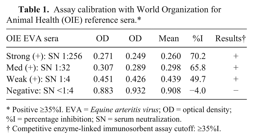
Positive ≥35%I. EVA = Equine arteritis virus; OD = optical density; %I = percentage inhibition; SN = serum neutralization.
Competitive enzyme-linked immunosorbent assay cutoff: ≥35%I.
Evaluation of assay repeatability
Intrarun repeatability
The evaluation of intrarun repeatability involved the analysis of CVs of OD calculated from replicates of the EAV cELISA positive control serum and replicates of the EAV cELISA negative control serum. The CV of the OD ranged between 6.1% and 11.8% when tested with 16 replicates of EAV positive control serum using 2 different kit serials performed by 3 different operators (Table 2). The CV of the OD ranged between 7.5% and 9.2% when tested with 56 replicates of EAV negative control serum using 2 different kit serials by 3 different operators (Table 2). No significant (P > 0.05) differences were observed between different operators or kit serials within each run using the Kruskal–Wallis test.
Intrarun repeatability of the competitive enzyme-linked immunosorbent assay.*
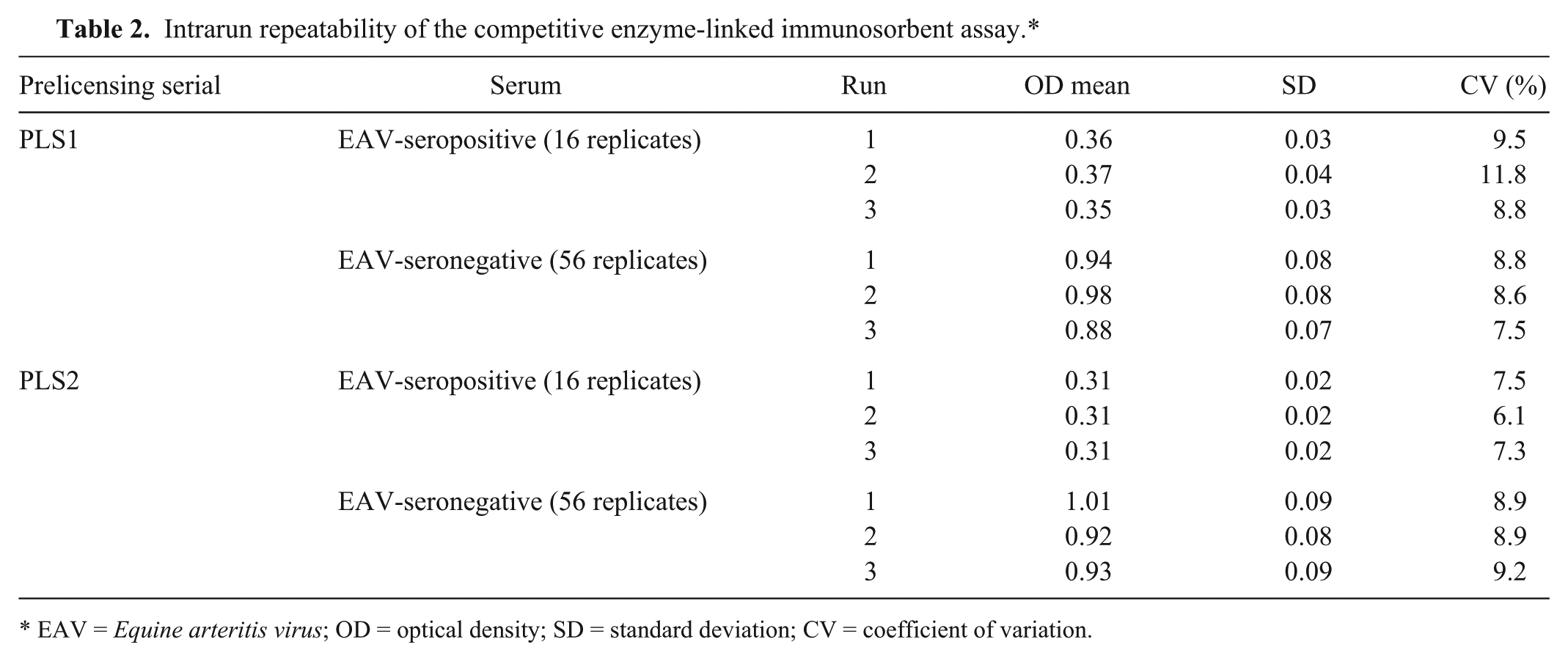
EAV = Equine arteritis virus; OD = optical density; SD = standard deviation; CV = coefficient of variation.
Interrun repeatability
The evaluation involved the analysis of the CV of the %I of 4 dilutions of a positive serum evaluated by different operators using different kit serials. The CV ranged between 7.1% and 10.6% when %I data from 38 runs by 3 operators using 3 kit serials at different times were evaluated (Table 3). No significant (P > 0.05) differences were observed between runs with different operators or kit serials using the Kruskal–Wallis test.
Interrun repeatability among 38 runs by 3 operators using 3 prelicensing kit serials.*
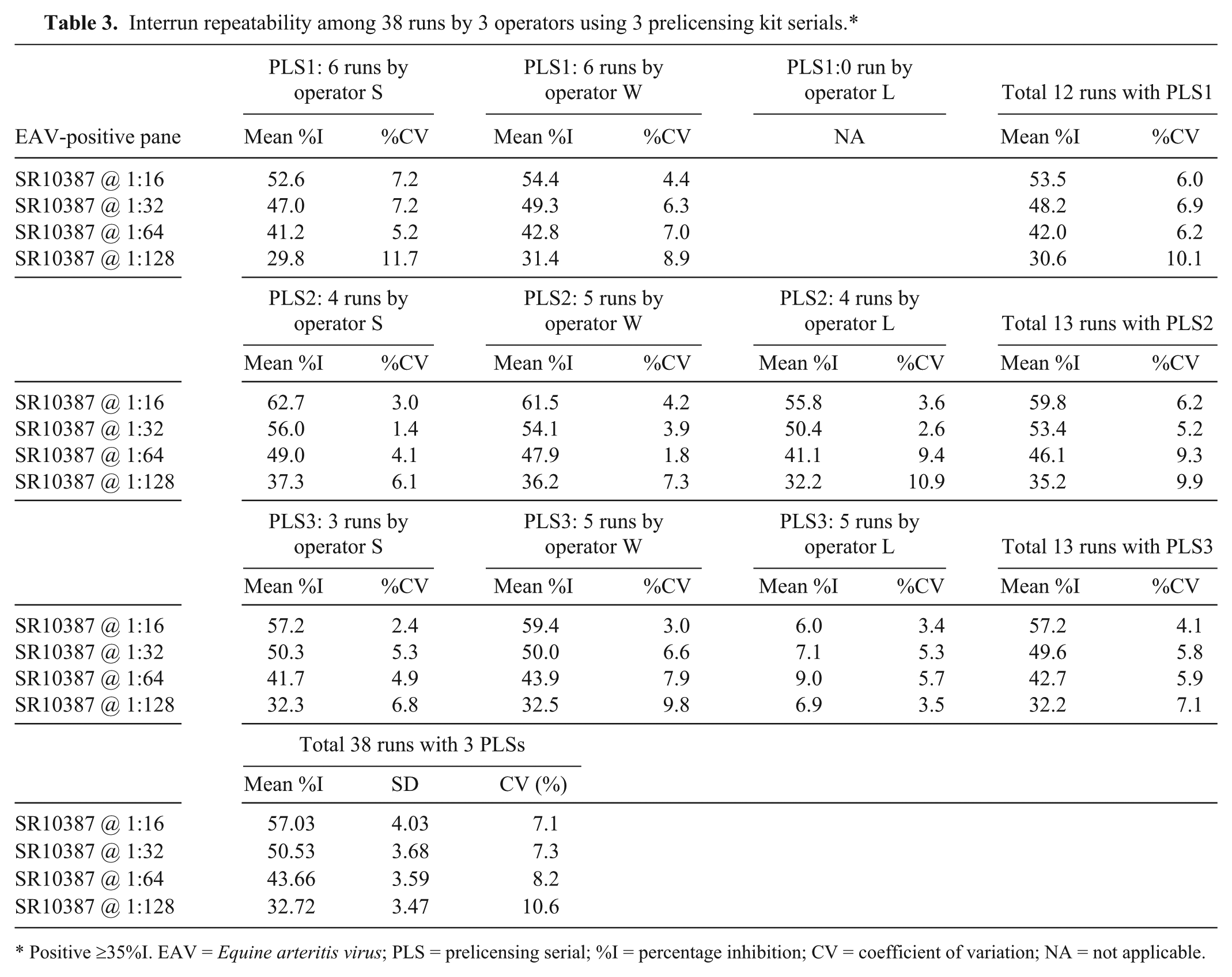
Positive ≥35%I. EAV = Equine arteritis virus; PLS = prelicensing serial; %I = percentage inhibition; CV = coefficient of variation; NA = not applicable.
Evaluation of assay analytical specificity
To evaluate the analytical specificity of the EAV cELISA, sera antibody positive and negative to the closely related arterivirus (neighbor), PRRSV, were evaluated. Both PRRSV antibody-positive and -negative sera were negative using the EAV cELISA (<35 %I) indicating no cross-reactivity with the epitope recognized by mAb 17B7.
Evaluation of assay analytical sensitivity
To determine the analytical sensitivity of the EAV cELISA, post-vaccination sera sequentially collected from 2 MLV-vaccinated horses were analyzed in both the SN and cELISA assays. The earliest time point with a SN assay-positive result with serum from both horses was 6 days post vaccination (DPV) and the earliest time point with a cELISA-positive result with serum from both horses was 8 DPV (Table 4). Spearman rank correlation analysis using the same data sets demonstrated that there was a highly significant (P < 0.0001) positive correlation between the cELISA and the SN assay results (r = 0.88).
Analytical sensitivity of Equine arteritis virus (EAV) competitive enzyme-linked immunosorbent assay (cELISA) and the serum neutralization (SN) assay tested with postvaccination sera collected sequentially from 2 EAV modified live virus–vaccinated horses.*
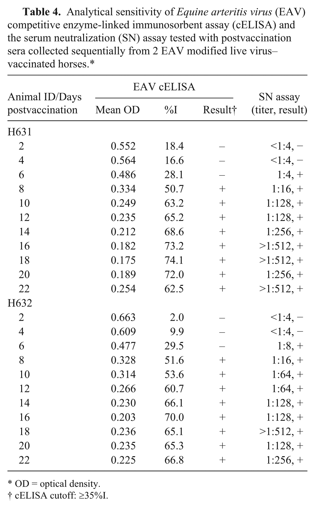
OD = optical density.
cELISA cutoff: ≥35%I.
Evaluation of duration of antibody detected by the EAV cELISA after vaccination and infection
To determine if antibody to the epitope recognized by mAb 17B7 in the cELISA persisted after EAV vaccination and infection, cELISA results were analyzed from sera sequentially collected for 436 DPV from 2 horses (H631 and H632) that received a single dose of MLV. All sera from H631 and H632 collected from 8 to over 400 DPV were positive, with a %I range of 50.9–76.6, which is clearly above the ≥35%I cutoff (Fig. 1). Sequential sera from H537, which received 4 vaccinations of MLV within 2 months of the first vaccination, and then a fifth dose at 1,964 DPV and a sixth dose at 2,001 DPV, were evaluated. Sera collected and tested between 1,923 DPV and 2,342 DPV were always cELISA positive, with a %I of 60.9 or higher (Fig. 1). Those sera collected between 1,923 and 1,964 DPV were of particular interest because they were all strong positives and were obtained more than 5 years after the fourth vaccination and before the fifth vaccination. In addition, persistent SN-positive responses (>1:256) were observed in long-term collected time points (data not shown).
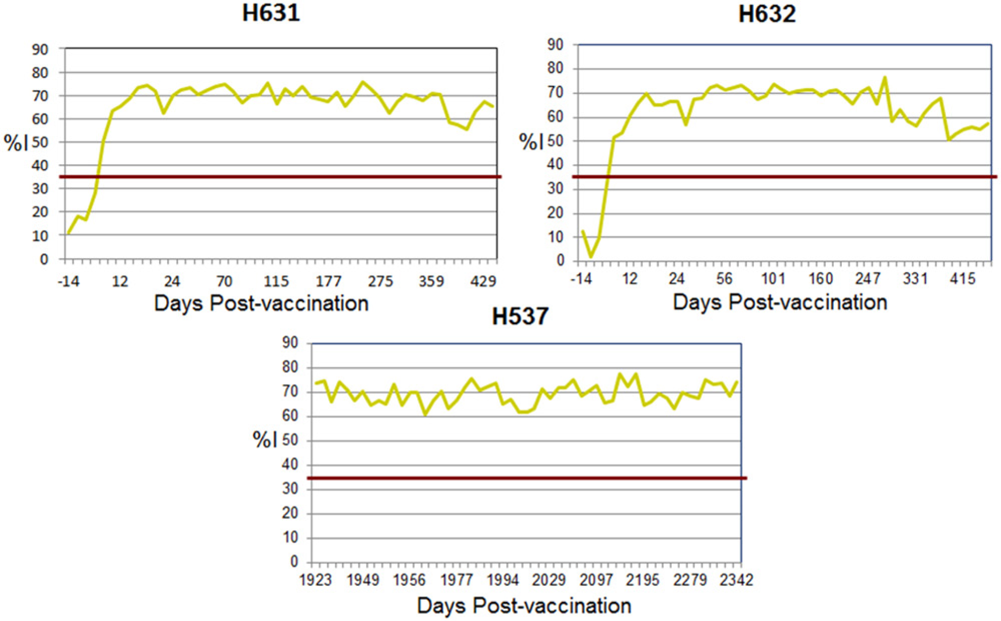
Duration of Equine arteritis virus antibody detectable by competitive enzyme-linked immunosorbent assay in horses vaccinated with modified live vaccine. The red line is at 35 percent inhibition (%I). Positive cutoff (≥35 %I); negative cutoff (<35 %I).
Horse C7 infected with EAV KY84 strain had the first positive cELISA result at 10 days postinoculation (DPI; Fig. 2). Horse F19 infected with an infectious complementary DNA clone of the EAV Bucyrus strain had the first positive cELISA result at 8 DPI. Both horses had consistent positive results until day 35, which was the last day tested (Fig. 2).
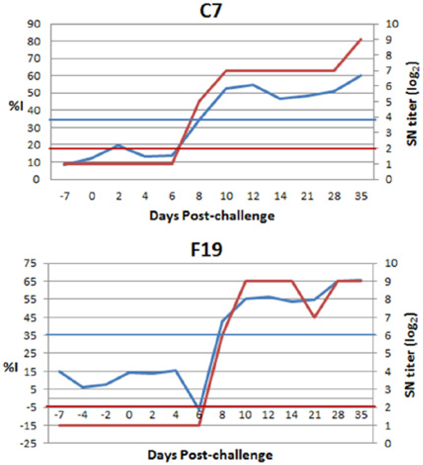
Detection of Equine arteritis virus (EAV) antibody by competitive enzyme-linked immunosorbent assay in EAV-challenged horses. Horse C7 was infected with EAV strain KY84. Horse F19 was infected with an infectious complementary DNA clone of EAV strain Bucyrus. The blue line is at 35 percent inhibition (%I). Positive cutoff (≥35%I); negative cutoff (<35%I). The red line is at 2 in serum neutralization (SN) titer (log2). Positive cutoff (≥2); and negative cutoff (<2).
Field trial to evaluate the assay diagnostic performance
Determination of robustness of cELISA with a defined interdependency reference serum panel
An interdependency panel including 24 EAV SN-positive sera and 26 EAV SN-negative sera was tested by cELISA in 3 veterinary diagnostic laboratories. The cELISA results from all 3 laboratories had a 100% concordance with SN test results. In addition, all 3 laboratories had the same cELISA results with the interdependency panel (Table 5).
Robustness of Equine arteritis virus (EAV) competitive enzyme-linked immunosorbent assay (cELISA) tested with an interdependency panel.*
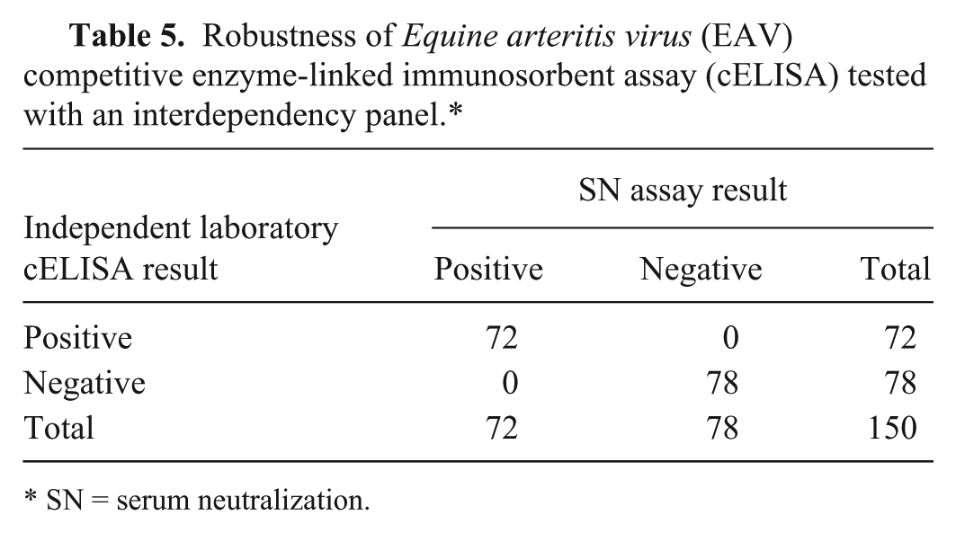
SN = serum neutralization.
Diagnostic sensitivity and specificity estimates using field serum samples
As the second part of the field trial, each of 3 participating laboratories prepared a field sera panel including 50–65 EAV SN-positive sera and 129–135 SN-negative sera. The SN results were obtained using each laboratory’s in-house SN assay. After cELISA testing of these field serum panels in each laboratory, diagnostic performance estimates of the cELISA were calculated based on comparison with the SN results. For the SN assay, samples with <1:4 titers were scored as negative and those with ≥1:4 titers were scored as positive. The diagnostic specificity of the cELISA was 99.5%, and the diagnostic sensitivity determined was 98.2% (Table 6).
Equine arteritis virus (EAV) competitive enzyme-linked immunosorbent assay (cELISA) diagnostic sensitivity and specificity estimates with diverse field sera.*
Sensitivity = (163/166) × 100% = 98.2%; specificity = (392/394) × 100% = 99.5%. SN = serum neutralization.
Reevaluation of cELISA cutoff resulting in best combination of diagnostic sensitivity and specificity
To confirm that using ≥35%I as the cutoff for defining seropositive samples and <35%I for defining seronegative samples in previous studies was optimal, data from cELISA and SN testing of field serum samples by the 3 diagnostic laboratories in the field trial were analyzed in a ROC curve. In this analysis, the best correlation between cELISA and SN results was observed at the cutoff used in the preliminary validation of the mAb 17B7–based cELISA. Thus, the field trial data derived from samples from various areas in the United States and Europe further support using a cutoff of ≥35%I for positive samples and <35%I for negative samples (Fig. 3).
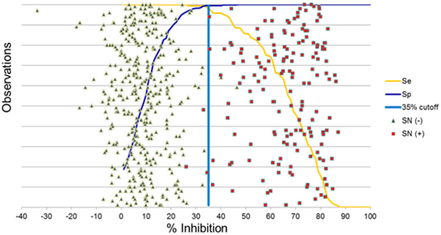
Receiver operating characteristic plot analysis of the competitive enzyme-linked immunosorbent assay field trial data. Blue line is at 35 percent inhibition (%I), which resulted in the best combination of diagnostic sensitivity and specificity. SN = serum neutralization.
Statistical analysis of the correlation between cELISA %I and SN assay titer in the field trial
The correlation of %I from the nonneutralizing mAb 17B7–based cELISA with the titer (log2) from the polyclonal neutralizing antibody response-based SN assay was analyzed using Spearman rank correlation. This analysis with the data from 3 field sample panels in field trial yielded a correlation coefficient (r) of 0.79 (P < 0.0001) indicating a strong and significant positive correlation. This significant positive correlation between the cELISA %I and SN titer data further confirms the significant correlation shown with the OIE EAV reference panel and post-EAV vaccination panel in Table 4.
Discussion
Equine arteritis virus is contagious among equids and occurs in countries from all continents, including the United States, Canada, France, Germany, The Netherlands, Denmark, Sweden, Hungary, Spain, the United Kingdom, Ireland, Australia, Mongolia, Slovenia, Argentina, and South Africa.3,4,11,12,16,17,21-27,29-31,33-37,40-42,44,45,47,48 The number of confirmed EAV cases is increasing,26,27,29,33,37,48 likely attributable to increases in global movement of horses, accessibility of carrier stallions and utilization of shipped, cooled or frozen, virus-infected semen. Each year approximately 3,000 major equestrian events occur internationally, requiring increasingly short quarantine time for entry to event-hosting countries and providing opportunity for virus spread. Therefore, rapid and accurate screening of horses for EAV involved in international as well as intranational movement is pivotal to controlling EAV transmission.
In March 2013, a cELISA detecting antibody response indicative of EAV infection or vaccination to a nonneutralizing epitope in EAV GP5 was developed with improved resolution and diagnostic performance 9 when compared to a previously reported cELISA 8 and several previously reported ELISAs.6,8,32,38 The improved cELISA reliably detects EAV-specific antibody responses without misinterpretation due to the presence of non–EAV-specific cytotoxic antibodies, which are a common occurrence and technical challenge using the SN assay. 32 In the current study, this mAb 17B7–based cELISA was further validated according to the OIE recommendations, including a field trial in 3 independent diagnostic laboratories. In the field trial, the diagnostic sensitivity of the cELISA was 98.2% and the diagnostic specificity was 99.5% when compared to results from the OIE-prescribed SN assay.
Three sets of data observed in the present study demonstrated that cELISA results correlate well with SN assay results, thus justifying the cELISA as an alternative to the SN assay. First, evaluation of the cELISA calibration using an OIE check-set panel, including strong, medium, and weak positive serum samples as well as a negative serum resulted in a significant (P < 0.05) positive correlation (r = 1.0) between SN titers and the cELISA %I results. Second, a strong (r = 0.79), highly significant (P < 0.0001) positive correlation occurred between the SN titer and cELISA %I of the field trial data. Third, there was a strong (r = 0.88) highly significant (P < 0.0001) correlation between SN titers and the cELISA %I, with sequential serum samples collected for early 22 DPV from horses vaccinated with EAV MLV.
The reliability of the improved cELISA was demonstrated in several different experiments. First, this cELISA, which detects antibody to a nonneutralizing mAb 17B7 GP5-specific epitope, was positive for up to 4.7 years after the last boost with no samples below the positive cutoff. Second, this cELISA had good analytical specificity and differentiated antibodies to a closely related arterivirus, PRRSV. Third, interrun and intrarun repeatability of this cELISA were excellent when tested with various EAV SN assay–positive and –negative serum samples. Fourth, robustness of the cELISA was clearly demonstrated from 100% concordance among testing results from 3 independent diagnostic laboratories and several operators. So far, there has been no evidence of variation of the EAV GP5 epitope recognized by mAb 17B7. It will be important to evaluate the performance of this cELISA in other countries and in additional laboratories in the United States using a larger number of equine serum samples collected from the field. Data unpublished by the authors suggest that the equine seroconversion curve for EAV is steep, transiting within 12 hr from antibody-negative to antibody-positive by both cELISA and SN. Considering the typical low background of the cELISA, samples producing high negative %I (25 > n < 35) are likely from horses in the process of seroconversion following EAV vaccination or infection. In the opinion of the authors, such individual horse should be resampled a few days subsequent to the initial sample and retested by cELISA.
In conclusion, the nonneutralizing mAb 17B7–based cELISA had a specificity and sensitivity of 99.5% and 98.2%, respectively, when compared with SN assay results in the field trial using 3 diagnostic laboratories. Furthermore, this cELISA has comparable analytical sensitivity and specificity to the OIE-prescribed SN assay. The results in the current study and a previous article 9 strongly support the use of this cELISA as a suitable alternative to the SN assay for serodiagnosis of EAV.
Footnotes
Acknowledgements
The authors thank Amanda Grimm, Chandima Bandara Bandaranayaka Mudiyanselage, Lorraine Tanaka, and Sara Schlee for technical support.
a.
ARVAC, Zoetis Animal Health, Madison, NJ.
b.
Stripwell High Binding 96-well plates (Costar 2592), Corning Life Sciences, Tewksbury MA.
c.
ELISA blocking buffer, VMRD Inc., Pullman, WA.
d.
ELISA antibody diluting buffer, VMRD Inc., Pullman, WA.
e.
Mylar Bags, IMPAK Co., Los Angeles, CA.
f.
Goat anti-mouse immunoglobulin G conjugated to horseradish peroxidase, VMRD Inc., Pullman, WA.
g.
TMB substrate solution, SurModics, Eden Prairie, MN.
h.
Stop solution, SurModics, Eden Prairie, MN.
i.
Multiskan MCC/340 ELISA reader, Titertek Instruments Inc., Huntsville, AL.
j.
Excel 2010, Microsoft Corp., Redmond, WA.
Declaration of conflicting interests
The author(s) declared no potential conflicts of interest with respect to the research, authorship, and/or publication of this article.
Funding
The author(s) disclosed receipt of the following financial support for the research, authorship, and/or publication of this article: This research was supported by the general research funding of VMRD (Veterinary Medical Research and Development) Inc.