
Abstract
A 2-month-old male Holstein calf showed clinical signs of abdominal bloating, melena, and pain and was suspected of having a perforating abomasal ulcer. Necropsy revealed a large mass located preferentially around the abomasum and a large perforating abomasal ulcer on the pyloric antrum. Milky white masses of various sizes were also found in the abdominal cavity that consisted of agglutinated nodules ranging in size from a pinhead to a golf ball and were distributed on the surfaces of the liver, reticulum, omasum, abomasum, and diaphragm. Microscopic examination revealed that the masses were composed primarily of hyaline matrices, epithelioid tumor cells, and large atypical cells with hyaline droplets and/or vacuoles. Stromal hyaline matrices and hyaline droplets of the large tumor cells stained positive with periodic acid–Schiff stain. Tumor cells showed a positive reaction to anti-human alpha-fetoprotein, which is a marker of yolk sac tumors. These findings strongly suggested that the masses found in the abdominal cavity were yolk sac tumor, a rare germ cell tumor in cattle.
Perforating abomasal ulcers are a common clinical problem in calves that cause septic peritonitis.3,7 Several factors, including feeding large volumes of milk, stress, nonsteroid anti-inflammatory drug administration, and hairball formation, are known causes of abomasal ulcer in calves. 7 Although tumors such as lymphosarcoma can cause abomasal ulcers in adult cattle, 6 there have been no reports of this in calves to our knowledge. Herein, a rare yolk sac tumor in a Holstein calf that caused a perforating abomasal ulcer is described.
A 55-day-old male Holstein-Friesian calf presented with bloating and anorexia. At initial examination (day 1), the calf had a body temperature of 40.0°C and a heart rate of 90 bpm and showed severe abdominal bloating. On day 2, clinical signs included lethargy and diarrhea. Melena was observed beginning on day 5. On day 7, the calf could not stand up by itself, and severely pale mucous membranes were observed. Despite fluid transfusion, hemostatic therapy, and administration of antibiotics and metoclopramide, the general condition of the calf did not improve. The calf was taken to the Teaching Veterinary Hospital at the Obihiro University of Agriculture and Veterinary Medicine (Obihiro, Japan) on day 9.
On initial physical examination at the hospital, body temperature (39.5°C) and heart rate (84 bpm) were within normal range, but the calf was unconscious and had cold extremities, severe dehydration (>5%), pale mucous membranes, abdominal bloating, and melena. Splashing sounds in the abdomen on shaking were recorded by abdominal auscultation. Aspiration of abdominal fluid using a fine needle recovered red serous fluid. Analysis of the fluid revealed neutrophils (700/µl), red blood cells (40,000/µl), and bacteria but no tumor cells. Hematologic examination of peripheral blood revealed severe anemia (red blood cell count: 2.41 × 10⁶/µl; hemoglobin concentration: 2.4 g/dl; and packed cell volume: 9%) and neutrophilia (40,400/µl [stab neutrophils: 2,020/µl; segmented neutrophils: 31,916/µl; lymphocytes: 5,252/µl; monocytes: 808/µl]). Abnormalities in blood chemistry included increased aspartate aminotransferase activity (543 U/l), alkaline phosphatase (1,022 U/l), and creatine kinase (1,657 U/L) and decreased levels of total protein (3.3 g/dl), albumin (1.5 g/dl), and total cholesterol (22 mg/dl). Based on the physical signs and laboratory findings, perforating abomasal ulcer with peritonitis was suspected. Despite administration of Ringer solution and dexamethasone, the calf died the next day, and a necropsy was performed.
At postmortem examination, the omentum was highly thickened, and the extended omental bursa (80 cm × 50 cm × 20 cm) contained a large amount of red viscous fluid with fibrin. Milky white masses of various sizes filled the abdominal cavity. The masses consisted of agglutinated nodules ranging in size from a pinhead to a golf ball and were distributed on the surfaces of the liver, reticulum, omasum, abomasum, and diaphragm (Fig. 1). Abdominal organs adhered to the peritoneum and to each other via these masses. In addition, a large perforating abomasal ulcer was found on the pyloric antrum. The largest mass, with a diameter of 10 cm, adhered to the outside of the abomasal ulcer. Contents of the abomasum were released from the ulcer into the adhered mass but not into the abdominal cavity.
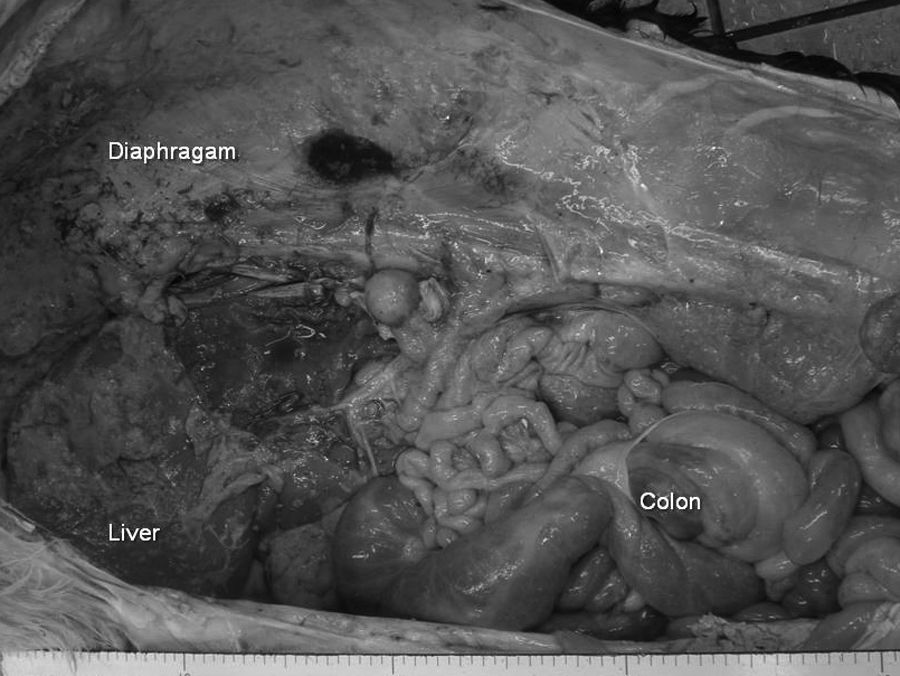
Holstein calf; abdominal cavity after removing rumen, reticulum, omasum, and abomasum. Milky white masses of various sizes filled the abdominal cavity. The masses consisted of agglutinated nodules that ranged in size from a pinhead to a golf ball and were distributed on the surfaces of the liver, reticulum, omasum, abomasum, and diaphragm.
For histopathological examination, the masses and major organs and tissues were fixed in 15% neutral buffered formalin and embedded in paraffin wax. Paraffin sections were routinely stained with hematoxylin and eosin. Selected sections were also stained with periodic acid–Schiff (PAS) reaction and immunostained with anti-human alpha-1-fetoprotein (AFP) polyclonal rabbit antibody a using a streptavidin–biotin method. b For the immunohistochemical evaluation, sections from a canine case of hepatocellular carcinoma and fetal calf liver were used as a positive control. Histological examination revealed that the masses were composed primarily of hyaline matrices, epithelioid tumor cells, and large atypical cells with hyaline droplets and/or vacuoles (Fig. 2). Stromal hyaline matrices and hyaline droplets of the large tumor cells stained positive with PAS stain (Fig. 3). Immunohistochemical examination revealed that tumor cells had a positive reaction to anti-AFP, which is a marker of yolk sac tumors (Fig. 4). These findings strongly suggested that the masses found in the abdominal cavity were yolk sac tumor. No abnormalities were found in the testis. The pathological diagnosis was yolk sac carcinoma and perforating abomasal ulcer.
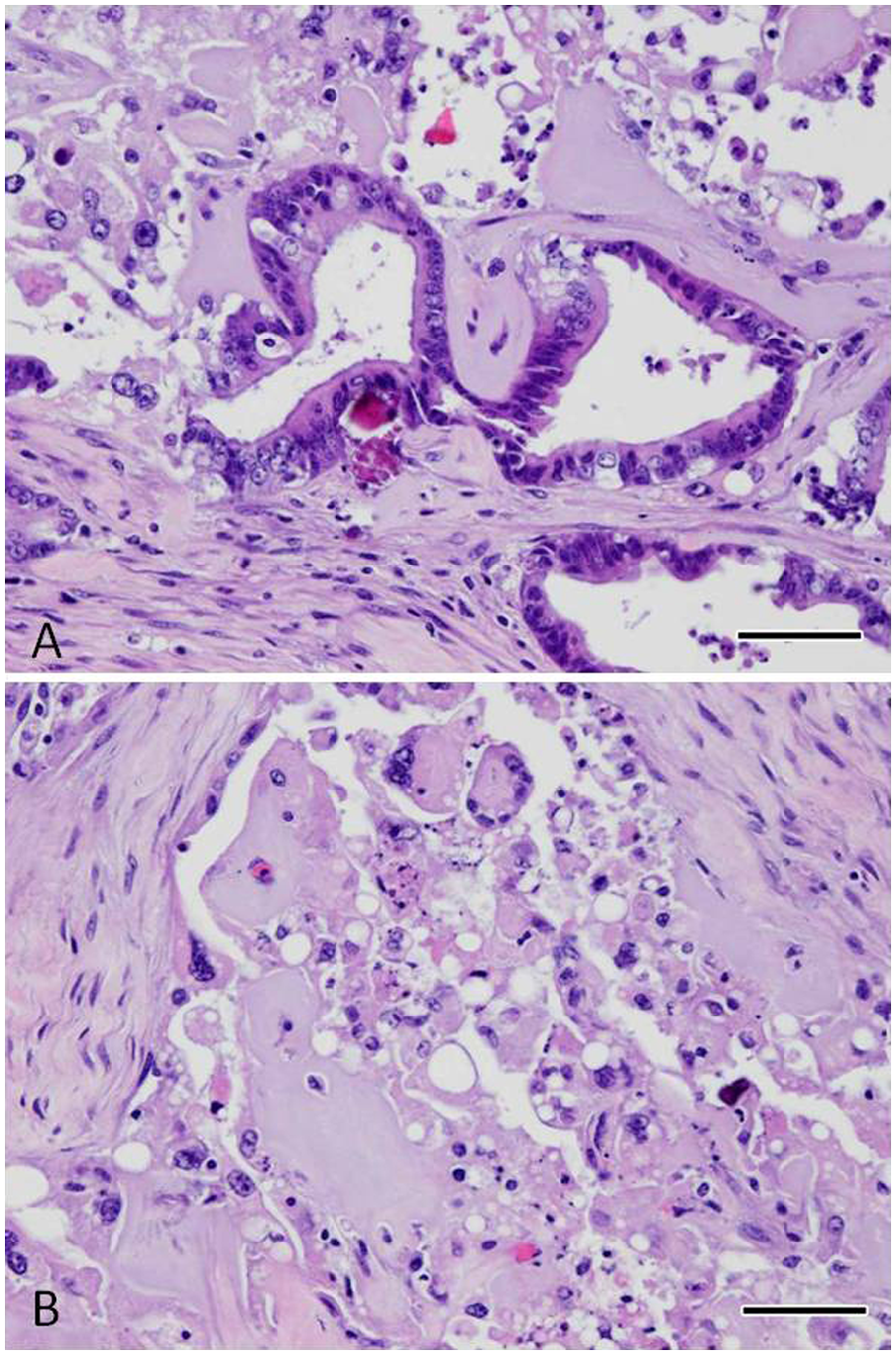
Holstein calf; histopathological analysis of masses distributed in the abdominal cavity.
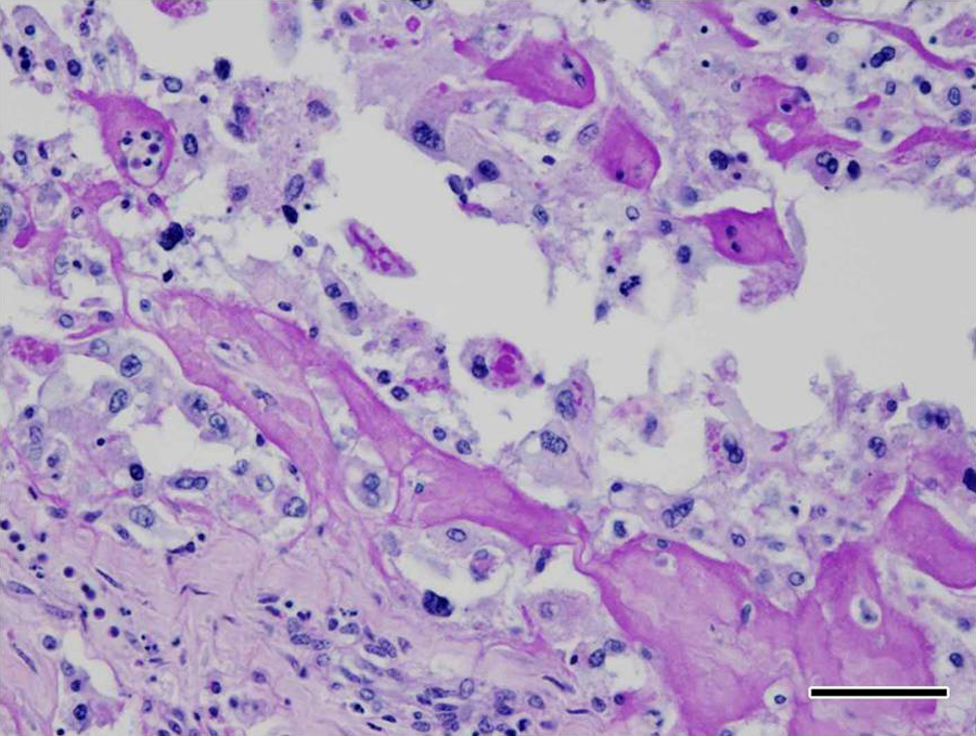
Holstein calf. Stromal hyalinous material and hyaline droplets of large tumor cells stained positive with periodic acid–Schiff stain. Bar = 50 µm.
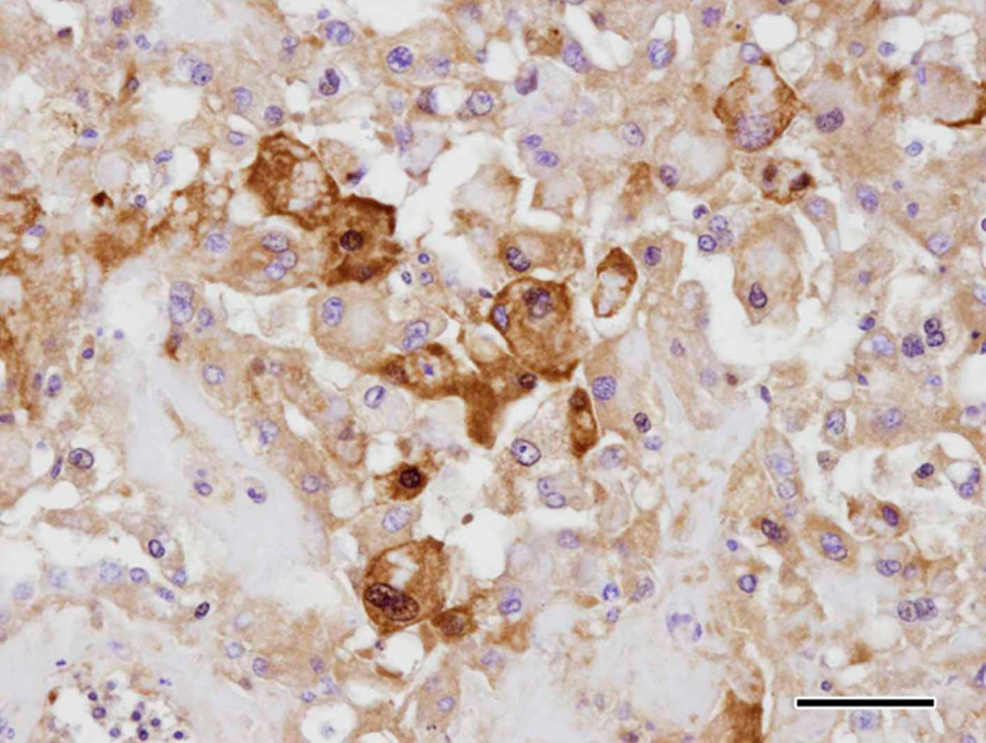
Holstein calf. Immunohistochemical examination revealed that tumor cells had a positive reaction to alpha-a-fetoprotein, which is a marker of yolk sac tumors. Bar = 50 µm.
Yolk sac tumor, alternatively endodermal sinus tumor, is classified as a germ cell tumor and generally originates from reproductive organs such as the testis and ovary; however, other organs, including the mediastinum, retroperitonium, sacrococcygeal region, pineal gland, and stomach, are involved in such tumors in human beings.2,5,8 The occurrence of germ cell tumor is very rare in animals compared to human beings. There have been only 2 case reports in cattle: a 28-day-old male Japanese black calf 4 and a 55-day-old male crossbred (Holstein-Friesian/Japanese Black) calf. 1 As in the present case of widespread intraabdominal metastasis, various sized white masses were found in the abdominal cavities in the previous reports.1,4 Although the masses were diffused, the abomasal wall was suspected as the primary tumor lesion in the present case because the largest mass was found at the pyloric antrum of the abomasum. The protective effect of the abomasal mucus membrane against gastric acid secretion might have been compromised with tumor invasion, leading to abomasal ulcer formation.
Although an abomasal ulcer caused by lymphosarcoma was reported in an adult cow, 6 there have been no reports of abomasal ulcers caused by tumors in calves to the authors’ knowledge. Accordingly, the present case is a rare case of perforating abdominal ulcer related to yolk sac tumor in a calf. In conclusion, yolk sac tumor can cause carcinomatous diffuse peritonitis and perforating abomasal ulcer in calves.
Footnotes
a.
Dako Japan Inc., Tokyo, Japan.
b.
Histofine SAB-PO kit, Nichirei Corp., Tokyo, Japan.
Declaration of conflicting interests
The author(s) declared no potential conflicts of interest with respect to the research, authorship, and/or publication of this article.
Funding
The author(s) disclosed receipt of the following financial support for the research, authorship, and/or publication of this article: This work was supported in part by the Tokachi Agriculture Mutual Aid Association.