
Abstract
A 12-year-old intact male Miniature Schnauzer dog with chronic diarrhea that was unresponsive to empirical treatment was presented to a referring veterinarian. A laparotomy was performed, and formalin-fixed biopsies of duodenum, jejunum, and colon were sent to Oklahoma Animal Disease Diagnostic Laboratory for evaluation. Histologic examination revealed a severe, diffuse, granulomatous enteritis and colitis with intralesional yeast and hyphal forms. Grocott methenamine silver stains revealed short, aseptate hyphae co-mingled with 2–8 µm, oval to round yeast organisms consistent with Histoplasma capsulatum. The atypical presentation of both yeast and hyphal forms prompted identification of the organism. Direct sequencing of a polymerase chain reaction product from paraffin-embedded intestinal samples confirmed the presence of Ajellomyces capsulatus with a homology over 99% to several sequences in GenBank. Ajellomyces capsulatus is the holomorphic name for H. capsulatum. Therefore, the mycelial form of a dimorphic fungus such as H. capsulatum can coexist with yeast cells within lesions of histoplasmosis. Following diagnosis, the dog was treated with itraconazole for 6 months and has improved.
Keywords
A 12-year-old, 9.1-kg, intact male Miniature Schnauzer dog with chronic diarrhea of 2 months duration was presented to a rural clinic. Prior empirical therapies included antibiotics, steroids, anthelmintics, and a diet change, which failed to resolve the symptoms. Blood work was performed, including a complete blood cell count and serum biochemical profile. The results of all tests were within normal limits. The referring veterinarian performed a laparotomy. No gross abnormalities were seen; therefore, survey biopsies were taken from the duodenum, jejunum, and colon and were submitted to Oklahoma Animal Disease Diagnostic Laboratory (Stillwater, Oklahoma).
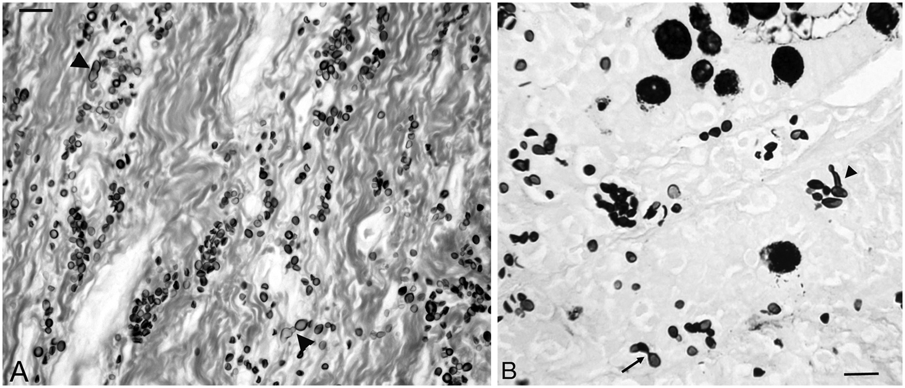
Formalin-fixed tissue samples were received and routinely processed, embedded in paraffin wax, sectioned at 5 µm, and stained with hematoxylin and eosin (HE). Additional recuts were stained by Grocott methenamine silver (GMS) method. Histologically, the colonic mucosa was expanded and regionally effaced by inflammatory cells consisting primarily of epithelioid macrophages, lymphocytes, neutrophils, and a few plasma cells. The inflammatory infiltrates separated colonic glands and, in some sections, extended into the submucosa (Fig. 1A). Embedded within all regions of the inflammation were yeast-type organisms characterized by a 1.0 μm, round to crescent-shaped yeast body surrounded by a variably sized (2.0–8.0 µm) unstained halo (Fig. 1B). By GMS stain (Fig. 2A, 2B), the organisms exhibited heterogeneous morphological features. Most typical were variably sized and shaped yeast forms (2.0–8.0 µm) with occasional narrow-based budding. Less frequently, the yeasts were accompanied by short, sometimes branching hyphal forms with either parallel or undulating walls. Rarely, there were straight to coiled chains of spherical structures with larger (5.0–8.0 µm) fruiting body-type structures (ascocarp) located terminally. The yeast forms were located within cells morphologically consistent with macrophages. The mycelial forms were always located extracellularly. Whereas some of the features seen on routine and GMS stains were consistent with Histoplasma capsulatum, the variation in yeast size along with apparent sexual reproductive forms in tissue suggested perhaps a different etiology. Therefore, identification of the organism by polymerase chain reaction (PCR) was attempted.
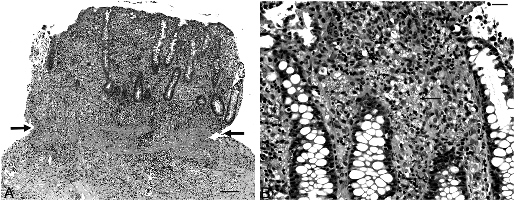
Dog, colon.
Dog, colon.
Total DNA was extracted from the formalin-fixed, paraffin-embedded tissue samples using a commercial kit a following the manufacturer’s instructions. The integrity of the extracted DNA was tested using mammalian universal 18S rDNA primers. 6 A 256–base pair fragment of ribosomal RNA gene of Ajellomyces capsulatus (the holomorphic name for Histoplasma capsulatum) was amplified using a newly designed, species-specific PCR primer pair. The specificity of the primers was confirmed by direct sequencing of the PCR product (GenBank accession no. JX534222). The forward and reverse primers were 5′-TCCGGCATC GATGAAGAACG-3′ and 5′-TCCAGAGCGGGTGGCAA AG-3′, respectively. The 25-μl PCR reaction contained approximately 150 ng of total DNA, 1× PCR buffer, b 1.5 mM MgCl2, 200 μM deoxyribonucleotide triphosphates, 400 nM of each primer, and 1.25 U of Taq DNA polymerase. c The cycling conditions were: 1 cycle of 4 min at 95°C, 30 sec at 63°C, and 30 sec at 72°C, followed by 34 additional cycles of 30 sec at 95°C, 30 sec at 63°C, and 30 sec at 72°C, with a final extension of 3 min at 72°C. The purified PCR product was directly sequenced in both strands. d The complete 256-bp sequence of the PCR product obtained from the biopsy material was deposited in an international databank (GenBank accession no. JX534222). The DNA sequence exhibits over 99% homology to several ribosomal RNA gene sequences of A. capsulatus (Fig. 3).
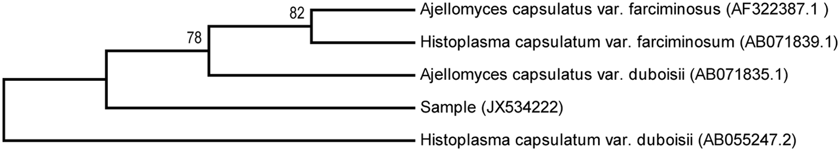
Evolutionary relationship to other known Histoplasma/Ajellomyces species. The evolutionary history of the isolated sequence and few other closely related sequences was inferred using the maximum parsimony method. Numbers adjacent to the nodes are bootstrap values ≥70%. Phylogenetic analyses were conducted in MEGA5. 14 GenBank accession numbers of the sequences are given in parentheses next to the sequence names.
The final interpretation included severe granulomatous colitis with intralesional yeast and fungal organisms consistent with A. capsulatus, the holomorphic name representing all stages and morphological forms that include for H. capsulatum. The yeast form (H. capsulatum) is classically oval or round, 2–4 µm in diameter, with narrow-based buds and a spherical, slightly basophilic central body surrounded by a clear halo.2,10 In the current case, HE stains showed yeasts that resembled H. capsulatum, but were atypically larger (up to 8 µm) in diameter with some hyphal forms present. The GMS stains were performed to clarify fungal morphology and revealed a mixture of yeast and hyphal forms. Because H. capsulatum does not typically have hyphal forms in tissue, 2 alternate etiologies were considered. Molecular biology was utilized with PCR and DNA sequencing to identify the organisms seen on histopathology. This identification confirmed that the yeast and hyphal forms seen microscopically were from a pure infection by A. capsulatus. Following diagnosis, the dog was treated with itraconazole for 6 months and has improved.
Histoplasma capsulatum is a dimorphic fungus that is the causative agent of histoplasmosis. The fungus is commonly found in soil that has been contaminated with bird or bat guano. 1 In the United States, it is frequently reported to infect mammals, including human beings, in the Mississippi, Missouri, and Ohio River valleys but can be found elsewhere in the Midwest. 4 The organism exists in the soil as a saprophytic mold that produces infective microconidia. 1 Once inhaled, microconidia convert to a yeast within the respiratory system 1 and replicate inside phagolysosomes of the mononuclear-phagocytic system.1,16 In immunocompetent human beings and dogs, initial infection is usually self-limiting; however, those hosts that are immunocompromised or received large doses of microconidia have a greater risk of developing the characteristic granulomatous disease. 16 The infection typically develops in the lungs and most commonly disseminates to the gastrointestinal tract, but can also disseminate to the eyes, skin, bone, and rarely the brain. 16 In 1973, the sexual stage or teleomorph of H. capsulatum was discovered and today is accepted as A. capsulatus.7,11 The parasitic yeasts most commonly seen in tissue infections represent the asexual or anamorphic stage of this dimorphic organism. 8 Although these different phenotypes (teleomorphic vs. anamorphic) have different nomenclature (Ajellomyces vs. Histoplasma), they are one in the same organism. For the mycologist, to identify the whole organism regardless of phenotype, holomorphic nomenclature is used, which in this case is A. capsulatus. However, in clinical laboratories, the name of the anamorph is typically used when describing infections by dimorphic fungi. 9 Thus in human and veterinary medicine, the disease is histoplasmosis (anamorph) instead of ajellomycosis (teleomorph or holomorph).
Histoplasma capsulatum is not the only dimorphic fungus reported to have both the hyphae and yeast present in the same sample. In 1979, hyphal forms of Blastomyces dermatitidis (named Ajellomyces dermatitidis) could be found concurrently with yeast forms on histopathology of a dog spleen. 5 More recently, A. dermatitidis hyphae and B. dermatitidis yeast cells were found in a cytologic smear from a subcutaneous mass at the base of the ear from a dog. 3 It was postulated that the rare mycelial state was artifactually created due to low environmental temperature and time delay between sample collection and slide preparation. 3 Artifactual induction of the mycelial phase is not likely in the current case because samples were collected surgically and immediately placed in formalin. In addition to H. capsulatum and B. dermatitidis, Cryptococcus neoformans was also reported to have the teleomorph (Filobasidiella neoformans) found with the yeast phase. Both hyphae and yeast of C. neoformans were cultured from the urine of a neonatal southern right whale (Eubalaena australis) and identified by PCR. 12 Finally, the mycelial phase of Coccidioides immitis was found in a section of a human lung. 13 Taken together, most of the important veterinary dimorphic fungal pathogens have been found, albeit rarely, to exhibit mycelial morphology in tissue sections.
Polymerase chain reaction amplification of paraffin-embedded tissues has been available for years to identify infections and certain cancers. Similar to the current case, the technique has previously been used to diagnose histoplasmosis in a subcutaneous infection in a dog from Japan. 15 Polymerase chain reaction identification or confirmation of etiology is extremely useful when tissue received on a biopsy service (formalin-fixed) is not suitable for culture or other diagnostic methods.
In summary, the mycelial form of a dimorphic fungus such as H. capsulatum can coexist with yeast cells in vivo and cause disease. In light of the current case and other reported cases, a diagnosis of histoplasmosis should be considered even if hyphae are seen in tissue sections. Additionally, the present report shows that an etiological diagnosis can be made from DNA obtained from formalin-fixed, paraffin-embedded tissue samples.
Footnotes
a.
QIAamp DNA mini kit, Qiagen Inc., Valencia, CA.
b.
PerkinElmer Inc., Waltham, MA.
c.
Promega Corp., Madison, WI.
d.
ABI 3130 Genetic Analyzer, Life Technologies Corp., Carlsbad, CA.
Declaration of conflicting interests
The author(s) declared no potential conflicts of interest with respect to the research, authorship, and/or publication of this article.
Funding
The author(s) received no financial support for the research, authorship, and/or publication of this article.