
Abstract
A 2.5-year-old intact male miniature lop rabbit (Oryctolagus cuniculus) was presented with multiple nodules surrounding the eyes, nose, mouth, and prepuce. Cytological evaluation of the periocular nodules revealed the presence of intracellular (within macrophages) and extracellular yeast organisms. The yeast organisms were approximately 3–5 µm in diameter, round to oval, with a thin clear capsule, and contained an eccentrically placed basophilic crescent-shaped nucleus. The clinical pathological interpretation was granulomatous inflammation with intralesional yeast of a morphology consistent with Histoplasma spp. The rabbit was treated with microsized griseofulvin (25 mg/kg, orally, once a day) for 12 days pending final cytological diagnosis of histoplasmosis. No significant improvement was noted during the treatment period, and humane euthanasia was performed. Postmortem examination revealed the presence of intracellular and extracellular yeast organisms in the small intestine, skin (antebrachium, perioral, palpebral, perianal, and pinnal), penis, penile urethra, rectum, axillary lymph node, and conjunctiva. Postmortem fungal culture yielded Histoplasma capsulatum. Based on clinical and postmortem findings, a definitive diagnosis of disseminated histoplasmosis was made. Disseminated histoplasmosis appears to be unreported in rabbits. Although the treatment used did not provide noticeable improvement, available information on histoplasmosis treatment in other species has been reviewed to provide useful information for future management of this condition in rabbits.
Histoplasmosis, also called Darling’s disease or spelunker’s disease, is a clinical condition that usually induces respiratory and systemic disease caused by Histoplasma capsulatum, a dimorphic fungus. 34 The saprobic mold form is commonly found in rich and acidic soils associated with bird and bat guano.5,14,21 However, H. capsulatum has not been isolated from the feces of birds; therefore, it is uncertain if birds are carriers of the organism.1,25 In addition to dogs, cats, and people, naturally occurring histoplasmosis has been reported in both captive and wild animals.28,35 Although it has a worldwide distribution, some serovars are considered endemic in the Mississippi, Missouri, and Ohio River Valleys in the United States, as well as certain areas of South and Central America, Mediterranean countries, Asia, Africa, and Australia.18,21,31 High endemicity appears to be associated with a moderate climate with constant humidity. 11 Endemic canine and feline histoplasmosis have been reported in El Paso, Texas. 16
While a case of naturally occurring, localized histoplasmosis has been reported in a pet rabbit, 9 disseminated disease does not appear to have been described. In the previously reported case, the anorectal region was the only region affected, and no clinical evidence of systemic disease was observed. 9 Although natural cases of histoplasmosis in rabbits appear to be underreported, experimentally induced histoplasmosis has been relatively well-reported in laboratory rabbits.6,12,27 In a previous report, intravenous injections of live yeast cultures in 15 albino rabbits resulted in nodular lesions of the pinnae in all of the test subjects and on the rim of the eyelids in 11 animals. 12 Fourteen of the animals developed conjunctivitis and rhinitis and, in 1 animal, cervical lymphadenitis; 11 of the 15 animals showed evidence of weight loss. 12 In that report, 7 of the 15 animals showed spontaneous clinical improvement after 2 weeks and had completely recovered spontaneously in 2–3 months with resolution of cutaneous and mucocutaneous lesions; most animals also had regained weight. 12 The other 8 animals died from causes not related to their disease, although 2 animals did not receive postmortem examination to determine definitive cause of death. 12
A 2.5-year-old intact male miniature lop rabbit (Oryctolagus cuniculus) was presented to the Kees Park Animal Clinic (Pineville, Louisiana) for periocular lesions. The animal had been rescued approximately 1 year before, during which time it was maintained indoors with a cat. The animal was rehomed 1 year after being found, at which time the new owner noticed several periocular nodules and the animal was immediately brought for medical assessment. No information on the duration of the clinical signs was available. The rabbit was kept isolated from other rabbits in the household.
On presentation, the rabbit was bright, alert, and responsive. The body weight was 2.12 kg with a body condition score of 3 out of 5 (ideal), and the rabbit had a 38.7°C rectal temperature. Multiple, 3–5 mm, pale to red periocular alopecic nodules were noted. Similar nodules were also detected surrounding the nose, mouth, and prepuce (Fig. 1). No other abnormalities were detected. Fine-needle aspirates were collected from the periocular nodules. Initial cytology results of the nodule aspirates revealed heterophils, macrophages, and an abundant number of yeast organisms, which were suspected to be Histoplasma spp. Dermatophytosis was also considered; therefore, treatment with microsized griseofulvin (25 mg/kg, orally once a day) a was initiated, pending final cytological evaluation. Cytology samples were submitted for evaluation by the Clinical Pathology Laboratory, Veterinary Teaching Hospital and Clinics, Louisiana State University (Baton Rouge, Louisiana).
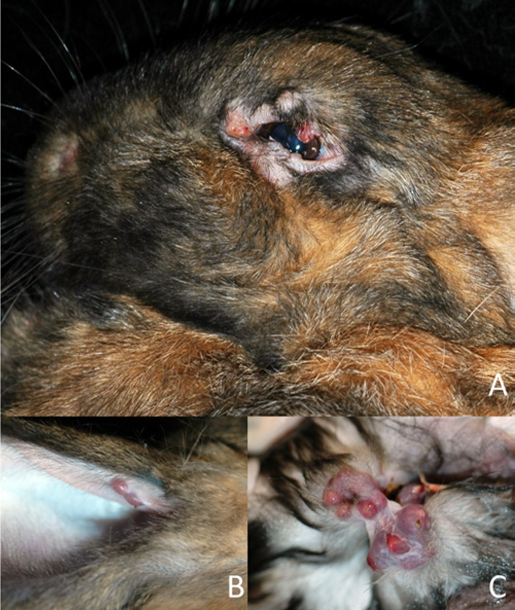
Cutaneous nodules in a rabbit with histoplasmosis. Multifocal granulomas are present in the periocular (
The nodule aspirates contained many well-preserved nucleated cells and low to moderate amounts of blood contamination. Nucleated cells consisted predominately of macrophages, with markedly lower numbers of heterophils and few multinucleated giant cells. Both intracellular (within macrophages) and extracellular yeast organisms were observed. Yeast organisms were approximately 3–5 µm in diameter, round to oval, with a thin clear capsule and contained an eccentrically placed basophilic crescent-shaped nucleus. The interpretation was granulomatous inflammation with intralesional yeast that showed a morphology consistent with Histoplasma spp. Based on the cytological examination results, a lack of response to treatment at 12 days, and potential propagation to other rabbits in the household, the owner elected for humane euthanasia. The body was submitted to the Louisiana Animal Disease Diagnostic Laboratory, Louisiana State University (Baton Rouge, Louisiana) for necropsy.
At the time of postmortem examination, the rabbit was noted to be in adequate body condition with mild postmortem autolysis. There were several multifocal to coalescing, pale tan to red-purple, firm cutaneous nodules along the margins of the pinna and around the mouth, nose, eyes, prepuce, and anus. The nodules ranged in size from 3 to 5 mm in diameter. On cut surface, the nodules were pale tan and occasionally contained caseous material. Similar nodules were present along the brachia and antebrachia, within the axillae, on the medial aspect of the right pelvic limb, and along the left flank. No other significant gross abnormalities were noted. Based on the clinical suspicion of histoplasmosis, approximately 2 cm × 2 cm sections of the dermal nodules from the left superior palpebra and the right axilla were submitted separately for fungal culture.
Histological examination revealed the presence of intracellular and extracellular yeast organisms in the small intestine, skin (antebrachium, perioral, palpebral, perianal, and pinnal), penis and penile urethra, rectum, axillary lymph node, and conjunctiva of the eye (Fig. 2). Yeasts were ovoid and approximately 3–5 µm in diameter with eccentric, round to crescent-shaped nuclei surrounded by a clear halo (consistent with Histoplasma spp.; Fig. 3). Organisms stained with Gomori methenamine silver and periodic acid–Schiff stains (Fig. 4). Multifocal foci of hepatic inflammation with rare multinucleated giant cells raised suspicion of hepatic involvement, but organisms could not be identified in the sections examined. Definitive evidence of pulmonary involvement was not observed. Postmortem samples collected for fungal culture (superior palpebral and right axillary nodule) confirmed identification of H. capsulatum.
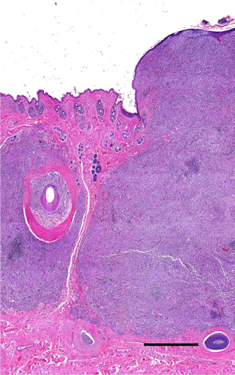
Histology of a cutaneous nodule from a rabbit with histoplasmosis. Multifocally expanding the dermis and elevating the epidermis are large granulomas. Bar = 1 mm.
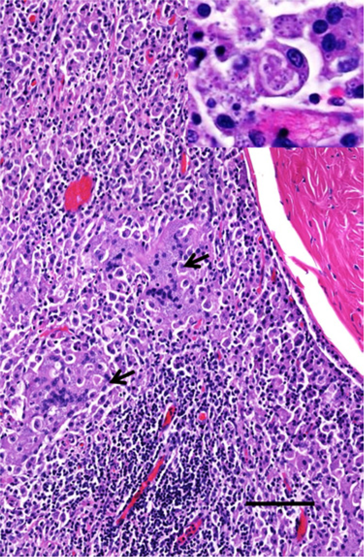
Histology of a cutaneous nodule from a rabbit with histoplasmosis. The granulomas frequently contain multinucleated giant cells (arrows) with myriad phagocytized yeasts. Bar = 200 µm. Inset: yeasts are round, 3–5 µm in diameter, with a basophilic center and a clear halo.
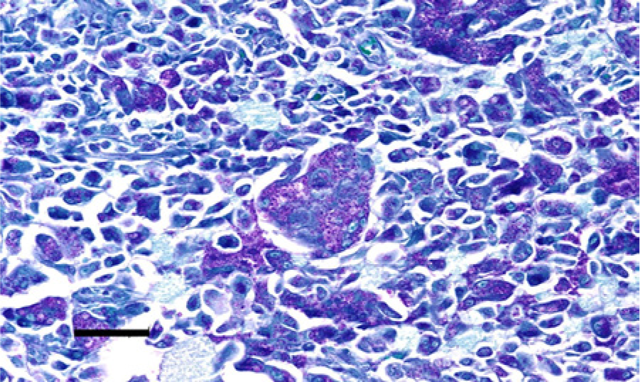
Histology of a cutaneous nodule from a rabbit with histoplasmosis. The yeasts stain pink with periodic acid–Schiff stain. Bar = 50 µm.
In the current case, the initial diagnosis was based on clinical findings and cytology of the periocular lesions. The presence of organisms consistent with Histoplasma spp. was highly suggestive of local histoplasmosis. The disease continued to progress despite medical therapy. Although the histological and cytological appearance of the organisms recovered from the dead rabbit was highly suggestive of H. capsulatum, isolation of the organism by fungal culture was performed to confirm the diagnosis. Culture and isolation of the organism from clinical and biological samples, including blood, is considered the definitive diagnosis of histoplasmosis in human beings. 11 Experimental histoplasmosis shares some similarities to the current case in lesion distribution (periocular cutaneous and conjunctival lesions as well as pinnal cutaneous lesions). However, anogenital and intestinal lesions were not described in the rabbits in the report of experimental disease. 12 Given the lesion distribution in experimentally induced histoplasmosis and the predominance of cutaneous and mucocutaneous lesions, it is possible that rabbits may have a greater predilection for developing lesions at cutaneous and mucocutaneous sites, rather than primarily respiratory and gastrointestinal systems, as diagnosed in cats and dogs, respectively.6,12 It is uncertain why the disease continued to progress and become disseminated. In human beings, immunosuppressed patients are considered to be at a greater risk for the development of disseminated and fatal disease.3,11,34 In immunocompetent individuals, the disease is usually self-limiting; however, cases of progressive disseminated histoplasmosis have been described in people with no apparent predisposing factors.17,36 In such cases, where a cause for immunodeficiency cannot be identified, it is considered possible that a “yet-to-be identified” immunodeficient condition may exist. 36 Although no cause for immunosuppression could be found in the current case, it is possible that this patient was immunocompromised. With the exposure to a new environment, which could have held a source of H. capsulatum, the animal may have become infected. Recurrence of an old and/or previous infection is another potential mechanism of pathogenesis that has been postulated in human medicine, although definitive proof of this disease scenario is somewhat lacking in the literature. 31
Treatment options for histoplasmosis include the use of antifungal medication. In the current case report, treatment for a possible dermatophytosis was attempted with microsized griseofulvin. This medication was previously considered to be the treatment of choice for dermatomycoses although it appeared to be ineffective against deep-seated mycotic infections.8,19,24 Triazoles (e.g., fluconazole, itraconazole) or imidazoles (e.g., ketoconazole) are commonly used drugs for the treatment of fungal diseases. 10 In human beings, H. capsulatum has been said to have acquired resistance to fluconazole, although 600–800 mg per day has been shown to be clinically efficient in 36 out of 37 cases. 26 However, in patients with acquired immunodeficiency disease syndrome, fluconazole induced resistance during treatment and was less effective than itraconazole. 32
The treatment of choice for histoplasmosis in dogs and cats is itraconazole.5,18 Itraconazole alone or in combination with ketoconazole or amphotericin B has been reported to be effective in clinical cases of feline histoplasmosis.2,13 In dogs and cats, the clinical use of voriconazole appears to be underreported although in vitro testing revealed good results for both the mycelial and yeast-like phase. 4 In the same study, caspofungin had the highest minimum inhibitory concentration against feline histoplasmosis mycelial and yeast-like phase when compared to amphotericin B, itraconazole, and voriconazole. 4 Posaconazole (approved by the U.S. Federal Drug Administration in 2006) has been shown to have a lower minimum inhibitory concentration against human H. capsulatum isolates, even against those isolates with low susceptibility to fluconazole and voriconazole. 20 Amphotericin B, commonly considered the gold standard antifungal drug to which all other antifungal drugs are compared, is not commonly recommended to be used in cases of histoplasmosis due to possible renal toxicity; however, its use for specific cases of histoplasmosis has been reported to provide good results.7,17,33 It is important to mention that some fungal isolates (e.g., Aspergillus terreus) have developed in vitro resistance to amphotericin B. 30
As the griseofulvin treatment was ineffective and an in vitro sensitivity study was not performed in the present rabbit case, it is unclear which medication would have been the most appropriate. The use of itraconazole (20 and 40 mg/kg, orally, once a day for 5 days) or amphotericin B (1.5 mg/kg, intravenously, once a day for 5 days) in a rabbit model of invasive aspergillosis did not cause mortality in the treatment groups and it allowed significant reduction of Aspergillus fumigatus in the affected tissues when compared with control groups in which more than 75% mortality occurred.22,23 In a rabbit model of coccidioidal meningitis, treatment with itraconazole (80 mg/kg/day, orally, once a day for 28 days) or fluconazole (80 mg/kg, orally once a day for 28 days) did not result in mortality associated with the medication. 29 Oral ketoconazole (approximately 35 mg/kg, orally, twice a day for 7 days) and fluconazole (20 or 80 mg/kg, orally, once a day for 7 days) did not cause mortality in a candidiasis rabbit model. 15 These studies do not provide information on the adequate treatment for rabbit histoplasmosis but do show the safety of these dosages and drugs while used in animal models for other fungal diseases. Future studies are needed to assess the optimal medication as well as the dosage for the treatment of rabbits diagnosed with histoplasmosis.
In conclusion, the current case report describes a case of a previously undescribed disseminated histoplasmosis in a pet rabbit diagnosed both antemortem via cytology and postmortem via histology and culture of H. capsulatum from the lesions. Although the treatment used did not provide noticeable improvement, available information on histoplasmosis treatment in other species has been reviewed in an attempt to provide useful information for future management of this condition in rabbits. Based on the information compiled herein, hypothetically, itraconazole may be an adequate treatment option for rabbits. This is based on the knowledge that itraconazole is the treatment of choice for histoplasmosis in dogs,5,18 and that the use of high doses of itraconazole in rabbits for several weeks did not cause mortality. 29 This suggested treatment option is a hypothesis that requires confirmation and validation.
Footnotes
Acknowledgements
The authors thank Dr. Alma Roy for assistance in mycology and the Louisiana State University histology laboratory for tissue processing.
a.
Griseofulvin oral suspension, Perrigo Co., Yeruham, Israel.
Declaration of conflicting interests
The author(s) declared no potential conflicts of interest with respect to the research, authorship, and/or publication of this article.
Funding
The author(s) received no financial support for the research, authorship, and/or publication of this article.