
Abstract
An aflatoxicosis outbreak affected 65 dogs from 9 different farms after they were fed diets with cooked corn meal as a common ingredient. Of the dogs, 60 died. Numerous dogs died on additional farms, but those dogs were not included in the study. The farmers acquired the contaminated maize products, in the form of whole corn grain or as corn meal, from the same supplier. The corn product was mixed with meat that was left over from home or commercial rations to form corn polenta, which was fed to the dogs. Necropsy was performed on 3 dogs. Two of the dogs died after a few days of refusing food, showing anorexia, polydipsia, icteric mucous membranes, hematemesis, hematochezia, or melena, and bleeding of the skin, eye, ear, and mouth. The primary necropsy findings included jaundice, hemorrhages in several organs, and yellowish enlarged liver with enhanced lobular pattern. The dog that experienced chronic ascites had a yellowish liver with reduced volume, irregular surface, and increased consistency. The main histological findings included hepatocyte fatty degeneration, biliary duct hyperplasia, cholestasis and, in the chronic case, hepatic fibrosis. High-performance liquid chromatography analysis of the corn meal from 2 affected farms revealed 1,640 ppb and 1,770 ppb of aflatoxin B1, respectively. The current study demonstrates an additional way that dogs can be exposed to, poisoned, and killed by aflatoxin.
Mycotoxins are fungal metabolites that are currently considered to be the most dangerous and widespread contaminants in food and animal feed worldwide. Aflatoxins, a group of mycotoxins mainly produced by Aspergillus flavus and Aspergillus parasiticus fungi, are of particular importance to human and animal health. These organisms invade crops and may grow on foods during storage if temperature and humidity levels are favorable. The major aflatoxins produced in feedstuffs are B1, B2, G1, and G2, among which aflatoxin B1 is the most pathogenic. 10 The major aflatoxins are hepatotoxic and can be immunosuppressive, nephrotoxic, and/or carcinogenic, and may induce hemolytic anemia and coagulopathies.7,14 The main hepatic effects of aflatoxins are hepatocellular steatosis and necrosis, bile duct proliferation, cholestasis, and fibrosis. Pigs and dogs are the most susceptible mammals to the effects of aflatoxins. 13 Mycotoxin contamination poses a serious threat to the pet food industry, and aflatoxins have been the most common cause of acute mycotoxicosis outbreaks related to commercial dog food. Maize products are the usual sources of aflatoxins in these cases. 5 In Brazil and in other countries, significant levels of aflatoxins have been detected in up to 50% of samples of commercial dog diets analyzed (Mallmann CA, Mürmann L, Almeida CAA, et al.: 2003, Níveis de aflatoxinas em rações caninas [Aflatoxin levels in canine commercial diets]. XIII Brazilian Congress of Toxicology, Londrina, Paraná, Brazil. In Portuguese).5,7 Such contaminations may be attributed to the exposure of the rations to high humidity and temperature, or to the use of contaminated ration components. Sporadic reports have linked animal mortality to high levels of aflatoxins in feed constituents offered to them. In Australia, several dogs were poisoned and killed by aflatoxins derived from moldy bread. 6 The clinical presentation of aflatoxicosis in dogs is related to injuries to the liver, and young dogs may die several hours after ingesting feed contaminated with high levels of aflatoxins. 13 The diagnosis of aflatoxicosis should be based on anatomical and pathological examinations of the liver and the presence of aflatoxins in the feed. 2 The current study describes the epidemiological and clinicopathological findings observed in a large aflatoxicosis outbreak in dogs, which occurred in 2011 in southern Brazil.
Epidemiological data were retrieved from the owners of the dogs and local veterinarians from the municipalities of Encruzilhada do Sul and Amaral Ferrador in the State of Rio Grande do Sul. The regional temperature range was 16–31°C in the period in which the dogs were poisoned. Information on storage conditions and a visit to the storage facility where the corn was stored were denied by the owners of the warehouse. Three dogs were submitted for necropsy, during which the tissues were sampled, fixed in 10% buffered formalin, processed by standard histological methods, and stained with hematoxylin and eosin. Commercial dog ration and corn meal samples from batches used in the diet of the dogs from 2 affected farms were submitted for mycotoxin analysis at the Laboratory of Micotoxicological Analysis (Federal University of Santa Maria, Santa Maria, Rio Grande do Sul, Brazil) by extraction, clarification, and 100% automated derivation, in addition to high-performance liquid chromatography.
According to local practice, the local farmers feed their dogs with mixes that include corn polenta as the main diet component. The corn meal was acquired by the farmers from the same commercial source, and then cooked and fed to the dogs as corn polenta. The dogs were also fed meat, leftover food from the home, and commercial rations. From March to June 2011, 60 dogs from 8 farms were affected and died spontaneously, after showing similar clinical presentation (Table 1). Clinical course of disease ranged from 1 to 15 days, and most dogs succumbed in 3 days; however, in 1 dog, clinical disease lasted for 45 days before the dog died. The first clinical sign observed in the dogs was the refusal of any diet in which the contaminated corn meal was present. Such behaviors motivated the farmers to increase the amount of other feed components, rather than to exclude the corn polenta from the diets. The dog owners also noticed that when the dogs were occasionally offered foods that did not contain the corn polenta, the dogs ate voraciously. The dogs presented with hematemesis, jaundice, hematochezia or melena, abdominal enhancement, wasting, polydipsia, prostration, anorexia, dried feces, inquietude, bleeding from the skin, eye, ear, and mouth, and orange urine. Most farms were located in a rural area, and all of the dead dogs were adults. Most of the surviving dogs did not eat the corn polenta but instead had consumed commercial diets and leftover food from homes. Despite ingesting the contaminated food, 5 dogs from 3 farms recovered after showing enhanced abdominal volume and polydipsia. Local veterinarians reported dog deaths from at least 6 additional farms, but the data were not included in the current study because no details of clinical signs or course of the disease were available to the authors.
Evolution and clinical signs in dogs affected with aflatoxicosis.*
+ = presence of clinical sign; − = absence of clinical sign.
With the exception of one dog that died after 45 days.
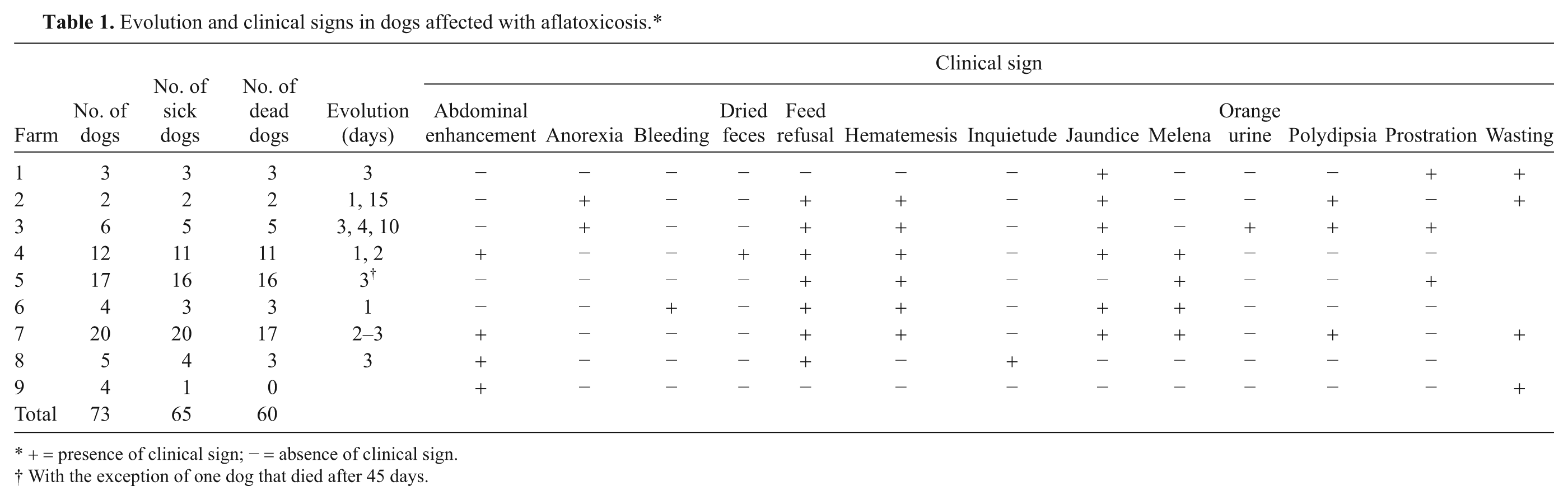
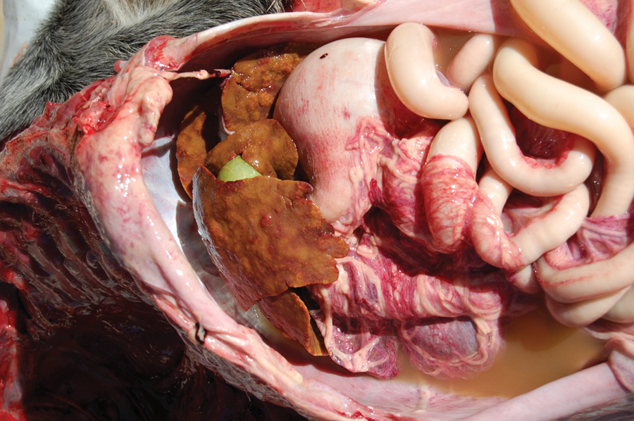
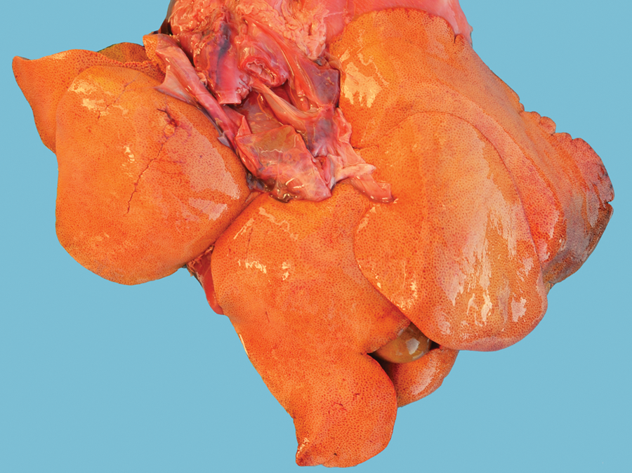
Three affected dogs were necropsied: a Border Collie (case 1: male, 1 year of age, farm 1), a mixed breed (case 2: female, 4 years of age, farm 6), and an Australian Cattle Dog (case 3: female, 1 year and 6 months of age, farm 5). The last dog survived for 45 days with the disease before dying. The necropsied dogs had regular (case 1) or bad (cases 2 and 3) body condition, jaundice (cases 1 and 2), general paleness (case 3), multifocal hemorrhages that were subcutaneous, muscular, in the parietal pleura, and in the pancreas (case 1), hemothorax (case 1), and severe pulmonary edema and congestion (cases 1 and 2). The abdominal cavities of the dogs contained a bloody fluid (case 1) or yellowish-white, cloudy (case 3) exudate (Fig. 1). Mildly enhanced, diffusely yellowish-orange livers with enhanced lobular pattern (cases 1 and 2; Fig. 2), or decreased liver with irregular surfaces bearing yellowish and pale random areas, with enhanced lobular pattern (Fig. 1), and increased consistency (case 3) were also observed. Dark red liquid contents were observed in the stomach (case 2) and small intestines (cases 1 and 2; Fig. 3) of the dogs. In case 3, a perforated ulcer in the duodenum that was 0.6 cm in diameter with dark red fluid in its edges and surroundings was also observed.
Aflatoxicosis epizootic in dogs. Abdominal yellowish-white fluid and decreased, yellowish liver with irregular surface and random pale areas. Case 3 is shown.
Aflatoxicosis epizootic in dogs. Yellowish-orange mildly enhanced liver showing an enhanced lobular pattern. Case 2 is shown.
Aflatoxicosis epizootic in dogs. Bloody contents in the whole extension of the small intestine. Case 2 is shown.
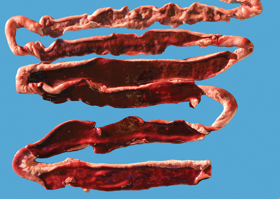
The main histological lesion was a severe and diffuse cytoplasmic vacuolization of the hepatocytes. This lesion was associated with bile duct proliferation, cholestasis (cases 1 and 2; Fig. 4), mild diffuse mononuclear inflammatory infiltration (case 1), and mild periportal fibrous connective proliferation (case 2). In the liver of the dog with chronic disease (case 3), there was severe proliferation of the fibrous connective tissue, isolation of lobules and hepatocytes, nodules of regeneration, intense bile duct proliferation, mild cholestasis, and mild hepatocyte vacuolization within the regeneration nodules (Fig. 5). The fibrous connective tissue was evidenced by Masson trichrome staining (Fig. 6). There was also moderate coagulative renal necrosis (case 1), mild vacuolization of the convoluted tubules (case 2), and mild hemosiderosis in the spleen (case 2). By high-performance liquid chromatography analysis of the corn meal samples, 1,640–1,770 ppb of aflatoxin B1, 105–154 ppb of aflatoxin B2, 10.5–27.8 ppb of aflatoxin G1, and 0–2.8 ppb of aflatoxin G2 were detected. No aflatoxins were detected in the samples from the commercial dog food.
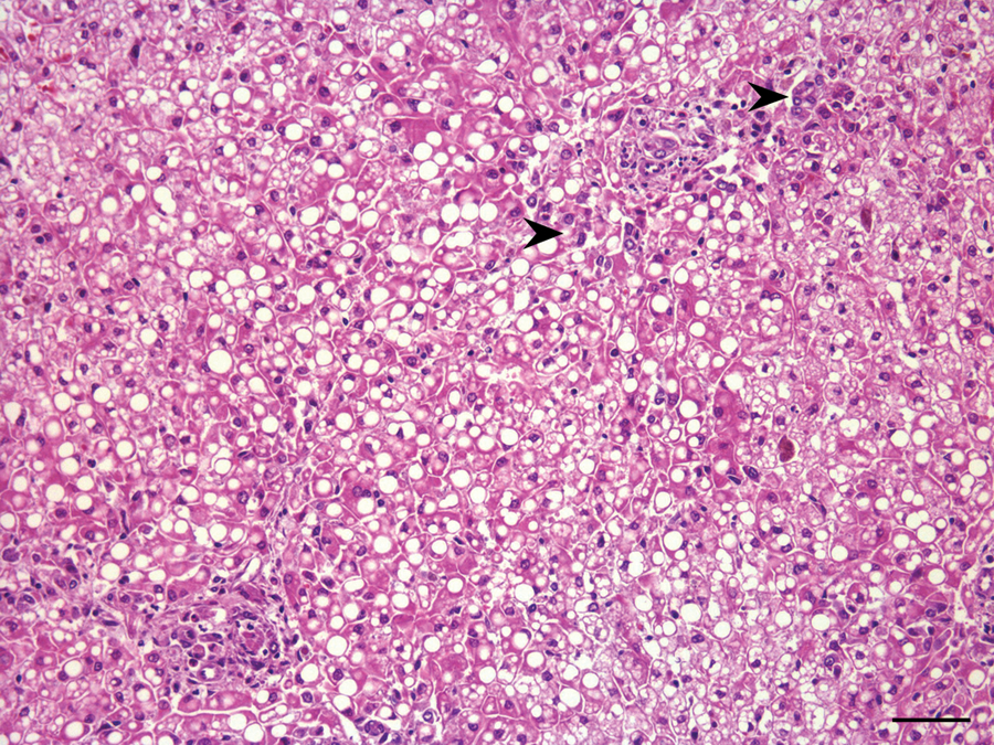
Aflatoxicosis epizootic in dogs. Liver showing severe and diffuse hepatocyte cytoplasmic vacuolization associated with bile duct proliferation (arrowheads). Case 1 is shown. Hematoxylin and eosin. Bar = 120 µm.
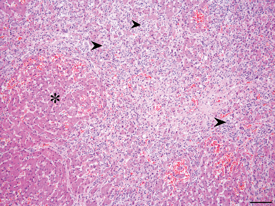
Aflatoxicosis epizootic in dogs. Liver showing severe fibrous connective proliferation, isolation of lobules and hepatocytes, nodules of regeneration (asterisk), and bile duct proliferation (arrowheads). Case 3 is shown. Hematoxylin and eosin. Bar = 200 µm.
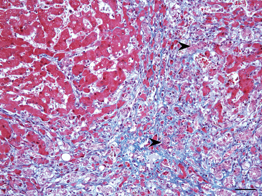
Aflatoxicosis epizootic in dogs. Liver stained with Masson trichrome method showing fibrous connective proliferation (in blue) and bile duct proliferation (arrowheads). Case 3 is shown. Bar = 120 µm.
The consumption of aflatoxin-contaminated commercial diets, in which the maize product is the source of the contaminant, has been the most common cause of the disease in dogs.9,10 In the outbreak described herein, the dogs were fed a homemade diet, which included aflatoxin-contaminated corn meal used to produce a cooked corn polenta, a local practice of dog owners. Although a cooking time of 30–45 min in boiling water (100ºC) is usual for preparing the polenta, the thermal stability of aflatoxin B1 is known to be disrupted at 160°C. 11 The corn meal samples had high levels of aflatoxin B1 content and were the sources of the toxin in these cases. The concentration of aflatoxin in the feed, time of exposure, affected species, amount of feed ingested, diet, nutritional state, and sex determine the effects of aflatoxin on animal health. 8 In Brazil, the maximum tolerated limit of total aflatoxin is 50 ppb (50 μg/kg) in feed ingredients submitted for ration formulation for animal consumption and 20 ppb in corn grain (whole, broken, crushed, ground), corn flour, or corn meal for human consumption (Brazilian Ministry of Health: 2002, Agência Nacional de Vigilância Sanitária. Resolução RDC 274. Regulamento técnico sobre limites máximos de aflatoxinas admissíveis no leite, no amendoim, no milho [Technical regulations on the maximum aflatoxin limits in milk, peanut, corn]. Available at: http://www.anvisa.gov.br/legis/resol/2002/274_02rdc.htm. In Portuguese).
Dogs are among the most aflatoxin-susceptible animal species and may be fatally affected by doses less than 1 mg/kg. 14 In the present cases, the possibility that the aflatoxin-contaminated component could have been included in the human food chain cannot be excluded. The clinical presentation of aflatoxin poisoning is mainly associated with the level of aflatoxin in the diet and the amount of food ingested, which affect the occurrence of acute, subacute, and chronic hepatopathy. 9 In this epizootic, 59 out of the 60 dead dogs suffered from acute clinical disease, and only 1 (case 3, farm 5) died of chronic aflatoxicosis. However, in 3 of the farms, some dogs developed ascites after surviving the acute stage of the disease. Feed refusal was observed in these cases, and such behavior has previously been linked to an altered palatability of the diet. 14 Conditioned taste aversion is an important biological component of the defense mechanisms against ingestion of food toxins in addition to being a valuable clinical sign.
The tendency for bleeding, which has been observed in the cases described herein and previously reported in many other cases of aflatoxin-poisoned dogs,9,14 may be linked to the prothrombin and fibrinogen deficiencies secondary to hepatic synthesis deficit and disseminated intravascular coagulation caused by the hepatic necrosis. Aflatoxicosis may be associated with hepatocellular degeneration, especially in acute cases. Biliary ductal proliferation is a common finding in subacute cases, and as severe fibroplasia in chronic cases. 4 Of the 3 dogs, 2 necropsied in the present study presented changes that are typical of subacute aflatoxicosis, despite the short clinical course. In previous reports, the acute 2 and subacute2,14 forms of the disease predominated. Hepatic steatosis was evident in the 3 necropsied cases in the current study. A previous study showed that hepatocyte degeneration was a consistent finding in canine aflatoxicosis and was independent of clinical evolution. 4 The previous study also observed hepatocyte megalocytosis that resembled the changes observed in pyrrolizidine alkaloid poisoning. Both agents inhibit hepatocellular regeneration and induce hepatic cirrhosis. 13
Previous descriptions of canine aflatoxin poisoning indicate that the species is highly sensible to the acute and subacute forms of the disease.4,9 Studies in small rodents have revealed the induction of hepatic carcinoma after prolonged ingestion of low aflatoxin doses, 3 which suggests that aflatoxins may also induce hepatic neoplasia in dogs. Aflatoxins are considered to be genotoxic and carcinogenic and have been associated with the development of hepatic cancer in human beings and animals. The induction of hepatic cancer is associated with the chronic consumption of low doses of aflatoxin. Therefore, with time, the actual number of dogs affected by aflatoxin outbreaks may be higher than that recorded during the short period (March–June 2011) of the current study. 1 Hepatic cirrhosis is an important degenerative disease that affects dogs in Rio Grande do Sul 12 ; however, in such cases, the etiology is often undetermined. The use of corn meal in the diet of dogs increases the risk of hepatic insufficiency secondary to aflatoxin poisoning in these animals. Chronic aflatoxicosis may represent a considerable cause of hepatic cirrhosis in dogs, a hypothesis that is reinforced by the local practice of feeding corn polenta to dogs. 3
Footnotes
Acknowledgements
The authors are grateful to Guilherme Vogg, Márcia Louzada, Pablo Ataide, and Rafael Barcelos for providing epidemiological data, and to Dr. Carlos A. Mallmann for the analytical toxicology.
Declaration of conflicting interests
The authors declared no potential conflicts of interest with respect to the research, authorship, and/or publication of this article.
Funding
The author(s) disclosed receipt of the following financial support for the research, authorship, and/or publication of this article: This work was financially supported by grants from the Conselho Nacional de Desenvolvimento Científico e Tecnológico (CNPq)–Brazil.