
Abstract
A 6-y-old, 3.5-kg, spayed female Toy Poodle was presented with left forelimb lameness of 2-d duration. Two months before the initial presentation, radiography showed osteolysis of the medial epicondyle of the left humerus, and the left forelimb was amputated. Grossly, the articular villi of the elbow joint were markedly thickened, and the articular cartilage surfaces of the distal humerus and proximal radius had partial erosion. Histologically, granulomatous arthritis and osteomyelitis characterized by the presence of abundant macrophages containing numerous fungi were observed. ITS and β-tubulin sequences amplified from the isolate from the specimen were 100% and 99% identical to type strain UTHSC D16-145T of Talaromyces georgiensis, respectively. Canine osteoarthritis caused by T. georgiensis has not been reported previously, to our knowledge.
Genus Talaromyces (family Trichocomaceae 12 ), known previously as an asexual genus of Penicillium, has been isolated from soil and insects worldwide. 3 Some Talaromyces spp. are pathogenic, and have been reported to cause serious systemic infections in humans and animals.9,25 The genus Talaromyces has ~ 110 species, and several new species, including T. georgiensis, have been discovered in 2017–2018.3,12 Herein, we report a case of arthritis caused by T. georgiensis in a dog.
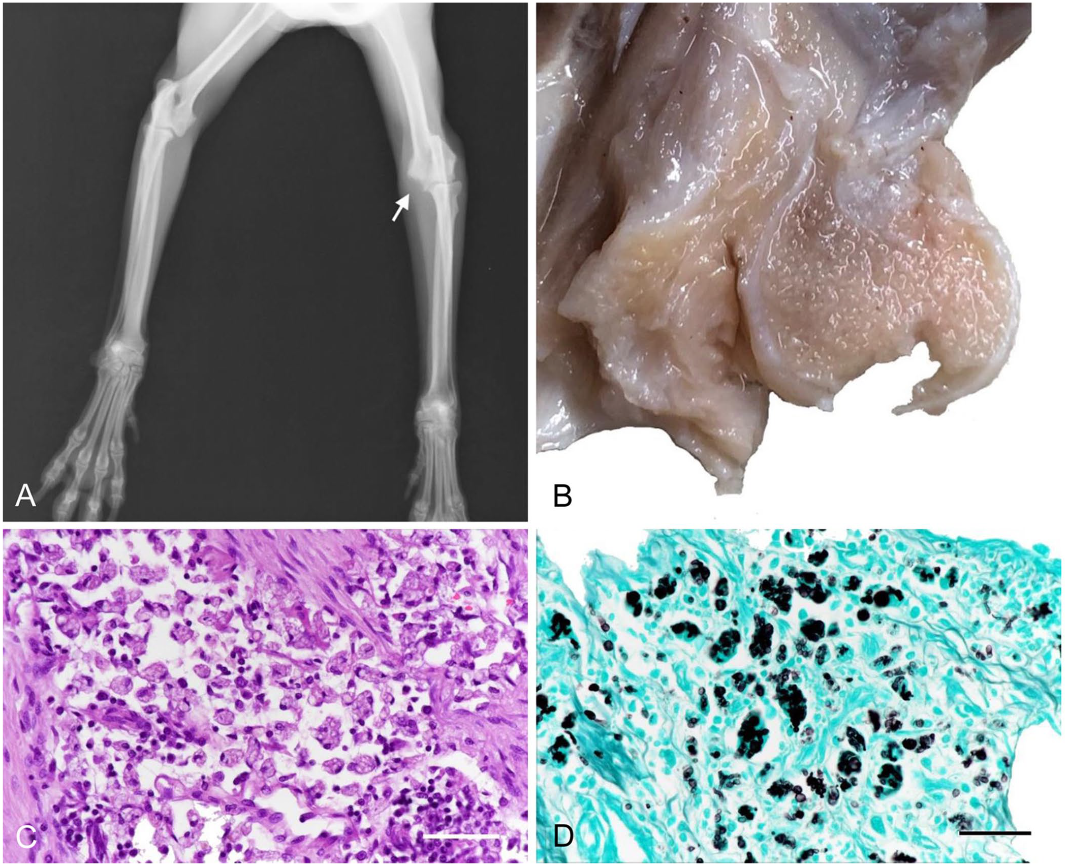
A 6-y-old, 3.5-kg, spayed female Toy Poodle was presented with left forelimb lameness of 2-d duration. Radiography showed mild periosteal reaction of the left distal humerus. Serum biochemistry revealed an increased C-reactive protein level (CRP; 60.2 mg/L). The dog was diagnosed with arthritis and was treated with carprofen (2.2 mg/kg PO q12h for 1 mo; Rimadyl; Zoetis). One month later, during the second consultation, swelling of the elbow joint was observed. Radiography (Fig. 1A) and computed tomography (CT) showed osteolysis of the medial epicondyle of the left humerus. Arthritis (caused by autoimmune or infectious diseases) and neoplasia (such as synovial sarcoma) were differential diagnoses. Bacterial cultures, performed at Hoken Kagaku Laboratory (Kanagawa, Japan), using sheep blood agar, fungal cultures using Sabouraud agar to detect filamentous fungi and Cryptococcus spp., and CHROMagar candida medium (BBL; Becton Dickinson) to identify yeast-like organs of the synovial fluid, were negative. At reexamination 1 mo later, clinical signs had not resolved, and the left forelimb was amputated because of ongoing pain and muscle atrophy. The dog was subsequently treated with terbinafine (35 mg/kg PO q24h for 3 wk; TRF, Lamisil tablets 125 mg; Sun Pharmaceutical).
Granulomatous polyarthritis caused by Talaromyces georgiensis in a dog.
The amputated forelimb was fixed in 10% formalin. Grossly, the articular villi of the elbow joint were markedly thickened (Fig. 1B). The articular cartilage surfaces of the distal humerus and proximal radius were extensively and partly irregular, and demonstrated erosion along with exposed subchondral bone (Fig. 1B); additionally, there was a 5-mm lytic lesion on the distal humerus. There were no ulcers or signs of trauma on the skin of the forelimb. Samples from the elbow joint were processed routinely and stained with hematoxylin and eosin, as well as Gomori methenamine silver (GMS) stain and periodic acid–Schiff (PAS) reaction. Histologically, severe granulomatous arthritis composed of infiltrates of numerous macrophages, lymphocytes, plasma cells, and a few neutrophils was observed in the articular villi of the elbow joint (Fig. 1C), underlying mild granulation tissue formation and edema. In addition, severe inflammation extended up to 3 mm into the bone marrow cavity of the distal humerus and proximal radius with mild granulation tissue; necrosis of the exposed distal humerus was observed. Most macrophages had cytoplasmic enlargement and contained abundant spherical yeast-like organisms 3–10 μm diameter, and 3-μm wide, septate, nonbranching hyphal-like structures. Both the yeast and hyphal-like organisms were positive with GMS (Fig. 1D) and PAS reaction. Fewer of these organisms were found extracellularly. Mild muscle atrophy around the joint was observed without inflammation. In the axillary lymph nodes, a few fungal-laden macrophages were found in the marginal sinus.
Four months after the amputation, the dog exhibited lameness of the right hindlimb. Radiography showed osteolysis in the medial aspect of the right stifle joint. Three weeks later, CT showed lysis and periosteal new bone production of the distal right femur medially, and mild enlargement of the right internal iliac lymph node. Tissues samples for histologic examination and fungal culture were taken from the distal right femur. Histologically, fungi with similar morphology to those observed previously in the left elbow joint were found within the macrophages.
Tissue samples were inoculated on a Sabouraud dextrose agar plate for 14 d at 28°C. Numerous flat, velvety, white and green powdery colonies were observed on the plate after 14 d incubation. On microscopic examination, septate, unbranching conidiophores and smooth-walled spherical conidia (2–3 μm diameter) were observed (Fig. 2).
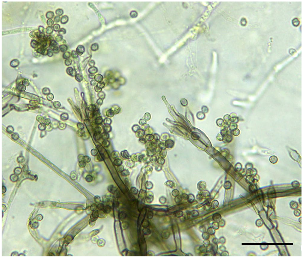
Septate, unbranching conidiophores and smooth-walled spherical conidia (2–3 μm diameter) are visible in fungal colonies cultured from the right distal femur in the dog. Bar = 20 μm.
Molecular identification of the fungus was conducted based on isolation of DNA genes using the colonies grown on the plate. The internal transcribed spacer (ITS) region of the DNA sample from the isolate was amplified using the universal fungal primers ITS5 (5′-GGAAGTAAAAGTCGTAACAAGC) and ITS4 (5′-TCCTCCGCTTATTGTAGC) 14 ; PCR amplification conditions and sequence analysis were performed as described previously. 2 The β-tubulin primers benA-F (5′-AATTGGTGCCGCTTTCTGG) and benA-R (5′-AGTTGTCGGGACGGAATAG) were used to amplify DNA from Aspergillus species. 1 Comparative nucleotide sequence analysis using the BLAST algorithm (http://blast.ncbi.nlm.nih.gov/Blast.cgi) showed that the ITS and β-tubulin sequences amplified from the isolate were 100% and 99% identical to type strain UTHSC D16-145T of Talaromyces georgiensis GenBank accessions LT558967 and LT559084, respectively. 12
In vitro susceptibility testing of the fungus was performed using the E test and M38-A microdilution methods. 22 The antimicrobial susceptibility tests for this organism showed that the minimum inhibitory concentration (MIC) of itraconazole (ITZ), voriconazole, TRF, amphotericin B, and fluconazole was < 0.002 mg/L, 0.012 mg/L, 0.06 mg/L, 0.25 mg/L, and > 256 mg/L, respectively. The dog was prescribed a 6-mo course of ITZ (7.5 mg/kg PO q24h; 50-mg tablets; Nichi-Iko Pharmaceutical) and TRF (35 mg/kg PO q24h) after diagnosis of the right hindlimb lesion. After 6 mo of antifungal treatment, the dog’s clinical signs had not progressed.
The dog had granulomatous arthritis associated with numerous intracellular and extracellular yeast and hyphae-like organisms. These organisms were confirmed to be T. georgiensis by fungal cultures, sequencing, and searching BLAST. T. georgiensis was isolated from joint fluid in an animal in 2017 12 ; details, including animal species and pathogenesis, are unclear. T. georgiensis has not been reported previously to cause disease in humans or animals, to our knowledge. T. georgiensis was found in multiple arthritic joints of our case, suggesting that T. georgiensis was the cause of fungal arthritis.
Some species of Penicillium have been reclassified as Talaromyces because the emphasis in taxonomy has shifted from morphologic features to phylogenetics via DNA sequencing technology. 24 With regard to Penicillium/Talaromyces spp., 3 cases of canine arthritis25,26,29 and 2 cases of canine osteomyelitis17,18 have been reported (Table 1). One of those cases was reported as a case of canine arthritis in the right cubital joint caused by a T. helicus infection. 25 T. helicus and T. georgiensis belong to the Helici section of Talaromyces. 3 The histologic morphology of T. georgiensis is similar to the cytomorphology of T. helicus, hyphae are 3-μm wide, septate, unbranched, and present both intra- and extracellularly; round structures were observed in both cases. With regard to the T. helicus infection, fungi were also observed in the skin, subcutaneous tissues, lymph nodes, and the spleen at autopsy. However, the fungi in our case were localized to multiple joints and the lymph nodes. These differences may be the result of variation in the original site of infection, associated pathogenicities, antifungal drugs, or host immunity. The other 2 case reports of Penicillium/Talaromyces spp. infection involving joints of dogs were associated with the presence of P. purpureogenum (renamed T. purpureogenus 28 ), which led to multiple lesions of diskospondylitis, 29 and P. verruculosum (renamed T. verruculosus 28 ), which led to osteomyelitis and arthritis of both hindlimbs. 26 Canine osteomyelitis caused by Penicillium/Talaromyces spp., including P. canis 17 and T. verruculosus, 18 have been reported; the specific involvement of joints was not described in these cases.
Penicillium/Talaromyces spp. infection in joints and bones of dogs.
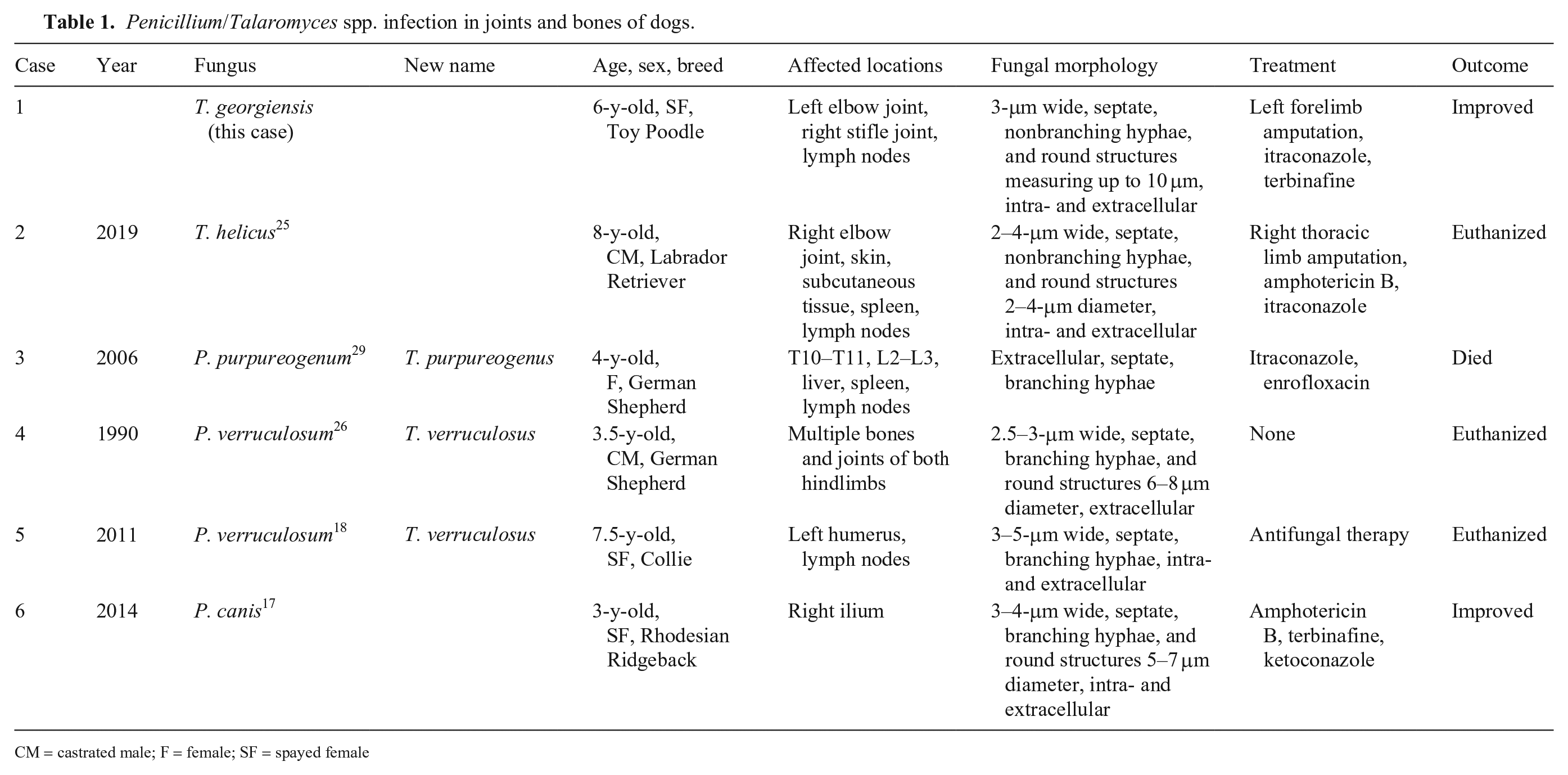
CM = castrated male; F = female; SF = spayed female
Although canine fungal arthritis is rare, the reported fungal causes include Aspergillus spp.,4,7,20,21,23,30 Cryptococcus spp.,5,8,13,16 Blastomyces dermatitidis,19,27 Candida guilliermondii, 6 Histoplasma capsulatum, 15 Paecilomyces sp., 10 and Sporothrix schenckii 11 (Table 2).
Summary data of cases in dogs of fungal arthritis reported in the literature.
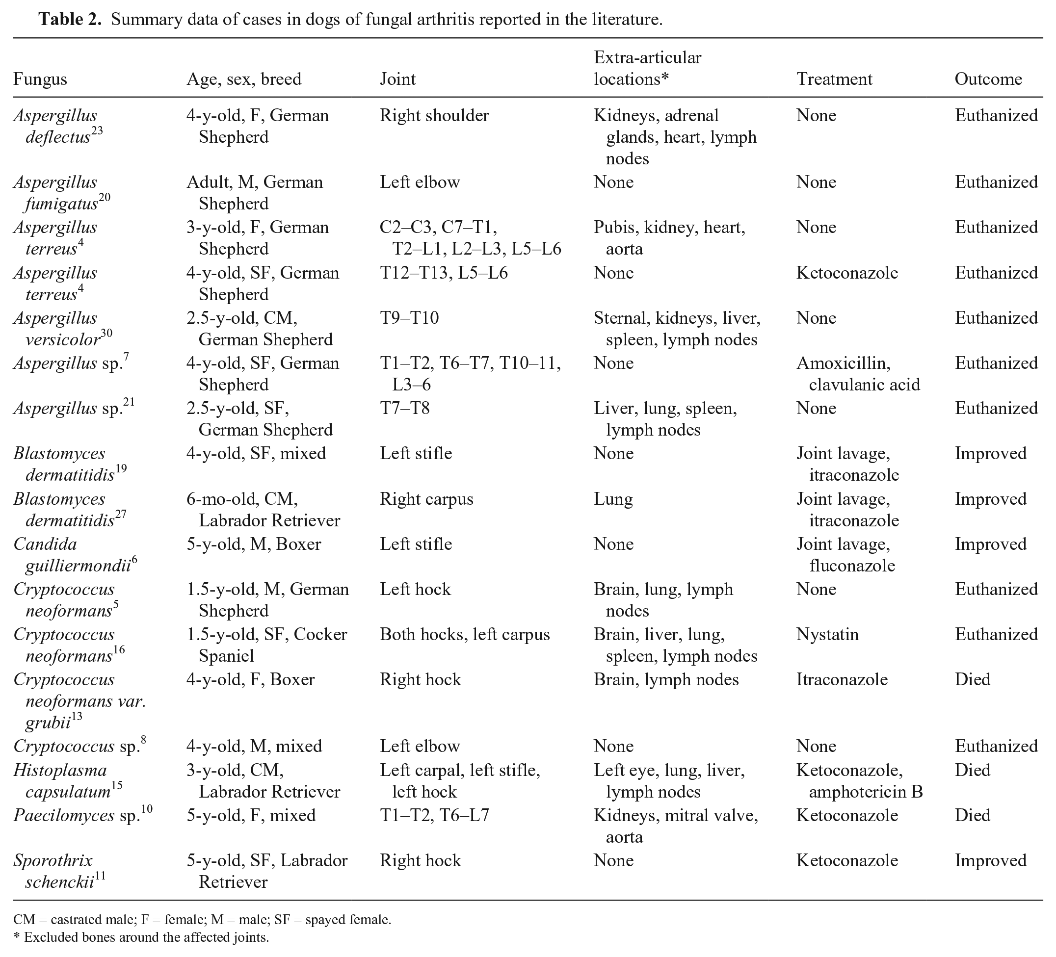
CM = castrated male; F = female; M = male; SF = spayed female.
Excluded bones around the affected joints.
Accurate identification of fungi by their histologic morphology alone is difficult. Molecular techniques such as DNA sequencing have made it possible to identify rare fungal species grown in culture or found in clinical specimens. Identification of the fungal agent is important for selecting appropriate antifungal therapy.
The most common routes of fungal infection are invasion from inhalation of spores, traumatic inoculation of spores, or hematogenous invasion by septicemia. In our case, no obvious immunologic abnormalities, or concurrent disease such as pneumonia or septicemia, were identified, thus it remains unclear how the fungus became widely disseminated.
Footnotes
Declaration of conflicting interests
The authors declared no potential conflicts of interest with respect to the research, authorship, and/or publication of this article.
Funding
The authors received no financial support for the research, authorship, and/or publication of this article.