
Abstract
A novel herpesvirus was detected in sun bears (Helarctos malayanus) with oral squamous cell carcinoma. Five captive sun bears from 4 institutions in the United States presented with oral lesions ranging from erythema and mild erosions to nodular, ulcerated masses. All 5 were diagnosed with squamous cell carcinoma. The tumors were treated with surgical resection but recurrence, local extension, or appearance of new lesions was noted in all cases. Intralesional chemotherapy was administered in 2 cases, and the nonsteroidal anti-inflammatory drug piroxicam was administered in 3 cases. Virus was detected in 4 of the 5 bears’ tissue samples using a consensus herpesvirus polymerase chain reaction. Nucleotide sequencing and phylogenetic analysis showed that this herpesvirus is in the subfamily Gammaherpesvirinae and distinct from other known herpesviruses. The association between the herpesvirus and squamous cell carcinoma is unknown. The current study presents a novel gammaherpesvirus within the order Ursidae, with the name Ursid herpesvirus 1 proposed.
Introduction
The smallest of the ursids, the sun bear (Helarctos malayanus) is an omnivorous, terrestrial bear inhabiting the tropical forests of Southeast Asia. Sun bear populations are threatened with extinction due to poaching for traditional medicine. 26 They are currently listed by the International Union for Conservation of Nature and Natural Resources (IUCN) Red List as vulnerable (Fredriksson G, Steinmetz R, Wong S, Garshelis DL: 2008, Helarctos malayanus. In: IUCN 2011. IUCN Red List of Threatened Species. Version 2011.1). In captivity, bears are generally considered long-lived and healthy species, with reported lifespans of over 20 years. The most frequently reported major health issues are tumors of the hepatobiliary system and dermatopathies. Dental problems, including periodontal disease and fractured canines, are also common. 22 There is 1 previous report of a mandibular squamous cell carcinoma in a Malayan sun bear. 20
The presence of herpesviruses in Ursidae, the family containing all 8 living species of bear, has been infrequently reported. Suid herpesvirus 1 (Pseudorabies virus) was isolated from an American black bear (Ursus americanus) and 4 captive European brown bears (Ursus arctos) with 100% mortality.25,33 Another outbreak of pseudorabies involved 3 Asiatic black bears (Ursus thibetanus), a Kodiak bear (Ursus arctos middendorffi), and 4 polar bears (Ursus maritimus) in a traveling circus; 4 of the 8 bears died and 1 was euthanized. 3 These infections were linked to the consumption of raw pork. Equid herpesvirus 9 was reported in a captive polar bear that was euthanized due to progressive meningoencephalitis. Zebra housed nearby were implicated as a possible source of the virus. 8 These cases likely represent cross-species transmission of a herpesvirus from its natural host reservoir to an aberrant host, with resultant severe disease and mortality. To our knowledge, an endemic herpesvirus has not yet been reported in an ursid.
Over 200 species of herpesviruses have been described. Herpesviruses tend to be host-specific and often appear to have co-diverged evolutionarily along with their hosts. Most vertebrate species examined have 1 or more endemic herpesviruses; the true number in nature is likely to exceed the 200 thus far discovered. All herpesviruses investigated to date are able to remain latent in specific cells of their natural host; they also frequently undergo active shedding without associated clinical symptoms, or with only mild symptoms. Fatal infections are usually rare in the natural, immunocompetent host. 21
Materials and methods
Animal information
Between September 2007 and August 2009, tissue samples from oral lesions of 5 captive sun bears at 4 institutions in the United States were submitted fresh frozen to the University of Florida Zoological Medicine laboratory (Gainesville, Florida) for viral polymerase chain reaction (PCR) panels, and submitted fixed in neutral buffered 10% formalin to Northwest ZooPath (Snohomish, Washington) for histopathology. Ages of the bears ranged from 8 to 19 years. Four were captive born, and 1 was wild caught.
Histopathology
All tissues were embedded in paraffin blocks, sectioned at 5 µm, and stained with hematoxylin and eosin. Hyperplasia of the oral mucosa or epidermis was characterized by hyperplasia of the epithelium with orderly maturation and no breach of the basement membrane. Dysplasia was characterized by hyperplasia with disorderly maturation, with mitotic figures appearing within the granular layers, and no breach of the basement membrane. Squamous cell carcinoma was characterized as orderly and disorderly proliferation of the epithelium into the submucosa or dermis, and breaching of the basement membrane, with varying degrees of inflammation and scirrhous response.
PCR amplification and sequencing
DNA was extracted from all of the tissue samples using a commercial kit. a Blank extractions were used as negative controls. Nested PCR amplification of a partial sequence of the herpesvirus DNA-dependent DNA polymerase gene was performed using previously described methods. 30 The PCR products were resolved in 1% agarose gels, excised, and purified. b To obtain additional sequence, a specific internal reverse primer was designed from the initial sequence (SunBearHV1rev: TCCCTGGAGCGTTACTGTCT). This was used with the forward pan-herpesviral primer DIEC (KNDSNTTYGAYATHGARTG). 17 Samples from cases 1 and 2 were also tested using a consensus papillomavirus PCR. 23 Sanger sequencing was performed c and analyzed. d Products were sequenced twice in each direction.
Phylogenetic analysis
The predicted amino acid sequence was compared to those in GenBank, as well as the National Center for Biotechnology Information Reference Sequence project, using BLASTP. 1 Predicted homologous 445-557 amino acid sequences of herpesviral DNA-dependent DNA polymerase were aligned using MAAFT. 14 Bayesian analyses of amino acid alignments were performed using MrBayes 3.1.2 24 on the CIPRES (Cyberinfrastructure for Phylogenetic Research) server (Miller MA, Pfeiffer W, Schwartz T: 2010, Creating the CIPRES Science Gateway for inference of large phylogenetic trees. In: Proceedings of the Gateway Computing Environments Workshop, pp. 1–8. November 14, 2010, New Orleans, LA), with gamma distributed rate variation and a proportion of invariant sites, and mixed amino acid substitution models. The first 10% of 2,000,000 iterations were discarded as a burn in. Maximum likelihood (ML) analyses of each alignment were performed using RAxML on the CIPRES server with gamma distributed rate variation and a proportion of invariant sites. 29 Psittacid herpesvirus 1 (GenBank accession no. NP_944403) was designated as the outgroup. The amino acid substitution model with the highest posterior probability in the Bayesian analysis was selected. Bootstrap analysis was used to test the strength of the ML tree topology, 9 with 1,000 bootstrap replicates.
Results
Animal information
Cases 1 and 2 were a 19-year-old female and an 18-year-old male sun bear, respectively, from the same institution. The female first presented with a swollen lower left lip that had mild spontaneous hemorrhage and multiple foci of erythema. This progressed over 1 month to become a 2-cm mass at the rostral aspect of the left lower lip, which was surgically removed with wide margins and diagnosed as squamous cell carcinoma (Fig. 1). Four months later, a new mass was noted on the ventrum of the oral cavity extending to the frenulum. Biopsies of this mass showed squamous cell carcinoma, which was marginally removed via laser surgery. The remaining tissue was infiltrated with 50 mg of carboplatin suspended in 30% sustained-release poloxamer. The right submandibular lymph node was removed at this time and showed no evidence of metastasis. Additional small areas of sublingual neoplasia were noted at the 1 month recheck and were treated with additional laser ablation and carboplatin–poloxamer sub-lesional injections.
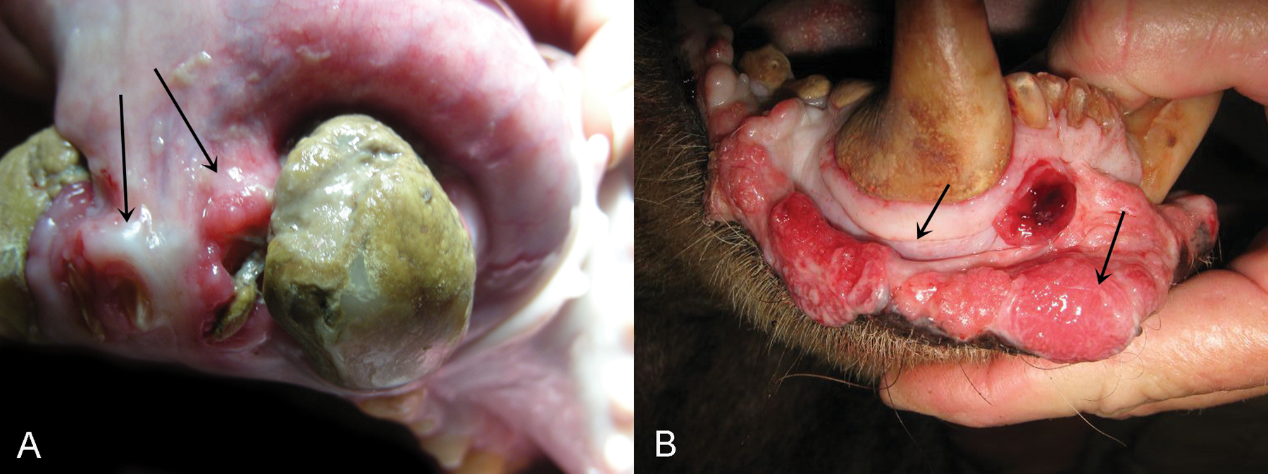
Sun bear (Helarctos malayanus); gross pathology of oral lesions.
The bear was started on piroxicam orally at 0.25 mg/kg/day. Both masses were noted to be slowly regrowing over the next month and were treated with additional intralesional carboplatin and laser ablation. For the next 2 years, the original sublingual and lip lesions appeared grossly normal. Hyperplastic gingiva was noted around the right maxillary and mandibular canines, and biopsies showed no evidence of neoplasia.
The following year, routine examination revealed inflammation and persistent gingival hyperplasia around the right maxillary and mandibular canines. Biopsies revealed chronic hyperplastic gingivitis and transformation to squamous cell carcinoma in one of the maxillary specimens. One year later (6 years after the original diagnosis), the former lip and sublingual lesions were grossly normal. Gingival biopsies revealed severe gingivitis with mild architectural atypia in 2 of the specimens, but no transformation to squamous cell carcinoma.
Three months after the onset of signs in the female, the male first presented with discharge from the right lower lip which progressed over 1 month to a noticeably raised and ulcerated mass. The mass was surgically removed at this time, and the bear was treated with piroxicam at 0.2 mg/kg orally once a day. Histologic examination revealed squamous cell carcinoma with neoplastic cells extending to all margins. Two years later, the tumor had spread; multiple biopsies of tissue adjacent to the upper right molars and lower right molars revealed squamous cell carcinoma. The bear also had gingivitis and periodontitis, as well as radiographic evidence of bilateral bone necrosis and lysis of the mandibles and left zygomatic arch. Biopsies of the oral lesions were repeated 6 months later and revealed hyperplasia and gingivitis, but no evidence of tumor in those samples. A computed topography scan revealed extensive bone changes in the mandibles and lytic lesions in the left zygomatic arch and along the right side of the palatine bone. Two years after the initial diagnosis of squamous cell carcinoma, the bear was euthanized due to head rubbing and a decrease in appetite attributed to oral pain despite daily piroxicam and tramadol therapy.
Case 3 was an 8-year-old male sun bear that first presented for a mass noted on the left lower lip. Upon examination under anesthesia, 2 friable, well-vascularized masses were present at the mucocutaneous junction of the left lower lip; the rostral mass was raised, red, 2.5 cm in diameter, and bled easily with slight pressure. The distal mass was pale tan, 2 cm in diameter, and contained a central depression corresponding to the location of occlusion with the left maxillary canine. Biopsies of the masses revealed squamous cell carcinoma (Fig. 2). A small, white, proliferative mass was also noted on the lateral corneal limbus of the right eye at this time.
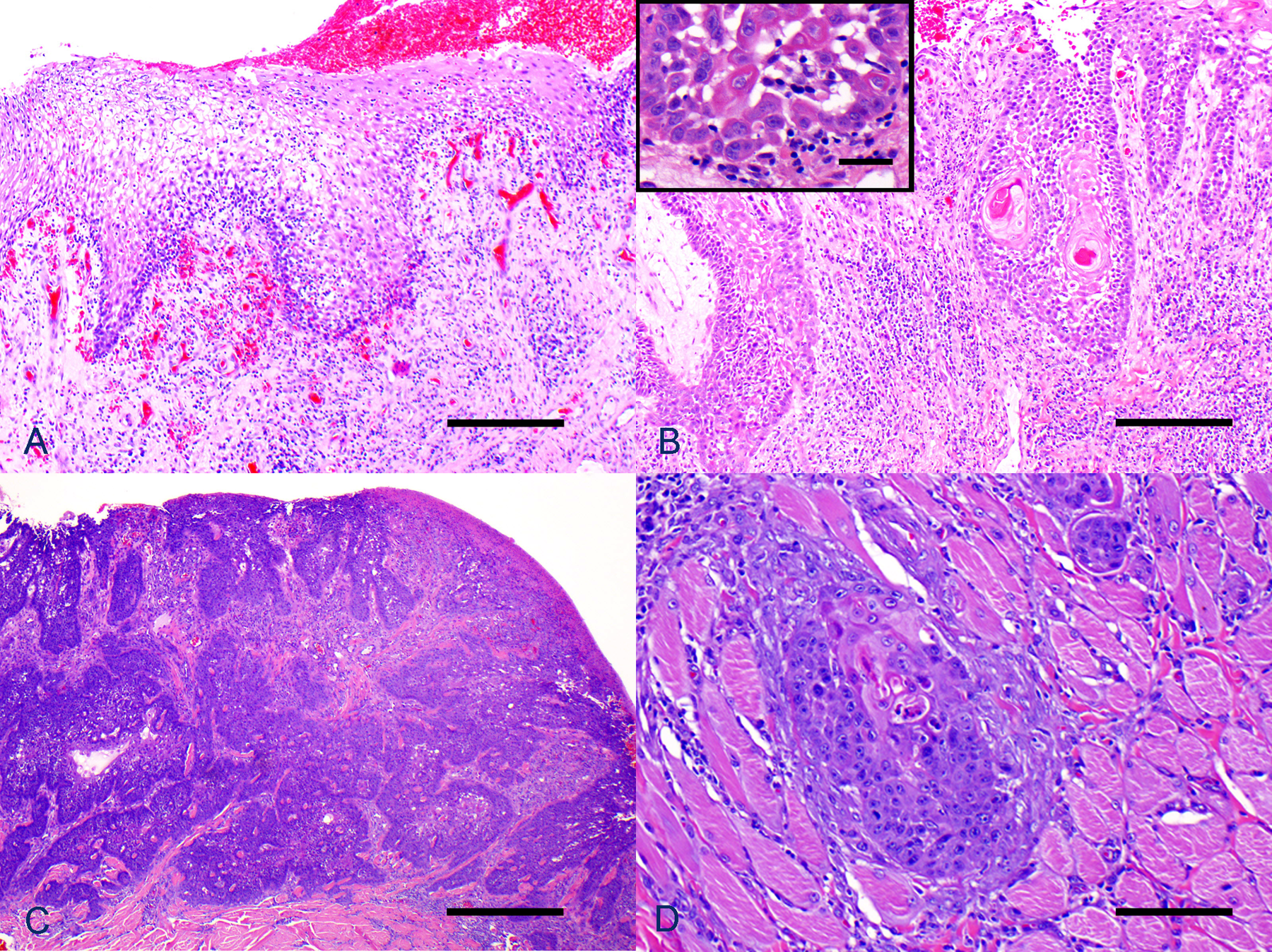
Sun bear (Helarctos malayanus); histology of oral lesions.
One month after biopsy, a full thickness wedge resection of the portion of the lower left lip containing the 2 masses was performed. The previously described mass on the right corneal limbus was still present; a similar but smaller mass was noted at the lateral limbus of the left eye. Vascularization was also noted on the left cornea at the edge of the mass. A biopsy of the right ocular mass revealed squamous cell carcinoma. Three months later, the ocular masses were debulked surgically followed by cryosurgery. Six years following the initial surgery, a rough, dark pink plaque was noted at midline of the lower lip in a new location relative to the previously resected squamous cell carcinomas Histologic examination revealed a squamous cell carcinoma, which was subsequently surgically removed via wedge resection. Histologic examination of the wedge revealed that one of the lateral margins had dysplasia suspicious for a preneoplastic change. The bear died 16 months later of fungal pneumonia, with no recurrence of the oral lesions found on gross necropsy.
Case 4 was a wild-caught, approximately 17-year-old female sun bear that initially presented for an ill-defined area of erythema on the left lower lip. The lesion was nonresponsive to antibiotics (a 10-day course of amoxicillin–clavulanic acid, 14 mg/kg, orally, twice daily), and antihistamines (hydroxyzine, 1 mg/kg, orally, twice daily). Over the course of 5 weeks, the appearance of the lesion varied, developing and resolving areas of ulceration and raised induration. At the time of resection, 3 lesions were present on the left lower lip: a patchy area of erythema close to midline; caudal to that an erythematous, slightly raised, indurated, and ulcerated round 1.5-cm lesion; and an erythematous, indurated, non-ulcerated, 3-mm lesion close to the left commissure. The 3 lesions were surgically resected and submitted for histologic exam, revealing squamous cell carcinoma. Tissue from the lesions was also submitted for herpesviral PCR. One month following resection, the area became erythematous again. Fifteen months later, another ulcerated lesion developed near the previous excision site. Excision and biopsy revealed dysplasia. One year later, an area of swelling and ulceration developed on the right lower lip; surgical resection and biopsy revealed hyperplasia.
Case 5 was an 18-year-old male initially presenting with a 2-cm, erythematous, ulcerated, and slightly raised lesion on the right lower lip. Two centimeters caudal to that lesion, the mucosa was focally irregular. Biopsies of both lesions revealed squamous cell carcinoma, which were treated with laser ablation. One and a half years later, mild erosive lesions were present in the same area; biopsies revealed hyperplasia and dysplasia. The lesions showed a waxing and waning course of severity over the next 4 years, during which time no treatment was administered. Samples were submitted for viral PCR testing. One year later, there was marked progression to nodular proliferations and more severe erosions (Fig. 1). One hundred fifty milligrams of 5-fluorouracil was administered intralesionally with initial improvement in both the erythema and swelling. A 6-week course of piroxicam (0.2 mg/kg, orally, once a day) and L-lysine (11 mg/kg, orally, twice a day) was instituted, with no change to the lesions. One hundred milligrams of 5-fluorouracil was administered intralesionally at 2-week intervals for 3 treatments, with no noticeable effect. Seven milligrams of cisplatin was then injected intralesionally every 3–4 weeks for 3 treatments, with minimal to no noticeable effect.
PCR amplification and sequencing
Cases 1–4 were positive for herpesviral DNA. Case 5 was negative. For cases 1–4, initial nested PCR amplification resulted in a product of 166 base pairs after primers were edited out. The sequence obtained from all 4 bears was identical. The modified second round produced a sequence length of 1,339 bp after primers were edited out. The sequence was submitted to GenBank under accession number JX220982. Pan-papillomaviral PCR on cases 1 and 2 was negative.
Phylogenetic analysis
Bayesian phylogenetic analysis found that the Whelan and Goldman (WAG) model of amino acid substitution was most probable, with a posterior probability of 1.000, which was used for ML analysis. 32 The Bayesian phylogenetic tree shows that the sun bear herpesvirus clusters with other members of the subfamily Gammaherpesvirinae (Fig. 3). Bootstrap values as percentages from ML analysis are shown on the Bayesian tree (Fig. 3). Based on naming conventions, the suggested name of this herpesvirus is Ursid herpesvirus 1 (UrHV-1).
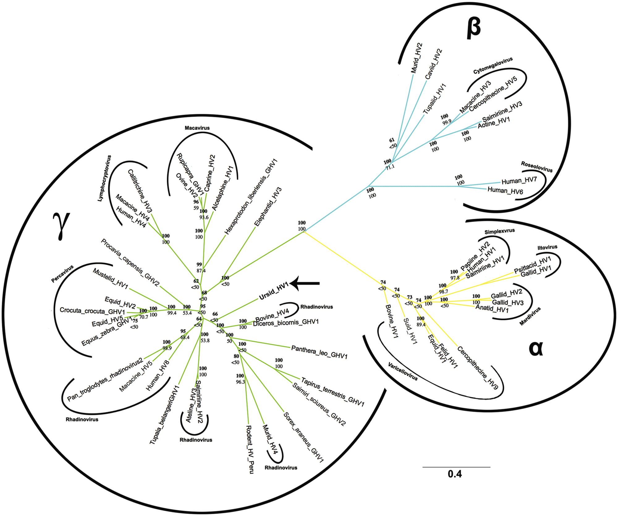
Bayesian phylogenetic tree of predicted 445-557 amino acid partial herpesviral DNA- dependent DNA polymerase sequences based on MAFFT alignment. Bayesian posterior probabilities of branchings as percentages are in bold, and maximum likelihood bootstrap values for branchings based on 1,000 re-samplings are given below. Ursid herpesvirus (HV) 1 is marked by a bold arrow. Psittacid HV2 (GenBank NP944403) was used as the outgroup. Sequences retrieved from GenBank include Alcelaphine HV1 (NP065512), Anatid HV1 (YP003084391), Ateline HV3 (NP047983), Aotine HV1 (YP004940081), Bovine HV1 (NP045328), Bovine HV4 (NP076501), Callitrichine HV3 (NP733857), Caprine HV2 (AAG13396), Caviid HV2 (YP002321247), Cercopithecine HV5 (YP004936030), Cercopithecine HV9 (NP077443), Crocuta GHV1 (ABI21852), Diceros Bicornis GHV1 (AAP42118), Elephantid HV3 (ABU52899), Equid HV1 (YP053075), Equid HV2 (NP042605), Equid HV5 (ACY71882), Equus zebra GHV1 (AAS75147), Felid HV1 (YP003331549), Gallid HV1 (YP182359), Gallid HV2 (YP001033959), Gallid HV3 (NP066862), Hexaprotodon liberiensis GHV1 (AAP42117), Human HV1 (NP044632), Human HV4 (YP401712), Human HV6 (NP050219), Human HV7 (YP073778), Human HV8 (YP001129355), Macacine HV3 (YP068180), Macacine HV4 (YP068007), Macacine HV5 (NP570750), Murid HV2 (NP064160), Murid HV4 (NP044849), Mustelid HV1 (AAM62282), Ovine HV2 (YP438136), Papiine HV2 (YP_443877), Pan Troglodytes Rhadinovirus 2 (ABU52897), Panthera leo GHV1 (ABI21851), Procavia capensis GHV2 (AEA39187), Rodent HV Peru (YP004207849), Rupicapra GHV1 (ABI21850), Saimiri sciureus GHV2 (AAN35123), Saimiriine HV1 (YP003933809), Saimiriine HV2 (NP040211), Saimiriine HV3 (YP004940227), Sorex araneus GHV1 (ABU52901), Suid HV1 (YP068333), Tapirus terrestris GHV1 (AAD30142), Tupaia belangeri GHV1 (AAP42119), and Tupaiid HV1 (NP116408).
Discussion
Herpesviral infections have been associated with neoplasia in several species, and the findings described herein are consistent with the clinical signs and lesions seen in other species. Varanid herpesvirus 1, an alphaherpesvirus, was found in 2 green tree monitors whose lesions developed from an initial ulcerative or proliferative gingivitis to marked progressive or recurrent oral squamous cell carcinoma. 31
Human herpesvirus 4 (HHV-4), a gammaherpesvirus in the genus Lymphocryptovirus, causes a variety of malignancies, including epithelial, mesenchymal, and lymphoid tumors that may be in the oral cavity. 28 Such tumors develop commonly in immunocompromised patients. 5 However, the role of HHV-4 in oral squamous cell carcinomas is controversial, with studies reporting its presence in 0–70% of these tumors. 13 The presence of HHV-4 DNA in oral squamous cell carcinomas was not associated with positive expression of transcripts or proteins. 6 A possible explanation lies in the hit-and-run theory, 2 which proposes that a virus may trigger malignant transformation of host cells and no longer be actively expressed in a tumor. Macacine herpesvirus 4, another member of Lymphocryptovirus, is associated with oral squamous proliferative lesions in macaques. 4
Human herpesvirus 8 (HHV-8), a gammaherpesvirus in the genus Rhadinovirus, has been established as a tumor virus by its presence in human malignancies, the transforming properties of several viral genes in vitro, and its ability to transform some primary cells in culture, although the full mechanism of its oncogenesis has not been worked out. 10 Among other disease syndromes, HHV-8 causes Kaposi sarcoma, a cancer of lymphatic endothelium. Otariid herpesvirus 1, an unclassified gammaherpesvirus, is strongly associated with metastatic carcinomas of urogenital origin in California sea lions. 16 Human herpesvirus 5, a betaherpesvirus in the genus Cytomegalovirus, has also been associated with proliferative gingivitis. 15
In people, a notable subset of squamous cell carcinomas affecting the oropharynx are caused by Human papillomavirus (HPV).11,18 This appears to be a distinct disease entity from HPV-negative squamous cell carcinoma, which is traditionally associated with the risk factors of alcohol and tobacco use. Human papillomavirus and HHV-4 co-infections have been implicated in oral squamous cell carcinomas. 28 Samples from the first 2 cases were tested using a consensus papillomavirus PCR, 23 which was negative.
More work needs to be done to demonstrate the etiology and risk factors for the development of oral squamous cell carcinomas in sun bears. The role, if any, of UrHV-1 is unclear. In case 5, tissue from the squamous cell carcinoma tested negative for UrHV-1. Squamous cell carcinomas can develop on a background of chronic inflammation caused by other factors, including excessive solar exposure, and alcohol and/or tobacco use in human beings. The location of these tumors inside the oral cavity makes a link to solar exposure unlikely. One bear also had bilateral squamous cell carcinomas at the lateral corneal limbus, a location with more solar exposure. The lesions demonstrated chronic but nonspecific inflammation, which could have been induced by solar exposure or viral infection.
In situ hybridization would be required to demonstrate the presence of the virus within the nuclei of cancer cells. Real-time PCR might be used to investigate a difference in viral load between cancer cells and unaffected tissue from the same animal.
All 5 bears in the case series had a history of periodontal disease. The most common clinical signs, present in all 5 bears, were moderate to severe dental calculus accumulation with associated gingivitis. Three bears had signs consistent with periodontitis, including gingival recession and formation of periodontal pockets. Two bears required dental extractions due to the severity of periodontitis, and one of these additionally had severe and extensive osteolysis of the mandible.
In humans, a causal link between herpesvirus infection and destructive periodontal disease has been proposed, on the basis that active infection causes immunosuppression and subsequent proliferation of periodontopathic bacteria. 27 The causal relationship between herpesvirus infection, periodontal disease, and squamous cell carcinoma development has yet to be elucidated.
One sun bear in the current study had a previous history of apparent stereotypic behavior of rubbing his lip with his claws; it is unclear if this behavior may have predated the development of the lesions or been in response to it. It is possible that the chronic trauma may have predisposed to squamous cell carcinoma.
The tumors described in the present study all had similar behavior and appear to represent a single clinical entity. The tumors are locally invasive though slow growing; complete excision appears difficult due to the long range of transition between normal tissue and cancerous cells. Recurrence at the same site is common, as well as local extension. Distant metastases have not been reported, though 1 bear presented with ocular as well as oral squamous cell carcinoma. Based on the findings, wide surgical margins are recommended; adjunctive local therapies such as radiation are recommended in dogs where surgical margins were incomplete, 12 and have been used in sun bears 20 including bears of the current study, one of which is in remission at the time of this report. In addition to the previously reported case and the 5 cases reported herein, between May 1995 and November 2008 there were 6 cases of sun bears with oral or nasal squamous cell carcinoma examined by one of the coauthors (MM Garner) suggesting that this is not an uncommon disease entity among captive sun bears. The herpesviral status of those sun bears is unknown.
Phylogenetic analysis demonstrated that UrHV1 has a relatively basal divergence within the subfamily Gammaherpesvirinae, without clear support for clustering within any of the known genera. Prior to 2009, Gammaherpesvirinae was divided into 2 genera, Lymphocryptovirus and Rhadinovirus. Macavirus and Percavirus were subsequently recognized as monophyletic clades within Rhadinovirus and split out into new genera. 7 Rhadinovirus contains the remaining viruses from the old genus and appears to be paraphyletic.
In this analysis, the Bayesian phylogenetic tree for subfamily Alphaherpesvirinae was not consistent with established trees generated from other data sets, 19 nor with the maximum likelihood alignment, wherein Gallid herpesvirus 1 and Psittacid herpesvirus 1 (used as the outgroup) represent a basal divergence among the alphaherpesviruses. Because the focus of the current study was a gammaherpesvirus, attempts were not made to resolve this discrepancy. The lack of support for the clustering is demonstrated by ML bootstrap values of less than 50%.
In conclusion, squamous cell carcinoma should be an important consideration in a clinical setting when a sun bear presents for oral lesions including erythematous macules, ulcers, or plaques, particularly if nonresponsive to treatment or if recurrent. A novel gammaherpesvirus, Ursid herpesvirus 1, was identified in 4 sun bears with oral squamous cell carcinoma. The prevalence of this virus in the captive and/or wild sun bear populations, as well as its pathogenicity and relationship to squamous cell carcinoma, is unknown and warrants further investigation.
Footnotes
a.
DNeasy kit, Qiagen Inc., Valencia, CA.
b.
QIAquick gel extraction kit, Qiagen Inc., Valencia, CA.
c.
BigDye terminator kit, Applied Biosystems, Foster City, CA.
d.
ABI 3130 automated DNA sequencer, Applied Biosystems, Foster City, CA.
Declaration of conflicting interests
The author(s) declared no potential conflicts of interest with respect to the research, authorship, and/or publication of this article.
Funding
The author(s) received no financial support for the research, authorship, and/or publication of this article.