
Abstract
A 16-year-old primiparous mare aborted an apparently normal fetus at 240 days of gestation. A large, oval mass, measuring approximately 20 cm × 30 cm × 20 cm, was detected attached to the umbilical cord of the fetus. On the cut surface, the mass showed multifocal cystic structures, foci of mineralization, and diffuse hemorrhages. Histological examination of the mass revealed haphazardly arranged cartilage, bone, mesenchymal stroma, adipose tissue, vascular structures, smooth muscle, ciliated epithelium, squamous cornifying epithelium, and undifferentiated germ cells with areas of necrosis and mineralization. The mass was diagnosed as an umbilical cord teratoma, which is an extremely rare tumor in human beings and, to the authors’ knowledge, has only described in the veterinary literature on one occasion.
Teratoma, a tumor arising from totipotential germ cells, contains at least 2, usually 3, different embryonic germ layers.9,11 Teratoma, as a true tumor, displays progressive autonomous growth, and can be composed of well-differentiated mature tissue, immature fetal-like tissues, or a mixture of both tissue types. 9 Tumor tissues are derived from endoderm, mesoderm, and ectoderm layers, and can be represented by different tissues such as digestive tract mucosa, muscle, and skin, respectively. 11 The present report describes a case of umbilical cord teratoma in a foal, an extremely rare tumor, with subsequent abortion.
A 16-year-old primiparous Italian saddle mare aborted a 240-day gestational age grossly normal fetus. Placenta and umbilical cord were submitted for examination. A large, well-demarcated, red to yellowish, oval mass was detected attached to the umbilical cord (Fig. 1). The mass measured approximately 20 × 30 × 20 cm and varied in texture from soft to hard. The cut surface showed multifocal cystic structures, scattered mineralized tissue, and diffuse hemorrhage. No other significant gross abnormalities were observed either in the placenta or in the umbilical cord. The length of the umbilical cord was within normal range.
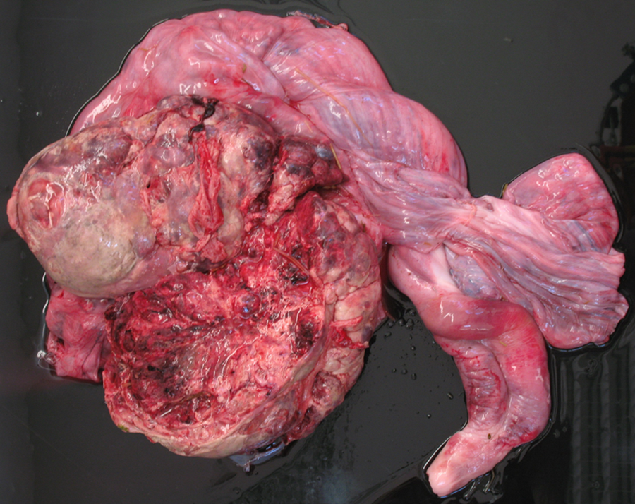
Foal; umbilical cord teratoma. Note the large, well-demarcated, red to yellowish, oval mass (20 cm × 30 cm × 20 cm) connected to the umbilical cord. The cut surface shows multifocal cystic structures and diffuse hemorrhage.
Samples from the mass, after fixation in 10% neutral buffered formalin, were processed routinely, embedded in paraffin, and sectioned for staining with hematoxylin and eosin. Histological examination of the mass revealed several different mature and immature tissues haphazardly arranged. The mass was lined by simple to pseudostratified ciliated epithelium and was characterized by various amounts of fibrous, adipose, and myxoid tissue, well-defined areas of mature cartilage, occasional foci of immature bone, vascular structures, and smooth muscle (Fig. 2). Epithelial tissues were mainly arranged in tubular and papillary projections lined by simple columnar ciliated epithelium. Small cystic and tubular structures lined by squamous keratinized epithelium, often filled with necrotic debris, were also present. Sheets and lobules of round cells, separated by fine fibrous stroma, admixed and surrounded by cartilage and epithelial tissue were present. The round cells were 20–30 µm in diameter, had distinct cell borders, a small amount of lightly basophilic cytoplasm, and large round centrally located nuclei with finely granular chromatin. Numerous mitoses (3–4 per high-power field) were present. These cells resembled germ cells. Wide areas of necrosis with numerous foci of dystrophic mineralization, and multifocal hemorrhages with numerous hemosiderin-laden macrophages were also scattered throughout the mass.
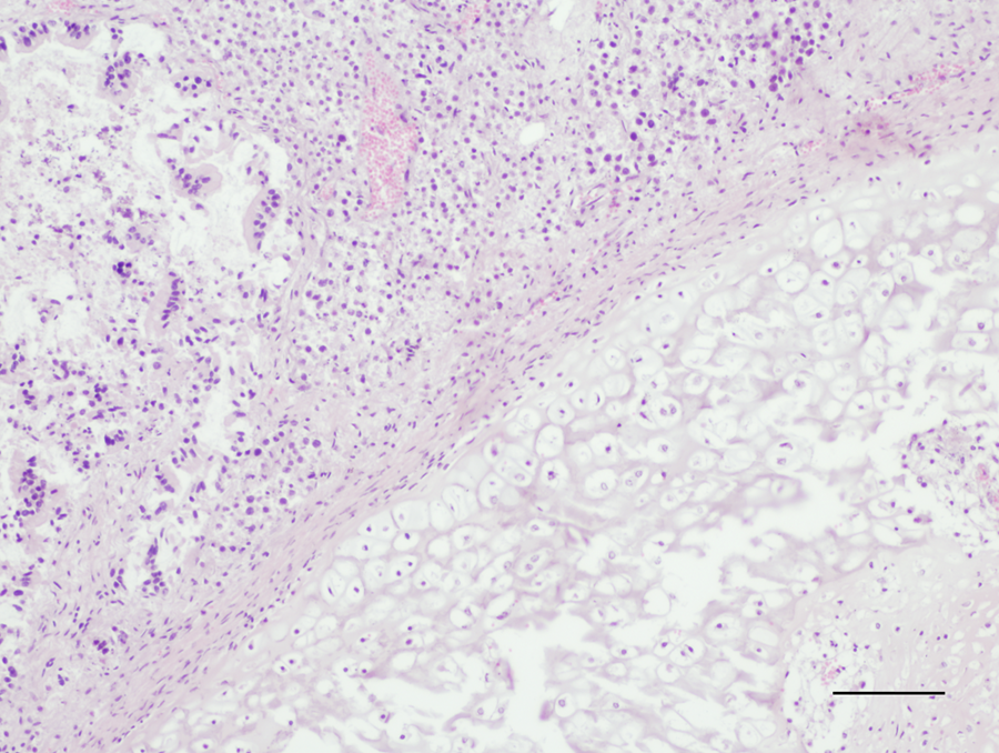
Foal; umbilical cord teratoma. The tumor is composed of mature cartilage and tubular structures lined by simple columnar epithelium surrounding by and admixed with round cells. Hematoxylin and eosin. Bar = 400 μm.
The umbilical cord mass was formed by different tissues, without organization and derived from different germ layers. The localization of the mass and its composition was consistent with the diagnosis of umbilical cord benign teratoma.
Histologically, the chorioallantois showed severe multifocal thrombosis, involving mainly small and medium size arteries. The lumen of these vessels was often obliterated by organized thrombi, and the vessel walls were infiltrated by numerous inflammatory cells, mainly composed of lymphocytes and plasma cells, fibrin, and extravasated red blood cells (vasculitis).
Teratoma is typically a benign tumor and is often composed of well-differentiated mature tissue; its malignant counterpart, teratocarcinoma, has been described in the literature. 11 Malignant teratomas are characterized by undifferentiated tissues with both mature and embryonal elements. 11 Teratomas are most commonly localized in the gonads, both ovary and testis, although extragonadal teratomas have been described. 11 Testicular teratoma has been reported most commonly in horses, and is extremely rare in dogs, cats, cattle, and pigs. 11 In stallions, this tumor occurs most commonly in cryptorchid testes; a congenital origin has been suggested, and the development of the tumor may prevent normal descent of the affected testis. 11 In horses, teratomas have also been described in ovaries, in the temporal bone, 12 and in the abdominal cavity. 2 A case of placental teratoma and one of placental teratocarcinoma have also been reported in horses.1,6 The gross appearance and localization of these placental tumors were totally different from the present case. In the report of placental teratoma, multifocal to coalescing nodules were disseminated throughout the fetal surfaces of the chorioallantois, vessels, and umbilical cord; the greatest density of nodules was in the placental horns, away from the insertion of the umbilical cord. 6 The report of a placental teratocarcinoma was of a small mass attached to the wall of the amnion. 1
In human medicine, teratomatous masses have been reported both in the placenta and in the umbilical cord. Although this type of tumor is extremely rare, teratoma of the placental plate seems to be the most common. 3 Only 12 cases of umbilical cord teratoma have been reported in the medical literature, 4 while in animals, only a single case of umbilical cord teratoma in a giraffe (Giraffa camelopardalis reticulata) has been reported. 13
In human medicine, solid masses of the umbilical cord may represent hemangioma (also termed “angiomyxoma”), hematoma, or the rare teratoma. Angiomyxomas are hamartomas that arise from proliferation of the primitive angiogenic mesenchyme of the cord. 14 Large solid teratomatous masses presenting in close association with the umbilical cord, as in the present case, need careful analysis to exclude the diagnosis of fetus acardiacus (acardiac fetus) or other types of twinning. 3 Fetus acardiacus is characterized by polarization and a rudimentary axial skeleton, presenting with a vertebral column associated with ribs and pelvic bones. 15 In the present case, the umbilical cord teratoma contained tissues derived from different embryologic layers, but lacked any degree of body organization and cephalocaudal polarity, thus ruling out the diagnosis of fetus acardiacus. The etiology of placental teratomas is controversial; some authors consider this entity to represent small acardiac twins, failed twin, or multiple gestations rather than a true neoplasm.5,14
Round masses with similar external gross appearance to teratomas, consistent with ossified remnants of the yolk sac, are a well-described finding in mares. 7 This malformation appears as a round bony structure attached to the umbilical cord with a fluid-filled central cavity and a thin outer wall of bone covered by connective tissue.7,16 In the present case, although the mass showed multifocal cystic structures and scattered foci of mineralization, the differential diagnosis of yolk sac remnants was ruled out based on gross and histological findings.
Usually, umbilical cord teratomas are benign and have been associated with apparently normal infants. Only a few cases have been described in association with severe fetal anomalies, such as anencephaly, abdominal wall defects, and intestinal anomalies. 3 In the present case, the mare aborted at the eighth month of gestation. The aborted foal was apparently normal, but severe damage to placental arteries was noted on histological examination. In the veterinary literature, there are described cases of umbilical cord compression due to remnants of the yolk sac, resulting in placental damage and subsequent abortion. 7 Although, in the case presented herein, the cause of the abortion could not be determined, it was speculated that the large mass in close association with the umbilical cord could have played a role in the mare’s abortion. Indeed, the mass could have partially occluded placental vessels, causing anoxia of the fetus and subsequent abortion.
Umbilical cord torsion is an important and frequent cause of abortion in horses, usually occurring between 6 and 9 months of gestation.8,10 Umbilical cord torsion is more common in umbilical cords of an excessive length, and the gross appearance includes severe hemorrhagic edema and engorged blood vessels. As no such findings were detected in the present case, umbilical cord torsion was excluded.
In conclusion, placental and umbilical cord teratomas are extremely rare neoplasms that can occur in horses. Careful obstetrical management during gestation is necessary to detect these rare conditions, which should be included in the differential diagnoses in cases of abortion. Moreover, a thorough histopathologic examination of the mass should be performed to define its composition.
Footnotes
Declaration of conflicting interests
The author(s) declared no potential conflicts of interest with respect to the research, authorship and/or publication of this article.
Funding
The author(s) disclosed receipt of the following financial support for the research, authorship, and/or publication of this article: This study has been funded by Prozoo project supported by Regione Lombardia, Fondazione Cariplo, Fondazione Banca Popolare di Lodi, Italy.