
Abstract
Reproductive failure represents an important cause of economic loss for the equine industry. We reviewed the cases of equine abortion and stillbirth submitted to the California Animal Health and Food Safety Laboratory System, University of California–Davis from 1990 to 2022. A total of 1,774 cases were reviewed. A confirmed cause of abortion was determined in 29.2% of the cases. Abortion or stillbirth was attributed to infectious agents in 18.7% of the cases, with Streptococcus spp., equine herpesvirus 1, and Leptospira spp. being the most prevalent. Noninfectious causes of abortion were established in 10.5% of the cases, with umbilical cord torsion being the most common. In 70.8% of the cases, a definitive cause of abortion could not be established. Our study demonstrated the difficulties in establishing an etiologic diagnosis, even when following a standard diagnostic work-up. New diagnostic approaches are needed to improve the likelihood of reaching a final diagnosis in cases of equine abortion and stillbirth.
Abortion is an important cause of economic loss for the equine industry given the costs associated with diagnosis, treatment, and loss of animals. Additionally, several infectious agents known to cause reproductive losses in horses are zoonotic and/or have a major impact on international trade. 15
Studies have shown that up to 40% of mares may suffer pregnancy failure (from fertilization to parturition).12,14,17,21,22 These reproductive losses are associated with infectious or noninfectious causes. Among the infectious causes are equine herpesvirus 1 (EHV1; Equid alphaherpesvirus 1), Streptococcus spp., Salmonella spp., and Leptospira spp.2,14,15,18 According to one study, the prevalence of noninfectious causes of equine abortions, such as torsion of the umbilical cord, has increased since 2018, surpassing the occurrence of infectious causes of abortion. 2
Surveillance of abortigenic conditions is critical to determine the most prevalent causes of abortion and to establish preventive measures.2,18 Also, increasing awareness of the presence of specific infectious causes of abortion in a particular geographic area is useful for local practitioners when assessing equine health. 18 The reported frequency of these causes varies over time as a result of changes in the capabilities of diagnostic laboratories, and changes in husbandry practices or climatic conditions of at-risk equine populations.13,14,22 Furthermore, correctly defining abortions, stillbirth, and perinatal mortality is also important, especially late in gestation.1,22
Our objectives were: 1) to identify the most common causes of equine abortion and stillbirth in cases submitted to the California Animal Health and Food Safety Laboratory System, University of California–Davis (CAHFS) laboratories between 1990 and 2022; 2) to establish which submitted specimens were most useful in establishing a cause of abortion or stillbirth; and 3) to characterize the main findings in cases of equine abortion or stillbirth.
Materials and methods
Case selection
We performed a retrospective analysis of equine abortions and stillbirth (fetuses and fetal membranes) submitted to the Davis, Tulare, and San Bernardino laboratories of CAHFS from January 1, 1990, to April 12, 2022. Abortion was defined as fetal loss before the 10th month of gestation. Stillbirth was defined as the delivery of a dead foal after the 10th month of gestation; stillbirths were foals considered to be capable of survival if they had been born alive.18,24 A case was defined as one or more fetuses, fetal tissues, or fetal membranes submitted at the same time. Cases consisting of intact fetuses or tissues from autopsies conducted in the field were included only if fresh and fixed heart, lung, liver, kidney, and intestine were available for evaluation.
For each case, the following information was incorporated into a spreadsheet: CAHFS laboratory branch (Davis, San Bernardino, or Tulare), accession number, login date, breed, age, sex (male, female, not recorded), state of postmortem preservation (good, moderate, or poor as interpreted by the pathologist in charge, or not recorded), placenta submission (yes or no), gross lesions, histologic lesions, final diagnosis, and etiology.
Criteria for inclusion and diagnostic testing
The gestational age of aborted fetuses or stillborn foals was determined using crown-rump length measurement, weight, and presence of hair in different locations over the body and/or incisor teeth eruption at the time of autopsy. 28 The aborted fetuses were clustered into 3 gestational age groups: 0–4 mo (first), 5–8 mo (second), and 9–10 mo (third). When available, umbilical cord length and placental weight were recorded.
All ancillary tests described below were performed following CAHFS standard operating procedures. Histologic examination was performed on H&E-stained sections of available tissues, including, in most cases, lung, liver, kidney, spleen, adrenal gland, thymus, lymph nodes, heart, skeletal muscle, stomach, intestine, brain, amnion, umbilical cord, and allantochorion.
Selected sections were also stained with Grocott–Gomori methenamine silver, Steiner, or periodic acid–Schiff stains, and immunohistochemistry (IHC) for Leptospira spp. (Leptospira multivalent rabbit antibody conjugate; National Veterinary Services Laboratories [NVSL], Ames, IA, USA) and EHV1/4 (mouse monoclonal cocktail anti-EHV1 [13B2, 16C2] and EHV4 [20F3, 21C5]; University of Kentucky, Lexington, KY, USA).
Bacteriologic tests included some or all of the following: selective culture for Campylobacter spp. and Salmonella spp. on stomach content and placenta; aerobic culture of stomach content, lung, liver, and placenta. The presence of Leptospira spp. and Salmonella spp. nucleic acids was tested by PCR on kidney and liver samples, respectively. A direct fluorescent antibody test (FAT) was performed on kidney and/or placenta for Leptospira spp. (multivalent rabbit conjugate; NVSL). Fungal cultures on placenta were performed in selected cases when considered necessary by the pathologist.
Virology testing included some or all of the following: FAT on frozen sections of liver and lung for EHV1 (monoclonal conjugated; VMRD); virus isolation and EHV1 PCR, with and without the neuropathogenic marker, on placenta, liver, and lung. When compatible lesions were present, PCR, viral isolation, or IHC for equine arteritis virus (EAV; Alphaarterivirus equid) was performed in a few selected cases.
Serologic testing was performed on fetal fluids for IgG, and for EHV1, EAV, and Leptospira spp. When available, serum from dams was also tested for EHV1, EAV, and Leptospira spp. antibodies. IgG was measured by a commercial ELISA (Triple J Farms). EHV1 and EAV serology were performed by virus neutralization. Leptospira spp. serology was performed by the microagglutination technique. Heavy metals (arsenic, manganese, molybdenum, lead, zinc, mercury, iron, copper), selenium, and vitamin E concentrations were measured in fetal liver of some cases.
The cause of abortion in each case was classified as confirmed, presumptive, or undetermined. Cases with a confirmed cause of abortion were those in which there were compatible gross and/or microscopic lesions coupled with detection of the cause of abortion in placenta and/or fetal samples by the corresponding ancillary testing. Mineral deficiencies (e.g., selenium values below RIs and associated with lesions) were included in this category. Dystocia was considered to be the cause of abortion in cases in which there were circulatory disturbances in the head and neck, rib fractures, intrathoracic hemorrhage, and/or ruptured liver, in conjunction with the absence of inflammatory microscopic changes and/or detection of significant pathogens. Umbilical cord torsion was considered to be the cause of abortion if the umbilical cord was >80 cm long and had more than 5 twists, coupled with circulatory disturbances, including ischemia, congestion with varicose dilation of umbilical vessels, hemorrhage, and/or edema.
The category of presumptive cause of abortion included cases in which gross and/or microscopic lesions in the placenta and/or fetal tissues were compatible with a particular entity but the agent was not detected. Cases with nonspecific or no detectable gross or microscopic findings and no detection of a significant agent were included in the category of undetermined cause of abortion; several congenital defects and mummified fetuses were included in this category.
Statistical analysis
We prepared contingency tables to study the association of variables among the cases of equine abortion. Pearson chi-square or Fisher exact tests were used in determining any statistically significant differences in the establishment of a final diagnosis (confirmed, presumptive, undetermined) and the following analyzed categories: period of gestation (first, second, third), state of postmortem preservation (good, fair, poor) and submission of placenta (yes, no). Likewise, the Pearson chi-square test was used to determine statistically significant differences between the period of gestation (first, second, third) and the most prevalent causes of abortion or stillbirth (Streptococcus spp., Leptospira spp., EHV1, fungi). Statistical analyses were performed using R for Mac (v.3.2.6; https://cran.r-project.org/bin/macosx/). A p-value ≤ 0.05 was regarded as statistically significant.
Results
Case selection
We included 1,774 cases of equine abortion and stillbirth submitted to CAHFS between January 1990 and May 2022. An average of 55 (±21) cases were received each year. A seasonal pattern was observed; most cases were received between September and April each year (n = 1,596; 90.0%) with a peak between November and February. The most prevalent equine breeds were Thoroughbred (n = 505; 28.5%) and Quarter Horse (n = 355; 20.0%), followed by Miniature Horse (n = 133; 7.5%), Arabian (n = 126; 7.1%), Paint (n = 73; 4.1%), and others (n = 582: 32.8%). Others included the following breeds: crossbred, Friesian, Andalusian, Warmblood Horse, Appaloosa, Morgan, Hanoverian, Standardbred, Warmblood, Tennessee Walking Horse, Peruvian Paso, Dutch Warmblood, Oldenburg, Saddlebred, Percheron, Missouri Fox Trotting Horse, Clydesdale, Holsteiner, Mustang, Gypsy Vanner, Przewalski, Welsh Pony and Cob, Icelandic, Norwegian Fjord, Paso Fino, pony, Belgian, Hackney, Lipizzaner, Pony of the Americas, Shetland pony, Swedish Warmblood, Trakehner, and Westphalian. The gestational age of the submitted cases was estimated to be mostly in the second (n = 694; 39.1%) and third (n = 763; 43.0%) terms, with a small number of cases (n = 78; 4.4%) in the first gestational period. In 239 (13.5%) cases, the gestational age was not recorded. Abortions (fetuses of <10-mo gestation) represented 1,036 of the 1,774 (58.4%) submissions; 499 submissions (28.1%) were considered stillbirths (≥10-mo gestation).
A total of 830 (46.8%) submissions were female and 672 (37.9%) were male. In 272 (15.3%) submissions, information about the sex was not available. The state of postmortem preservation was good in 594 (33.5%), fair in 330 (18.6%), and poor in 211 (11.9%) of the cases. In 639 (36.0%) cases, information about the state of postmortem decomposition was not available. In 1,371 (77.3%) submissions, placental tissues were received with the fetuses.
Gross abnormalities were observed in 457 (25.8%) submissions; no significant gross abnormalities were observed in 1,214 (68.4%) submissions. No gross description of tissues was available in 103 (5.8%) submissions. Microscopic abnormalities were observed in 681 (38.4%) submissions.
Causes of abortions and stillbirths
A definitive etiology for the cases of abortion and stillbirth was established in 518 (29.2%) cases. Most were infectious causes (n = 331; 18.7%). Noninfectious abortions or stillbirths occurred in 187 (10.5%) cases. Bacterial agents represented the most frequent infectious cause of abortion, followed by viral, mycotic, and coinfections (Table 1). Briefly, bacterial abortions were mostly characterized grossly by placentitis, and occasionally inflammation of several fetal tissues. EHV1 was the most prevalent cause of viral abortions, and gross lesions, when present, included random white pinpoint foci throughout the liver parenchyma (Fig. 1) and fibrin plugs within the tracheal lumen (Fig. 2). Mycotic abortions were characterized by severe fibrinonecrotizing placentitis (Fig. 3) and funisitis. The most common noninfectious cause of abortion was umbilical torsion (Table 2; Fig. 4).
Infectious causes of equine abortion and stillbirth diagnosed at the California Animal Health and Food Safety Laboratories, 1990–2022.
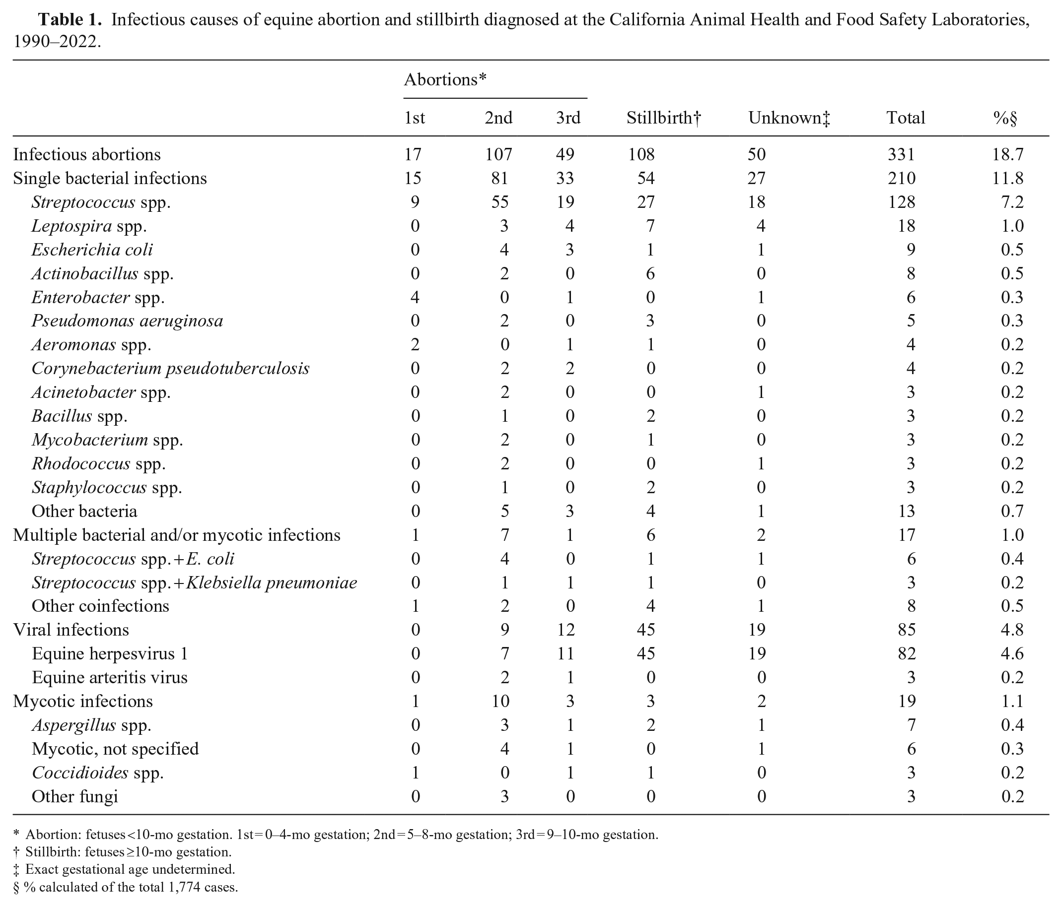
Abortion: fetuses <10-mo gestation. 1st = 0–4-mo gestation; 2nd = 5–8-mo gestation; 3rd = 9–10-mo gestation.
Stillbirth: fetuses ≥10-mo gestation.
Exact gestational age undetermined.
% calculated of the total 1,774 cases.
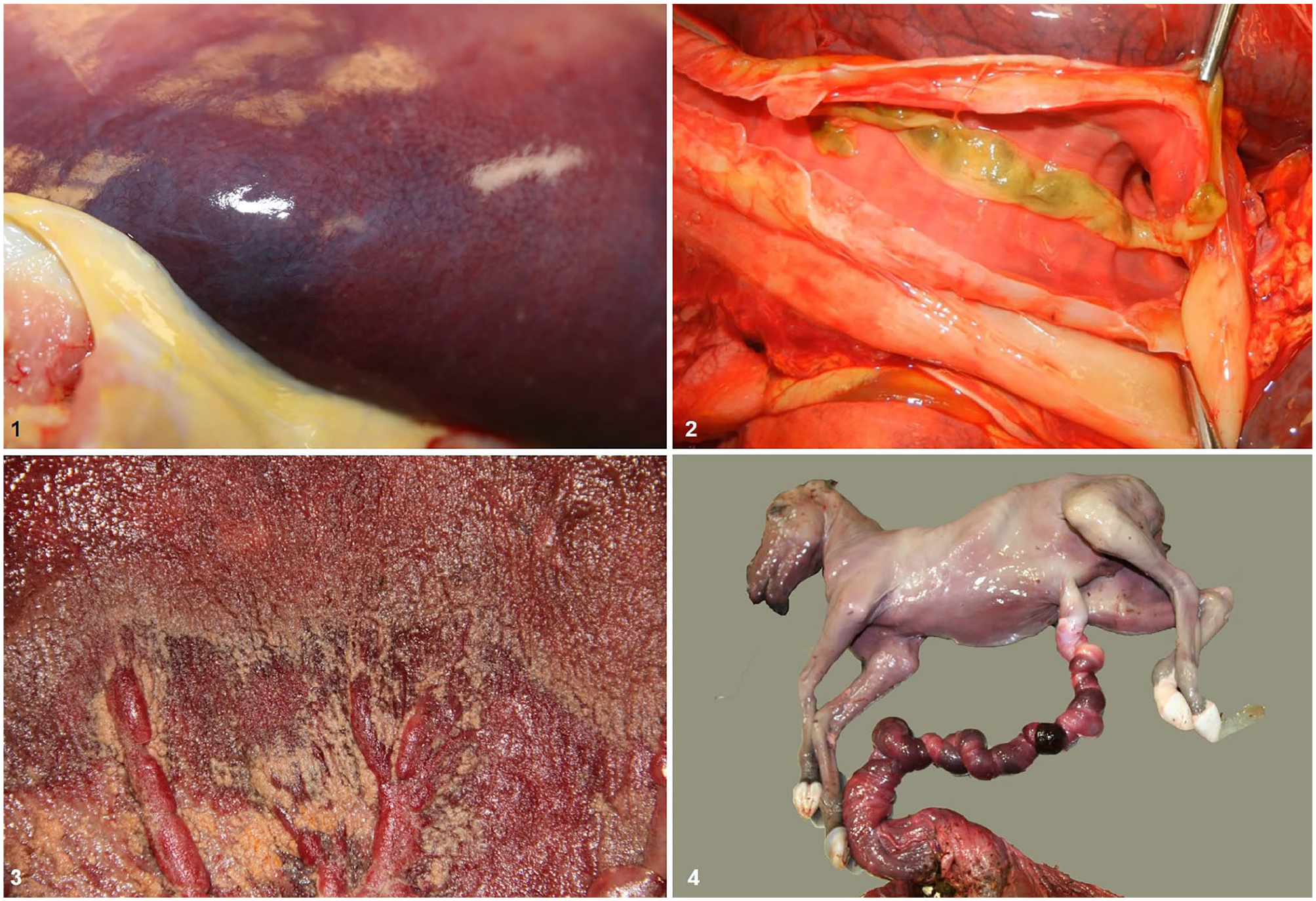
Gross findings in equine abortions and stillbirths.
Noninfectious and undetermined causes of equine abortion and stillbirth diagnosed at the California Animal Health and Food Safety Laboratories, 1990–2022.
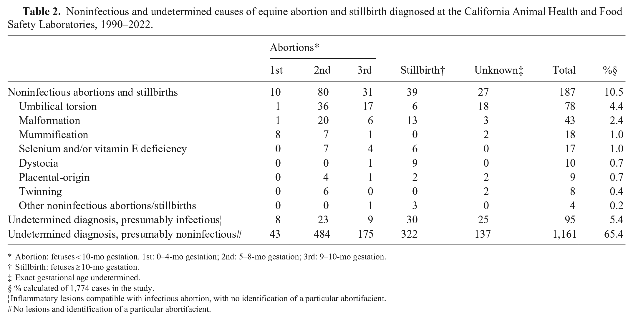
Abortion: fetuses <10-mo gestation. 1st: 0–4-mo gestation; 2nd: 5–8-mo gestation; 3rd: 9–10-mo gestation.
Stillbirth: fetuses ≥10-mo gestation.
Exact gestational age undetermined.
% calculated of 1,774 cases in the study.
Inflammatory lesions compatible with infectious abortion, with no identification of a particular abortifacient.
No lesions and identification of a particular abortifacient.
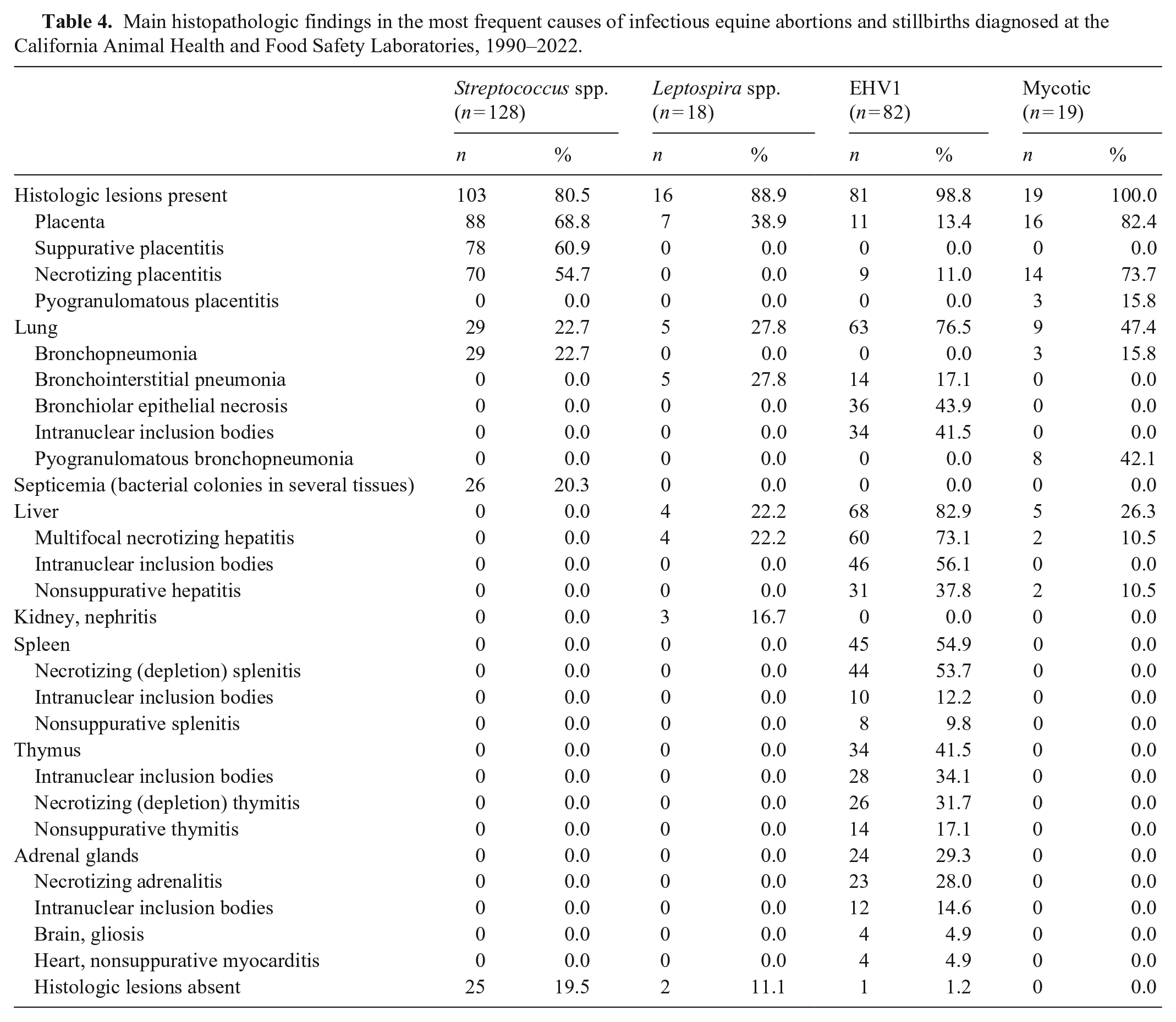
Briefly, histologic findings in equine abortions and stillbirths caused by most bacterial infections were characterized by necrosuppurative placentitis with inflammation of several other organs with intralesional bacterial colonies (Tables 3–5; Fig. 5). In mycotic abortions, the microscopic lesions consisted of necrosis and granulomatous inflammation with intralesional fungal hyphae; lesions were most frequently seen in placenta (Fig. 6), lung, and liver. In abortions caused by EHV1 infection, the most characteristic lesion was necrosis in various organs with prominent eosinophilic intranuclear inclusion bodies; the organs most frequently affected were liver (Figs. 7, 8), lung (Fig. 9), spleen, and thymus.
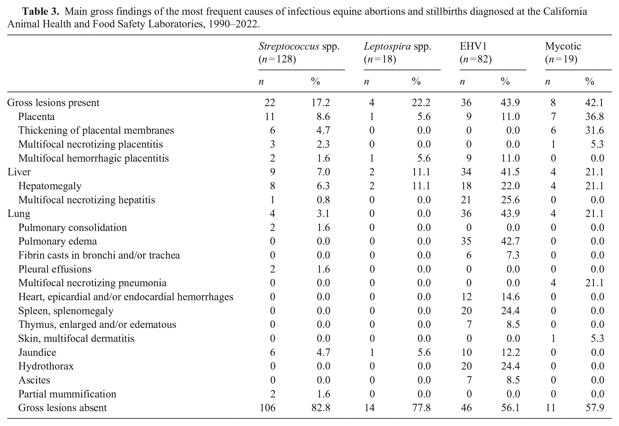
Main gross findings of the most frequent causes of infectious equine abortions and stillbirths diagnosed at the California Animal Health and Food Safety Laboratories, 1990–2022.
Main histopathologic findings in the most frequent causes of infectious equine abortions and stillbirths diagnosed at the California Animal Health and Food Safety Laboratories, 1990–2022.
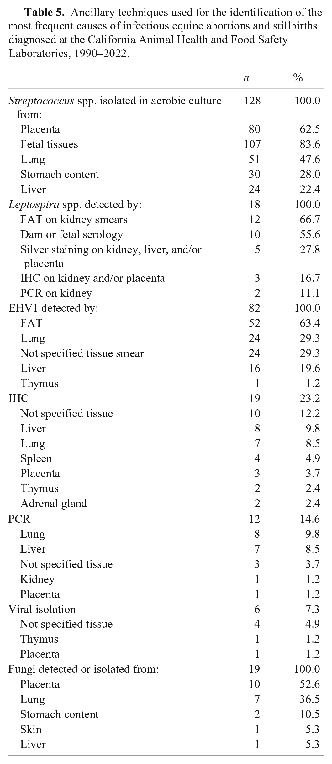
Ancillary techniques used for the identification of the most frequent causes of infectious equine abortions and stillbirths diagnosed at the California Animal Health and Food Safety Laboratories, 1990–2022.
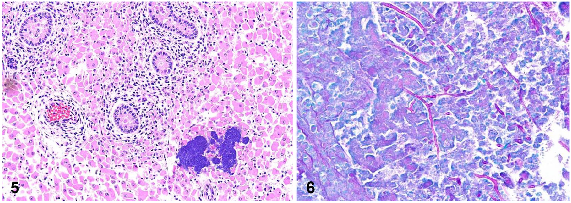
Microscopic findings in bacterial and fungal equine abortions.
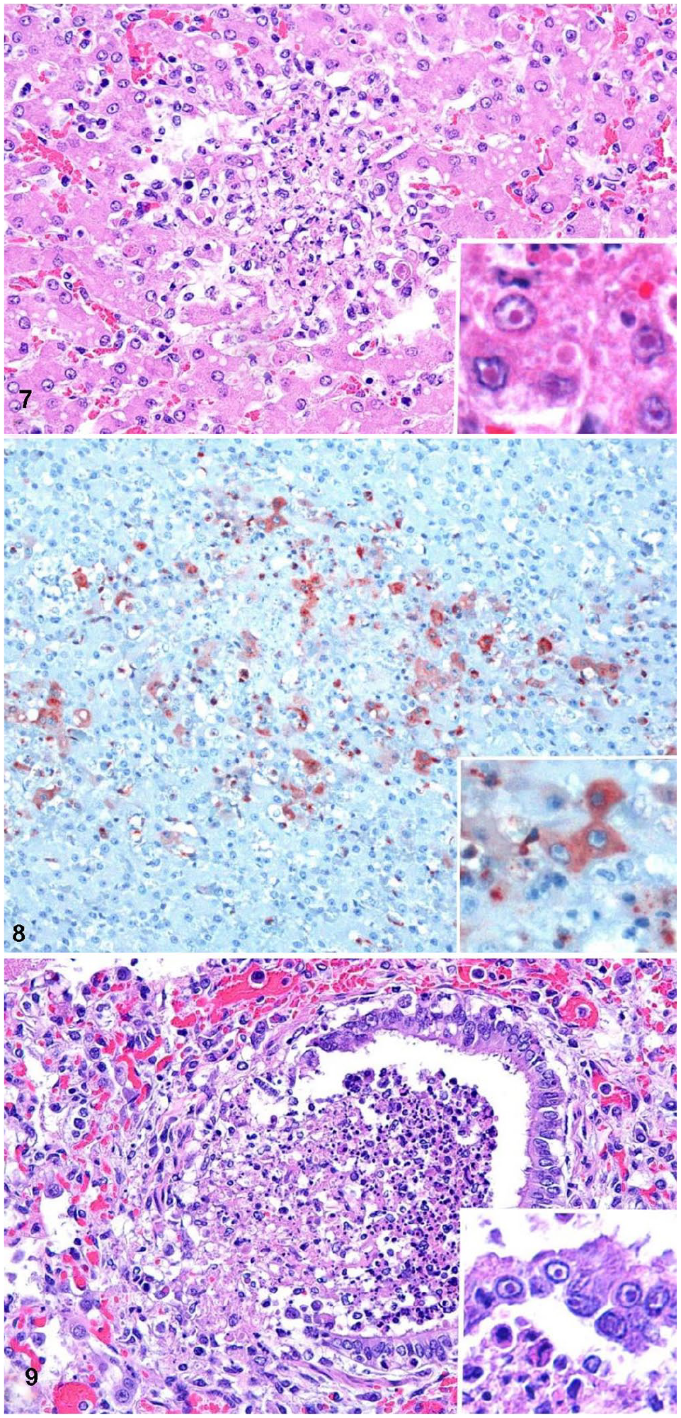
Microscopic changes in equine herpesviral abortion.
Statistical analysis
No association between the period of gestation or the inclusion of placenta among the submitted samples and the establishment of a confirmed diagnosis was detected (p >0.05). When the association between the state of postmortem preservation and the establishment of a final diagnosis was evaluated, a higher percentage of confirmed diagnoses was achieved in cases with a good state of postmortem preservation (p = 0.01). Abortions caused by Streptococcus spp. and EHV1 occurred most frequently in the second and third period of gestation (p <0.01). No association between period of gestation and abortion caused by Leptospira spp. or mycotic abortions (p >0.05) was detected.
Discussion
In agreement with previous studies,2,14,17,18,24 most cases submitted to CAHFS during this time frame were predominantly late-term abortions (third period). Although this could indicate that equine abortions occur more frequently in the second half of the pregnancy, it is more likely that earlier abortions were underestimated, 23 probably because most of them were more difficult to find on the farm given the size of the fetus.14,17
Seasonal variations in the frequency of abortions have been described. Abortions occur most commonly between November and May in the Northern Hemisphere, in concordance with the seasonal reproductive cycle of the mare,14,24 as observed in our study in California. Furthermore, most of the fetuses submitted to CAHFS were Thoroughbred, some of them probably used for racing, which may affect breeding schedules, particularly in the United States.
Despite advances in tests, a definitive cause of abortion was not determined in most of the cases. Comparison with other retrospective studies worldwide is difficult because different methods and diagnostic criteria were often used, and diseases and management conditions vary around the world. 2 In some studies from the United Kingdom and United States, the cause of equine abortion remained undiagnosed in ~10% of the cases21–23; in other studies, the undiagnosed cases reached up to 60% of submissions.2,6,14,18 In another study, an etiologic diagnosis was obtained successfully more frequently in late-term abortions compared to abortions in earlier stages of gestation.14,23 We found no statistical differences in detection of the etiology at different stages of pregnancy. Advanced autolysis usually jeopardizes the chances of determining an etiologic diagnosis in abortions,6,14 and, in our study, the degree of postmortem decomposition affected the success of establishing a final diagnosis.
In our study, the most common causes of infectious abortions were bacterial infections, in accordance with previous data.12,14 Gross or microscopic pathologic findings are not evident in many bacterial equine abortions. 17 It has been hypothesized that, in some cases, a septicemic process could be so rapid that there is no time for the development of inflammatory or necrotizing lesions.
Among the bacterial causes of abortion identified, Streptococcus spp. were the most prevalent, which agrees with previous reports.14,15,17,19,24 Streptococcus spp. are part of the normal microbiota of the genital canal, but they can invade the uterus and cause endometritis, ascending placentitis, and fetal death opportunistically. 23
Leptospiral abortions were not detected or were uncommonly diagnosed in several previous reports.2,6,9,22,27 However, Leptospira spp. has been detected frequently in other studies in the United States.10,11 The differences among these studies could be related to geographic and climatic variations associated with the characteristic of this spirochete, or the use of systematic vaccination in certain locations.6,25 Leptospira spp.-placentitis/funisitis with no fetal lesions has been reported as a frequent cause of equine abortion, 13 as detected in our study. This finding emphasizes the importance of placental submission in equine abortion to improve the chances of reaching an etiologic diagnosis.
EHV1 was the most common viral cause of abortion in our study. Despite the availability of vaccines and the implementation of good husbandry conditions, EHV1 continues to be one of the most commonly reported causes of equine abortion worldwide.7,14,15,18,26 In our study, information about the vaccination status of the dam was not available in most cases.
The other cause of viral abortion in our case series was EAV. Cases of equine viral arteritis (EVA) were, however, very rare; only 1 confirmed case and 2 presumptive cases were found in our case series. In the presumptive cases, compatible microscopic lesions were observed, but EAV was not detected. EVA in horses is associated with respiratory and reproductive disease, although the vast majority of EAV infections are subclinical 3 and abortions are detected only rarely. 14 A diagnosis of EVA-associated abortion may be challenging given that detection of the virus and lesions in fetal tissues is rare.4,8 In most cases of EVA, the abortion is associated with uterine lesions (e.g., myometritis), with a subsequent decrease in blood supply to the placenta and fetus. 4
Fungal infections were diagnosed uncommonly in our study. Aspergillus spp. was the most frequently detected fungus, which agrees with previous reports.12,14 Although some authors mentioned that fetal fungal infection is rare, 20 in our retrospective analysis, both placenta and fetal tissues (particularly the lungs and the liver) were affected.
In several studies, the most frequent causes of abortions were noninfectious.14,18,22 Among them, umbilical cord torsion with vascular compromise represents the most prevalent postmortem finding associated with equine abortion, stillbirth, and perinatal death.2,23,24 A confirmed diagnosis of umbilical cord torsion must be based on circulatory disturbances in the cord, such as edema, hemorrhage and/or thrombosis leading to placental vascular compromise, as well as the detection of >5 umbilical twists and an excessively long cord (>80 cm).2,13,14,21
Congenital malformations were observed most frequently in the CNS and the skeletal system, which agrees with previous reports.12–14,23 As in our study, the cause of such malformations is rarely established. Twinning-related abortion was rare, in agreement with previous studies.2,12,15 The occurrence of abortion of twins has decreased over time, which can likely be attributed to the use of early ultrasonography and appropriate management of multiple pregnancies.16,23
Selenium and/or vitamin E deficiency has been mentioned as a cause of equine gestational losses and perinatal mortality. 24 In our study, selenium and vitamin E deficiency with compatible histologic lesions was detected as the cause of 1% of the abortions. Selenium deficiency was detected in several other fetuses in which no compatible gross or microscopic lesions were observed (data not shown). Although considered an incidental finding, we cannot rule out that these deficiencies acted as factors predisposing to other causes of abortions and perinatal mortality. 5
Pathologic findings compatible with dystocia-perinatal asphyxia syndrome were detected in 12 late-term fetuses in our study. This syndrome is usually associated with malpresentation of foals, maiden mares, and/or large foals. 13 In our study, no clinical information about malpresentations was available in these fetuses, and their size was estimated to be appropriate.
Placental submission is key to reaching an etiologic diagnosis of abortion in several animal species, including the horse.17,21,23 Placental-origin abortion has been associated with idiopathic and infectious causes. In several studies, placentitis was one of the most important causes of equine reproductive loss.13,23,24 In our study, a presumptive or confirmed diagnosis of abortion was achieved in 172 (9.7%) of the fetuses, based on observation of placental lesions in association with detection of an etiologic agent in only the placenta. Complete placenta and umbilical cord were key for the confirmation of umbilical cord torsion, bacterial and mycotic abortions or stillbirths, as well as premature placental separation, as the cause of abortion. However, our statistical analysis suggests that submission of the placenta with the fetuses did not increase the chances of achieving a confirmed diagnosis.
Our study was carried out using the fetuses and/or fetal and placental tissues submitted for laboratory examination; therefore, this may not reflect the actual incidence at the population level. Consequently, it is challenging to accurately compare our results with other regions and/or studies about risk factors. Given that a cause of abortion was not identified in a large percentage of the submitted equine abortion and stillbirth cases included in our study, a more comprehensive approach should be considered to improve the chances of achieving an etiologic diagnosis. For instance, other factors, such as hormonal or endocrine deficiencies in the dam, toxicoses, and other nutritional and husbandry conditions, 12 may also play a role in equine reproductive losses and should also be considered.
Footnotes
Acknowledgements
We thank pathologists and technicians of the 3 branches of CAHFS, who conducted the postmortem examinations and ancillary testing (UC Davis). This research was supported by the U.S. Fulbright Program and INTA (Argentina).
Declaration of conflicting interests
The authors declared no potential conflicts of interest with respect to the research, authorship, and/or publication of this article.
Funding
The authors received no financial support for the research, authorship, and/or publication of this article.