
Abstract
Porcine circovirus-2 (PCV-2) serology is commonly used for PCV-2 herd status determination and optimal timing of PCV-2 vaccination programs. The objectives of the current study were to develop an in-house indirect enzyme-linked immunosorbent assay (ELISA) using a recombinant nuclear localization signal truncated capsid (rntCap) protein expressed in an Escherichia coli system and to determine the diagnostic performance of the developed rntCap indirect ELISA in comparison with immunoperoxidase monolayer assays (IPMAs). Based on a receiver operating characteristic (ROC) curve analysis of the rntCap indirect ELISA (n = 90), an optimum cutoff optical density (OD) of 0.330 was determined, which resulted in diagnostic sensitivity, diagnostic specificity, and accuracy of 98.33%, 93.33%, and 96.67%, respectively. Average OD values of the positive (n = 8) and negative sera (n = 8) tested by either purified glutathione-S-transferase (GST) protein or the rntCap protein as the coating antigen revealed that the mean OD values tested by the rntCap indirect ELISA were significantly different from using GST alone (P < 0.005). The correlation between the established rntCap indirect ELISA and the IPMA results demonstrated as the linear regression (Spearman correlation coefficient = 0.772, P < 0.005) indicated that the OD ratio obtained from the rntCap indirect ELISA could be used to predict the levels of the IPMA titers. More samples are needed for enhancing the diagnostic sensitivity, specificity and accuracy. In conclusion, the establishment of the rntCap indirect ELISA could be used as a serodiagnostic assay for large-scale detection of PCV-2 antibodies in swine and has the capability to be produced commercially for routine use in diagnostic laboratories.
Porcine circovirus is a nonenveloped, single-stranded, circular-genome DNA virus belonging to the family Circoviridae. Porcine circovirus-1 (PCV-1) was first isolated as a nonpathogenic contaminant virus from a porcine kidney cell line. 17 In contrast, Porcine circovirus-2 (PCV-2) was first recognized as a causative agent of postweaning multisystemic wasting syndrome (PMWS), a multifactorial disease in swine first identified in Canada in 1991. 6 Subsequently, PCV-2 has been reported in most intensive pig-producing countries worldwide including Thailand. 16 The genome of PCV-2 contains 3 major open reading frames (ORFs): ORF1, ORF2, and ORF3. ORF1 encodes the Rep proteins of 35.7 kDa involved in viral replication, ORF2 encodes the structural capsid protein of 30 kDa involved in immunogenicity of PCV-2, 12 and ORF3 protein is involved in PCV-2–induced apoptosis. 9
The most reliable serological diagnostic assays for antibody detection against PCV-2 consist of immunoperoxidase monolayer assays 1 (IPMAs) and indirect fluorescent antibody tests 2 (IFATs). However, these conventional assays are expensive, highly time-consuming, and carry the risk of virus contamination, whereas commercially available enzyme-linked immunosorbent assays (ELISAs) based on a recombinant capsid protein expressed in baculovirus expression systems 3 or bacterial expression systems11,15 are more convenient. Furthermore, the production of recombinant proteins in the eukaryotic expression system is costly. It should be noted that several antigenic epitopes of PCV-2 capsid protein were demonstrated at amino acid residues 47–85, 165–200, 230–233 8 and 65–87, 113–147, 157–183, 193–207, 10 whereas a 2011 report 5 demonstrated that the recognition epitope was also located in the nuclear localization signal (NLS) of capsid protein at amino acid residues 26–36. Nevertheless, most information of antigenic epitopes of PCV-2 suggested that the NLS region might not be the major domain for the conformational epitopes. As a result, in the current study, a recombinant NLS truncated capsid (rntCap) protein of PCV-2 expressed in an Escherichia coli system was used as an antigen for indirect ELISA development. The objectives of the study were to establish and validate the rntCap indirect ELISA diagnostic assay to detect anti–PCV-2 antibodies.
To construct the rntCap in expression plasmids, genomic DNA was obtained from purified PCV-2–infected porcine kidney epithelial cell (PK15) lysate of the PCV-2 isolate THA_07NP88 (genotype 2b); GenBank accession number JQ866913 7 was used as template polymerase chain reaction (PCR). The PCR product was amplified to generate the NLS truncated capsid (ntCap) fragment of PCV-2. The forward (5′-GCGGCCGCAATGGCATCTTCAACGC-3′) and reverse (5′-GCGGCCGCTTAGGGGTTAACTCGGGGGTC-3′) primers were designed to incorporate the NotI restriction sites (underlined) at both ends. Subsequently, the expression plasmid, pGEX-5x-3, a was digested with NotI endonuclease b and then the ntCap fragments were transferred into the expression plasmid, which resulted in the rntCap plasmid. Consequently, the rntCap plasmids were transformed into E. coli BL21-Rosetta 2. c For protein expression and purification, the rntCap proteins were produced and purified by affinity chromatography as previously suggested 18 with some modification. The purified rntCap proteins were separated by sodium dodecyl sulfate–polyacrylamide gel electrophoresis, and Western blot was performed. The rntCap protein was used as the coating antigen for the indirect ELISA. A final antigen concentration, serum dilution, conjugate-antibody concentration, and incubation period were determined by the checkerboard titration procedures. 4 Ninety field serum samples (30 IPMA-negative and 60 IPMA-positive sera) were used for the rntCap indirect ELISA validation. The optical density (OD) values obtained from 90 field serum samples were compared with the IPMA results. To determine a positive–negative cutoff and the diagnostic performance for this assay, a receiver operating characteristic (ROC) curve analysis was performed. A cutoff value was determined using maximized diagnostic sensitivity (DSn) and specificity (DSp) and minimized the number of false-negative and false-positive results. Evaluation of the assay repeatability within and between assays was performed using 8 positive and 8 negative serum samples. Mean OD ratio, standard deviation, and coefficient of variation (CV) of each test were analyzed. In addition, the rntCap indirect ELISA OD values obtained from 10 serum samples of known PCV-2–infected pigs were compared with the antibody titers tested by IPMA to determine the Spearman correlation coefficient.
Consequently, the amplified 579 nucleotides encoding the 42–234 peptide of ORF2 protein were sequenced and cloned into the expression vector pGEX-5x-3. Then, the rntCap protein was expressed as glutathione-S-transferase (GST)-tagged fusion protein, and the presence of the rntCap protein in the bacterial cell lysate after induction and purification was revealed (Fig. 1). Approximately, 40 kDa of rntCap protein from the purified protein on lane 4 (Fig. 1) strongly reacted with the monoclonal mouse anti–PCV-2 antibodies in Western blot. In addition, the reactivity of rntCap protein with swine anti–PCV-1 serum was not detected (data not shown), which is similar to results from a previous report. 13 The results indicated that this rntCap protein expressed in E. coli could be used as an antigen for the detection of specific antibodies against PCV-2 capsid protein. Additionally, to determine whether the GST tag interfered with the rntCap indirect ELISA, the established indirect ELISA was performed using either purified GST protein or rntCap protein as the coating antigen. The statistical analysis (paired t-test) between average OD values of the positive and negative sera tested by both assays revealed that the mean OD values of the positive and negative sera tested by the rntCap indirect ELISA was significantly different compared to the indirect ELISA using GST alone (P < 0.005). Moreover, the average OD values tested on the indirect ELISA using solely GST was not significantly different among the positive and negative sera (P > 0.05).
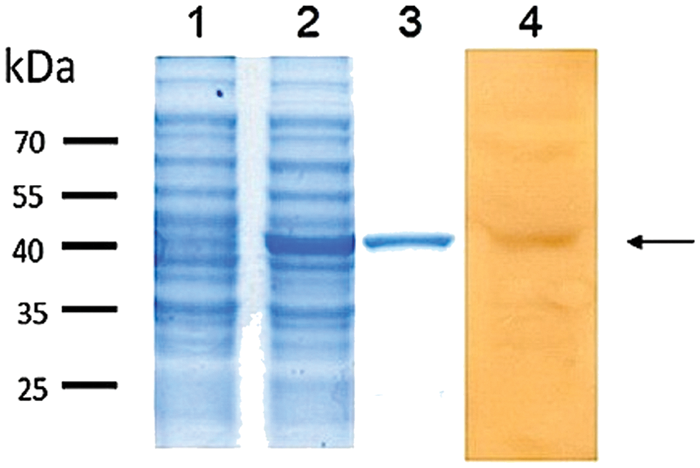
Expression of the recombinant nuclear localization signal truncated capsid (rntCap) protein was analyzed by sodium dodecyl sulfate–polyacrylamide gel electrophoresis (lanes 1–3) and Western blot (lane 4) with monoclonal mouse anti–Porcine circovirus-2 (PCV-2) antibody. A clear band of 40 kDa (arrow) after purification is demonstrated. Lane 1: crude protein from noninduced BL21 cell lysate; lane 2: crude protein from isopropylthio-β-galactoside (IPTG)-induced BL21 cell lysate; lane 3: purified rntCap protein from IPTG-induced BL21; lane 4: purified rntCap proteins reacting with monoclonal mouse anti–PCV-2 antibodies.
Based on the ROC curve analysis of the rntCap indirect ELISA, the OD value of 90 field serum samples varied from a minimum of 0.105 to a maximum of 0.551 for negative sera (n = 30) and from 0.312 to 1.091 for positive sera (n = 60). An optimum cutoff OD of 0.330 was determined, with a DSn of 98.33% and a DSp of 93.33%, whereas, the accuracy was 96.67% (Table 1). The results from 8 negative sera revealed that the intra-plate CV ranged from 0.37% to 19.86%, with a median value of 3.74%, whereas, those of the positive serum samples ranged from 0.91% to 7.68%, with a median value of 3.82%. The inter-plate CV for the negative sera was between 5.55% and 30.60%, with a median value of 21.10%, while, the CV of the positive serum samples was between 7.18% and 17.69%, with a median value of 12.21%. The results indicated that the rntCap indirect ELISA was repeatable. The correlation between the IPMA titers and the OD ratios of the rntCap indirect ELISA revealed the linear regression (Spearman correlation coefficient = 0.772, P < 0.005) within a minimum and a maximum limited range, respectively (2.90 and 3.80 for the IPMA, and 0.31 and 0.94 for the OD ratios). As a result, the OD ratios obtained from the rntCap indirect ELISA could be reliably used to estimate the levels of IPMA titers. Unfortunately, results were not compared with the commercially available competitive ELISA because the levels of the antibodies could not be estimated from the commercial competitive ELISA.
Comparison of area under the receiver operating curve (AUC) values (± standard deviation), diagnostic sensitivity, specificity, and accuracy between the recombinant nuclear localization signal truncated capsid indirect enzyme-linked immunosorbent assay and the immunoperoxidase monolayer assay on different cutoff optical density (OD) values.
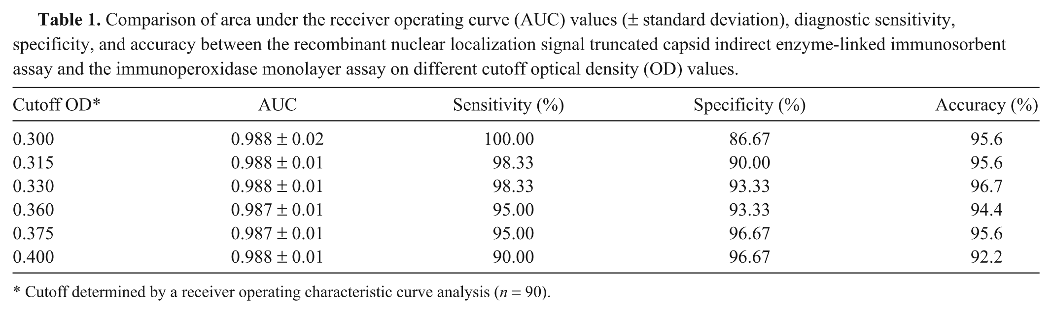
Cutoff determined by a receiver operating characteristic curve analysis (n = 90).
In the current study, the rntCap gene was expressed in the E. coli system as the rntCap with GST-tagged protein and was used as an antigen for the developed rntCap indirect ELISA. Because the rtCap protein was a recombinant GST-tagged protein, this possibly interfered with the results. To resolve the matter, the mean OD values of 10 positive and 10 negative serum samples were tested, and the results demonstrated that the GST-tagged protein in the rntCap protein was not interfering with the assay. It should be noted that both DSn and DSp of the developed assay yielded higher values than those of previous reports.15,19 However, the high sensitivity obtained from the rntCap indirect ELISA might have been derived from using the nonpurified rntCap protein antigen, which could possibly generate false-positive results. Nevertheless, the results from the established rntCap indirect ELISA demonstrated the appropriate accuracy rate (96.67%) and correlated well with the IPMA results when tested with the 90 field swine serum samples. More field samples are needed to confirm the reliability of this newly developed indirect ELISA.
The repeatability of the assay described herein also revealed a low variability. The intra-plate test showed minor different values when compared to the inter-plate test. However, the CV of the negative and positive serum samples in both assays indicated that the variability of the rntCap indirect ELISA was acceptable. The correlation between the rntCap indirect ELISA and the IPMA demonstrated linear regression indicating that the OD ratios from the rntCap indirect ELISA could be used to predict the levels of IPMA titers. Again, more samples are needed to enhance the DSn, DSp, and accuracy. Additionally, based on the results from 59 positive sera collected from vaccinated and nonvaccinated pigs (data not shown) and tested by the rntCap indirect ELISA (cutoff 0.330), the established rntCap indirect ELISA could detect antibodies against PCV-2 both from naturally infected pigs and vaccinated pigs. It would be of interest to observe whether the rntCap indirect ELISA could be used for PCV-2 antibody detection in oral fluid samples as oral fluid sample is currently widely used for major swine pathogen detection, including PCV-2, in the United States. 14 Another advantage of using the rntCap protein is the possibility for the development of other diagnostic tests such as a rapid strip test.
In conclusion, a newly developed rntCap indirect ELISA for PCV-2 antibody detection by using the rntCap protein expressed in the E. coli system in this report can be used as a serodiagnostic tool for PCV-2 antibody detection in swine herds. The applications of using the ELISA described herein include seromonitoring of NLS capsid of PCV-2 antibodies from maternal-derived antibodies, determining the time of infection in the production cycle, and possibly measuring the levels of the antibody levels after vaccination or infection. Such advantages can be analyzed in the future after gathering more data from field uses. In addition, this assay could be a valuable test for the routine diagnosis of large-scale samples and could possibly be produced for commercial purposes.
Footnotes
a.
GE Healthcare Ltd., Buckinghamshire, UK.
b.
New England Biolabs Inc., Ipswich, MA.
c.
Merck KGaA, Darmstadt, Germany.
Declaration of conflicting interests
The author(s) declared no potential conflicts of interest with respect to the research, authorship, and/or publication of this article.
Funding
The author(s) disclosed receipt of the following financial support for the research, authorship, and/or publication of this article: This study was financially supported by grants from the National Research University from Chulalongkorn University (Health Cluster-HR1164A7) and partially from the National Research Council of Thailand (2011). The authors would like to thank the Royal Golden Jubilee PhD Program from the Thailand Research Fund and Chulalongkorn University for financial support for Suphattra Jittimanee (PHD/0252/2550) PhD Program in Veterinary Pathobiology.