
Abstract
The current study describes a naturally occurring cluster of cases of Wedelia glauca intoxication. Seven of 14 axis deer (Axis axis) and 1 of 8 llamas (Lama glama) in a zoo of Buenos Aires province, Argentina, died suddenly after ingestion of a new batch of alfalfa (Medicago sativa) hay bales contaminated with the hepatotoxic plant W. glauca. Necropsies of 1 deer and 1 llama were performed. Pathological findings in both animals included severe diffuse acute centrilobular hepatocellular necrosis and hemorrhage, and clear yellowish translucent gelatinous edema on the wall of the gall bladder and the serosa of the choledochoduodenal junction. Fragments of W. glauca plants were identified in the hay based on the botanical characteristics of the leaves. Samples of gastric contents were examined by microhistological analysis, which identified epidermal fragments of W. glauca based on the presence of characteristic uniseriate glandular hairs (trichomes), confirming recent ingestion of W. glauca in both cases. The fragments were quantified and represented 5% of all examined vegetal fragments in the deer and 10% in the llama.
Wedelia glauca (Ortega, Hoffmann) is a herbaceous, invasive, perennial plant of the family Asteraceae native to South America. In Argentina, Wedelia glauca is known by the local names of “sunchillo,” “yuyo sapo,” and “asolador,” among others.1,7 The species is considered an agricultural weed 1 that causes acute lethal hepatotoxicity when ingested. The disease has been experimentally reproduced in cattle, sheep,5,17 pigs,14,17 and rats. 23 In Argentina, Brazil, and Uruguay, naturally occurring W. glauca intoxication has been diagnosed and reported extensively in bovine, 18 equine, 7 porcine,6,14 caprine, 7 and once in South American camelids (Lama glama). 12 The current report describes a naturally occurring outbreak of W. glauca intoxication in an axis deer (also known as chital, Axis axis) and a llama (Lama glama) held in captivity at a zoological garden in the province of Buenos Aires, Argentina.
In the late summer of 2010, 14 axis deer and 8 llamas were housed in 2 separate pens at a zoological garden in the Argentinean province of Buenos Aires. The animals were fed corn grain (Zea mays) and alfalfa (Medicago sativa) hay twice a day (morning and evening). In late February (day 0), zoo staff began feeding the animals with a new batch of hay bales. The following day, 2 deer were found dead and the mortality continued to occur intermittently during the next 7 days in the deer pen, killing 7 of the 14 animals. Additionally, 1 of the 8 llamas was found dead on the last day of the outbreak (day 8). The llamas had been fed the contaminated hay for the last time the evening of day 7.
Field necropsies were performed on a 3-year-old male axis deer and a 2-year-old female llama, both in fair state of postmortem preservation. In both cases, representative tissue samples including liver, spleen, gastrointestinal tract, kidney, heart, lung, and brain, were fixed by immersion in 10% buffered formalin and processed by standard histological techniques for the production of 5-µm thick sections stained with hematoxylin and eosin. Rumen content (deer) and content of the proximal gastric compartment (llama) were collected in both cases for microhistological analysis.
At necropsy, the llama had gross lesions limited to the liver, which showed a diffuse markedly enhanced reticular pattern (“nutmeg liver”) visible both on the capsular and cut surfaces (Fig. 1). In the axis deer, the liver was swollen, diffusely dark red with rounded edges, and there was focally extensive yellow, translucent, gelatinous edema of the gall bladder wall and serosa of the common bile duct and duodenum around the insertion of the common bile duct (choledochoduodenal junction). In addition, there were moderate multifocal petechiae and ecchymoses scattered throughout the abomasal mucosa, endocardium, epicardium, and some areas of the subcutis of the torso. The most significant histopathological finding in both animals was widespread, severe, hemorrhagic, centrilobular, and midzonal hepatocellular necrosis characterized by marked disruption of the hepatic cords surrounding central veins, which were separated by hemorrhage and edema, and had individualized, shrunken, hypereosinophilic hepatocytes with angulated cell borders and pyknotic, karyorrhectic, or absent nuclei (Fig. 2). Periportal and portal histoarchitecture were well preserved.
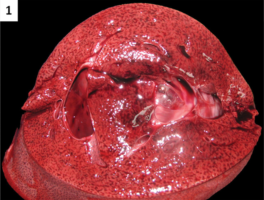
Llama (Lama glama). Diffuse enhancement of the reticular pattern due to severe diffuse centrilobular hepatocellular hemorrhagic necrosis (“nutmeg liver”) is seen on cut surface of the liver parenchyma.
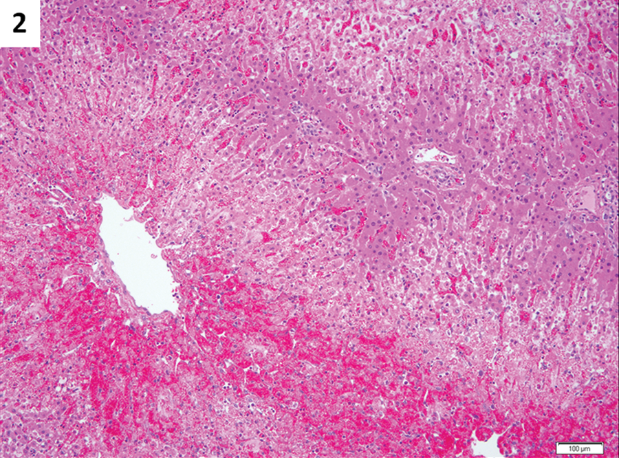
Llama (Lama glama); histopathology of the liver. Severe acute centrilobular and midzonal hepatocellular necrosis and hemorrhage with preservation of the periportal histoarchitecture. Hematoxylin and eosin. Bar = 100 µm.
Rumen content (deer) and content of the proximal gastric compartment (llama) were submitted refrigerated to the School of Agricultural Sciences at the National University of Mar del Plata (FCA-UNMDP; Facultad de Ciencias Agrarias, Universidad Nacional de Mar del Plata, Balcarce, Buenos Aires, Argentina) for plant fragment identification by microhistological analysis, as previously described.4,22,24 Briefly, gastric contents were washed with warm (30–35°C) tap water over a 100 mesh size sieve (100 openings per linear inch of mesh), dried at 60°C for 24 hr or until samples were totally dry, ground with a Willey-type grinder (1-mm mesh), and completely homogenized. From each sample, a 0.2-g subsample was obtained for optic microscopic examination. All samples were bleached by immersion in a 50% aqueous sodium hypochlorite solution for 2 min and rinsed over the 100 mesh size sieve. Each 0.2-g subsample was distributed onto 5 glass slides (24 mm × 48 mm) using glycerinated gelatin as a mounting media. Finally, the density of W. glauca fragments was quantified at 100× over 100 fields. The quantitative results (proportional amount of W. glauca ingested) were expressed in percentages. Wedelia glauca fragments were observed in the samples of rumen and gastric content, and were differentiated from other vegetal fragments based on the amber color and the presence of characteristic and specific trichomes (uniseriate glandular hairs; Fig. 3). The fragments of W. glauca represented 5% of all vegetal fragments in the deer sample and 10% in the llama. No vegetal fragments characteristic of other plants known to produce acute toxicity present in this region of Argentina, such as Cestrum parqui and Baccharis coridifolia, were observed.
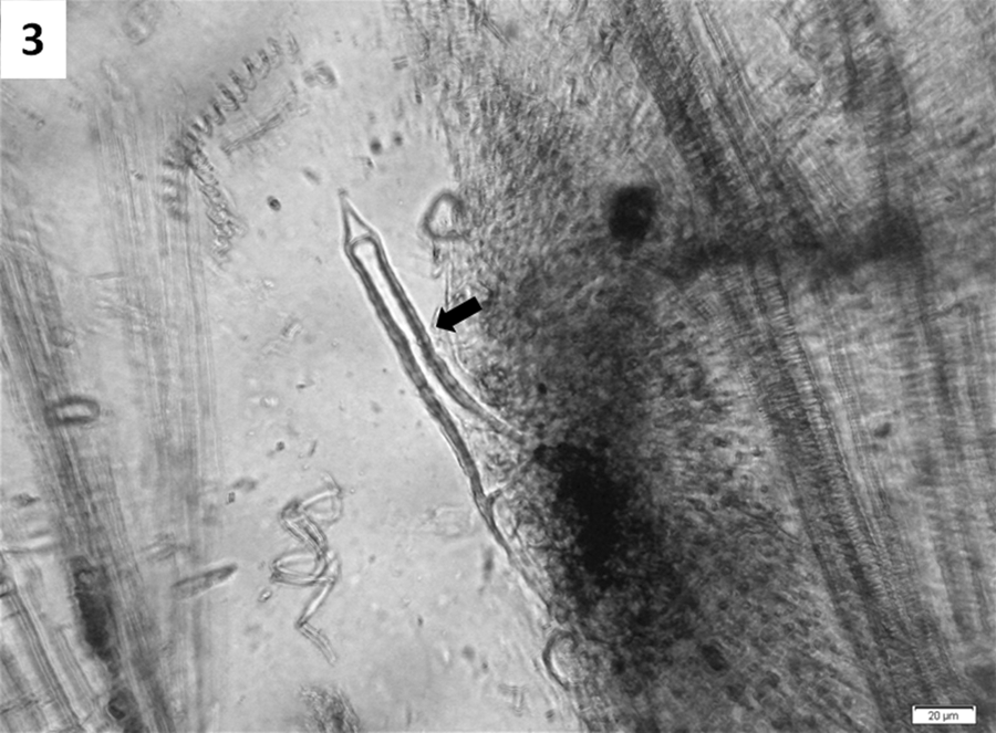
Axis deer (Axis axis); microhistological analysis of rumen content. A single uniseriate glandular hair (trichome) characteristic of Wedelia glauca is seen in the center of the image (arrow). Bar = 20 µm.
After the necropsies were performed, the pens where the animals were housed and 4 of the alfalfa hay bales that were being fed at the time the mortality occurred were thoroughly searched for toxic plants. Vegetal fragments found in the hay bales that did not resemble alfalfa were collected and submitted to the FCA-UNMDP for botanic identification. Wedelia glauca fragments were recognized based on their characteristic smell and distinct plant morphology, such as erect, pubescent, simple or slightly branched stems and opposite, 2 to 3-cm wide, lanceolate leaves with 3 main veins spreading from the base and 2 or 3 basal teeth. The morphologic characteristics allowed for the unequivocal identification of W. glauca and its differentiation from other common toxic plants from central and northern Argentina. 13 No toxic plants were found in the animal pens, and no other deaths occurred after the contaminated hay was discontinued.
The gross and microscopic pathological findings, the botanic identification of W. glauca in the alfalfa hay bales the animals were eating at the time of the mortality, and the detection of fragments of the plant in gastric contents by microhistological analysis confirmed the diagnosis of W. glauca intoxication in both animals.
Wedelia glauca is widely distributed in Argentina, Chile, Uruguay, Paraguay, Bolivia, and Brazil (http://www2.darwin.edu.ar/Proyectos/FloraArgentina/FA.asp), and a single report mentions its presence in the Spanish provinces of Madrid and Valencia. 3 The toxic principle is an atractyloside commonly named “wedeloside” that belongs to the diterpene glycosides group,9,16,21 which stops oxidative phosphorylation in the mitochondria of hepatocytes by inhibiting the action of adenosine triphosphate–adenosine diphosphate transporter proteins (translocases) in mitochondrial membranes,2,9 leading to centrilobular hepatocellular necrosis. 15 The toxic dose is considered low, with some authors reporting toxic doses for bovine and ovine animals of 4–5 g of dry matter per kg of body weight (gDM/kgBW),5,18 and others reporting even smaller toxic doses of 1.5–4 gDM/kgBW for sheep.17,24 In Argentina, numerous cases of cattle mortality due to the ingestion of W. glauca in free-grazing or feed-lot operations have been documented for many years, and this intoxication is considered one of the main toxic causes of mortality in adult cattle in the country according to the records of the Specialized Veterinary Diagnostic Service of the National Institute of Agricultural Technology (Servicio de Diagnóstico Veterinario Especializado, Instituto Nacional de Tecnología Agropecuaria, Balcarce, Buenos Aires, Argentina; E. Odriozola, personal observation, 2012), causing major direct economic losses every year.
In naturally occurring intoxications, clinical signs are rarely observed due to the peracute or acute course of the disease, with most animals found dead. 11 In experimental reproductions in sheep, clinical signs appeared 12–40 hr after the ingestion of total toxic doses of 4–10 gDM/kgBW and were characterized by depression, anorexia, muscle fasciculations, increased respiratory and cardiac frequencies, sternal and lateral recumbency with opisthotonus, terminal paddling movements, coma, and death. 5 No clinical signs were observed in any of the affected animals in the outbreak, as the animals were found dead. This is consistent with what has been described previously in naturally occurring cases of W. glauca intoxication in several species.11,12,14 Due to the peracute course of the disease, premortem serum samples could not be collected and therefore the serum activity of liver enzymes such as aspartate aminotransferase, lactate dehydrogenase, and gamma-glutamyl transferase, which have been found increased in experimental W. glauca intoxication in sheep, cattle and rats,5,23 were not able to be determined.
Necropsy findings in cases of W. glauca intoxication include a diffuse, markedly enhanced, hepatic reticular pattern (“nutmeg liver”), edema of the gall bladder wall, and duodenal serosa around the insertion of the common bile duct, petechiae and ecchymoses in the serosal and mucosal surfaces of the gastrointestinal tract, endocardium, and epicardium, and sometimes fluid hemorrhagic intestinal contents.5,17,18 Microscopically, lesions are characterized by diffuse, severe, hemorrhagic, centrilobular (periacinar) hepatocellular necrosis, and occasional vacuolar degeneration of adjacent midzonal hepatocytes.5,17,18,23 The pathological findings in the llama and axis deer in the current report, most notably the severe acute centrilobular hemorrhagic hepatic necrosis, were similar to that described by other studies of natural and experimental W. glauca intoxications in many animal species, including rats, 23 sheep, cattle, 5 horses, 7 pigs, 6 goats, 7 and llamas. 12 Pulmonary edema, described in experimentally intoxicated sheep and cattle, 5 and in naturally intoxicated llamas, 12 was not observed in the axis deer or the llama presented in the present report. Although not pathognomonic, the hepatic lesions are very characteristic, and their presence along with the detection of W. glauca in the feed and gastric contents allowed for the diagnosis of W. glauca intoxication in both animals.
Wedelia glauca ingestion was confirmed with the identification of specific W. glauca epidermal trichomes by microhistological analysis of gastric contents. As it has been demonstrated experimentally in sheep in which the exact amount of ingested W. glauca was known, microhistological analysis of the gastrointestinal contents allows not only for the detection of plant fragments but also for the quantification of W. glauca ingested in relation to other vegetal species. This technique is considered very accurate, especially when the digestion correction factor is applied.4,24 The digestion correction factor is calculated in ground, suspect samples comparing the vegetal fragments found and quantified before and after an in vitro digestion. Applying the correction factor is important when the diets are based on graminaceous plants as, generally, digestion affects the ability of agronomists to correctly identify dicotyledonous plants more so than graminaceous plants.8,10,20 In the current study, the digestion correction factor was not applied. However, because the hay samples were composed of 2 dicotyledonous plants (alfalfa and W. glauca), the percentages of W. glauca in the rumen content of the deer (5%) and gastric content of the llama (10%) are considered representative and fairly accurate. No other potentially toxic plants common in Argentina, causative of acute death and identifiable by microhistological analysis of gastrointestinal contents, such as Centrum parqui and Baccharis coridifolia,4,24 were identified by this method in either case. The identification of W. glauca in the hay bales was performed using plant identification keys and was based on the characteristic morphology of the leaves, which allows differentiation from other common hepatotoxic plants that are frequently found in central and northern Argentina. 13
Both leaves and stems of the standing green or dry W. glauca plants are toxic; the capability of W. glauca to maintain its toxicity even when dry in hay bales or rolls has been experimentally demonstrated, 11 and there are several descriptions of naturally occurring intoxications in livestock feeding on contaminated alfalfa and foxtail millet (Setaria italica) hay,12,19 as well as in free-grazing herds. 14 However, none of these previous reports describe the use of microhistological analysis of the gastrointestinal contents as an ancillary diagnostic test to identify and quantify fragments of W. glauca to confirm recent plant ingestion. This technique has been described as a useful diagnostic tool to demonstrate W. glauca ingestion and also estimate the proportion of this plant ingested in experimental reproductions in ovines.4,24 Given the fact that W. glauca has a small toxic dose (it is highly toxic) and that ingestion of small quantities of the plant are enough to cause death, its identification within the gastrointestinal tract of affected animals along with compatible gross and microscopic lesions allow for the diagnosis of W. glauca intoxication. In 2011, W. glauca intoxication was reported in llamas housed in pens and being fed alfalfa hay; 12 however, search of the veterinary literature yielded no report of W. glauca intoxication in cervids.
It was concluded that dry W. glauca, as can be found in hay bales or rolls, is toxic for axis deer and llamas, and intoxication by this plant should be considered a differential diagnosis in cases of sudden or unexplained death in these animal species. A presumptive diagnosis of W. glauca intoxication is based on the clinical history (acute death), gross and microscopic pathological findings, and the presence of defoliated W. glauca in the grazing pastures and/or within the rolls or bales of hay being fed to the animals. The microhistological analysis of gastrointestinal contents is considered a useful diagnostic tool to demonstrate recent consumption and quantify the amount of W. glauca ingested in herbivores and thus confirm intoxication in the presence of consistent pathological findings.
Footnotes
Acknowledgements
The authors thank Drs. Joanne and Mark Anderson for critical revision of this article.
Declaration of conflicting interests
The author(s) declared no potential conflicts of interests with respect to the research, authorship, and/or publication of this article.
Funding
The author(s) received no financial support for the research, authorship, and/or publication of this article.