
Abstract
Hemorrhagic splenic masses diagnosed as hemangioma or hemangiosarcoma were reviewed. Lymphoid hyperplasia was present in none of the hemangiosarcoma cases and in 27% of the hematoma cases. Siderotic nodules in the capsule or trabeculae were present in 25% of hemangiosarcoma cases and in 36% of hematoma cases. Hemoabdomen was noted in the clinical history of 54% of hemangiosarcoma cases and in 22% of hematoma cases. The average age (10.3 and 9.6 years, respectively), sex ratios (slightly more males), and most common breeds (Labrador Retriever, Golden Retriever, and German Shepherd Dog) were similar for the hemangiosarcoma and hematoma cases. Since lymphoid hyperplasia is much more common in cases of hematoma, the presence of this feature lends support to a diagnosis of hematoma rather than hemangiosarcoma. Signalment, history of hemoabdomen, and presence of siderotic nodules do not point to one diagnosis over the other.
Hemorrhagic splenic masses that are either hemangiosarcoma or hematoma constitute approximately 1.6% of the canine cases submitted to the Colorado State University Veterinary Diagnostic Laboratory (CSUVDL; Fort Collins, Colorado). Identification of the underlying cause of hemorrhage is important for prognosis and therapy. When hematoma is diagnosed, there can be concern that a hemangiosarcoma may have been undetected because hematomas that arise in a hemangiosarcoma could obscure the sarcoma both grossly and histologically. When an initial examination of a hemorrhagic splenic mass leads to diagnosis of hematoma but the submitting clinician suspects hemangiosarcoma, the usual method to try to increase confidence in the hematoma diagnosis is to examine more histologic sections of the tissue. In addition to that approach, if any features could be found that could be associated more with one condition than the other, then those features could be useful for strengthening confidence in a diagnosis.
Two common histologic changes in the canine spleen that might be associated with hemorrhagic masses are siderotic nodules and lymphoid hyperplasia. Siderotic nodules are areas of iron accumulation, mineralization, and connective tissue degeneration in the capsule and trabeculae (Fig. 1). The nodules are thought to be foci where bleeding had occurred previously. It is possible that hematomas might arise in these areas of altered connective tissue, but there is no known association of siderotic nodules with hemangiosarcoma or hematoma. Lymphoid hyperplasia is characterized by increased size and number lymphoid white pulp foci (Figs. 2, 3). It has been suggested that hematomas can arise in areas where the pattern of blood flow has been altered by the changes in the lymphoid tissues. 4 The current study was conducted to summarize characteristics of cases of hematoma or hemangiosarcoma in canine spleen and to investigate any patterns of signalment, clinical history, presence of lymphoid hyperplasia, or presence of siderotic nodules that might be associated more with hemangiosarcoma or with hematoma.
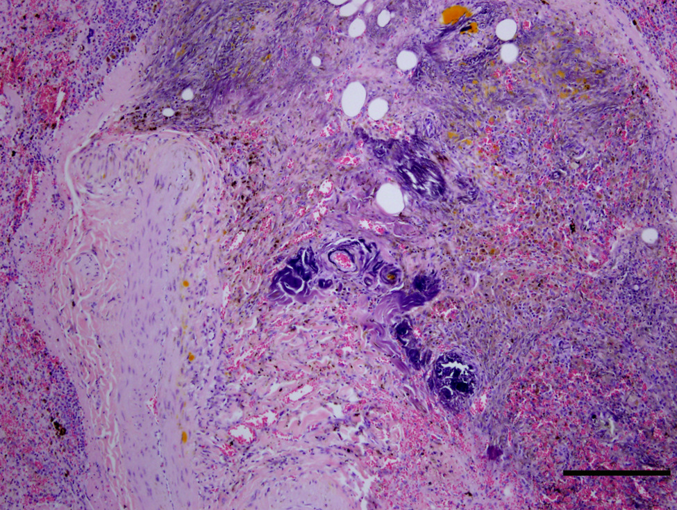
Canine spleen with a siderotic nodule in the connective tissue. Mineralization of the connective tissue, deposits of hematoidin pigment, and hemosiderin-filled macrophages are present. This spleen has hemangiosarcoma, not illustrated. Hematoxylin and eosin. Bar = 500 µm.
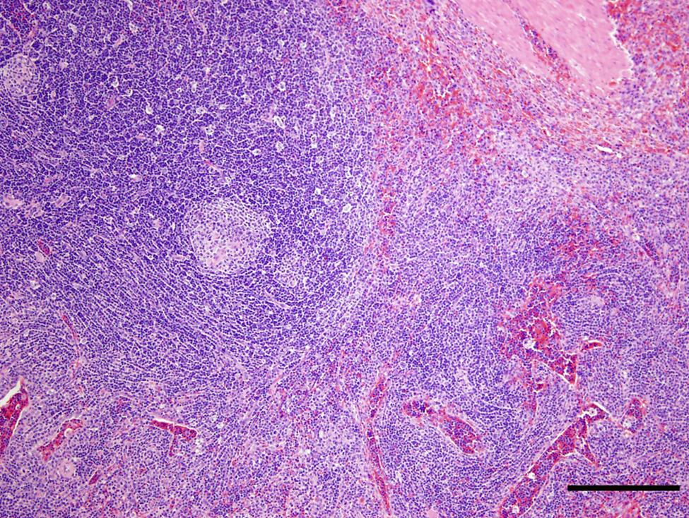
Canine spleen with lymphoid hyperplasia. A portion of a 4 mm in diameter nodule lymphoid tissue is present near the capsule of the spleen. This spleen has a hematoma located approximately 10 mm away, not illustrated. Hematoxylin and eosin. Bar = 500 µm.
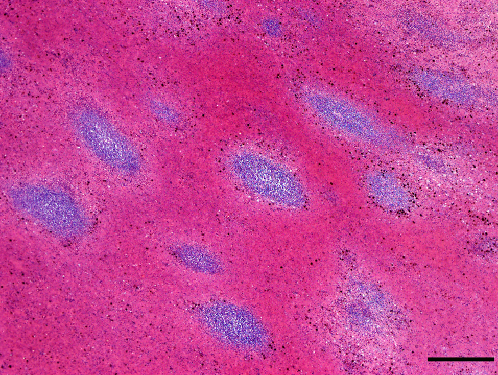
Canine spleen with lymphoid hyperplasia. Multiple closely spaces nodules of lymphocytes are present within a hematoma. Hematoxylin and eosin. Bar = 350 µm.
In the current study, 120 cases of splenic hemangiosarcoma and 100 cases of splenic hematoma were evaluated. Histologic sections and the information about age, sex, breed, and clinical history provided by the submitting veterinarian were reviewed. The named breeds include pure breeds or predominant crosses as interpreted by the veterinarians. These cases were submitted in a 30-week period and were selected in consecutive order. Hemorrhagic lesions caused by splenic torsion, trauma, or other types of tumors were not included in the study. Also, splenic masses interpreted as hemangiomas were excluded because this diagnosis represented only approximately 2% of the canine splenic masses evaluated at CSUVDL during the year of the study. The submissions included either the whole spleen or portions selected by the submitting veterinarian. Information about gross appearance was not available for all cases, but many hemangiosarcomas were a single nodule, and some of the hematomas were over 6 cm in diameter. At least 3 histologic sections had been prepared from each case with particular emphasis on sampling areas at the junction between hemorrhagic and solid tissue. Tissues were processed routinely, sectioned at 5 µm, and stained with hematoxylin and eosin (HE). The histologic sections were reviewed to confirm the diagnosis and identify siderotic nodules in the capsule and connective tissue trabeculae, as well as lymphoid hyperplasia. When lymphoid hyperplasia was identified in these cases, it was located within or adjacent to hematomas (Figs. 2, 3). In some cases, hyperplastic nodules were several millimeters in diameter and could be detected on hematoxylin and eosin–stained histologic slides without microscopic examination. Because histologic sections were prepared from areas near hemorrhagic lesions, it is not possible to know if any lymphoid hyperplasia existed at unexamined sites distant from the hemorrhagic lesions. Variations in the sizes of the samples prevented grading the extent of siderotic nodule formation.
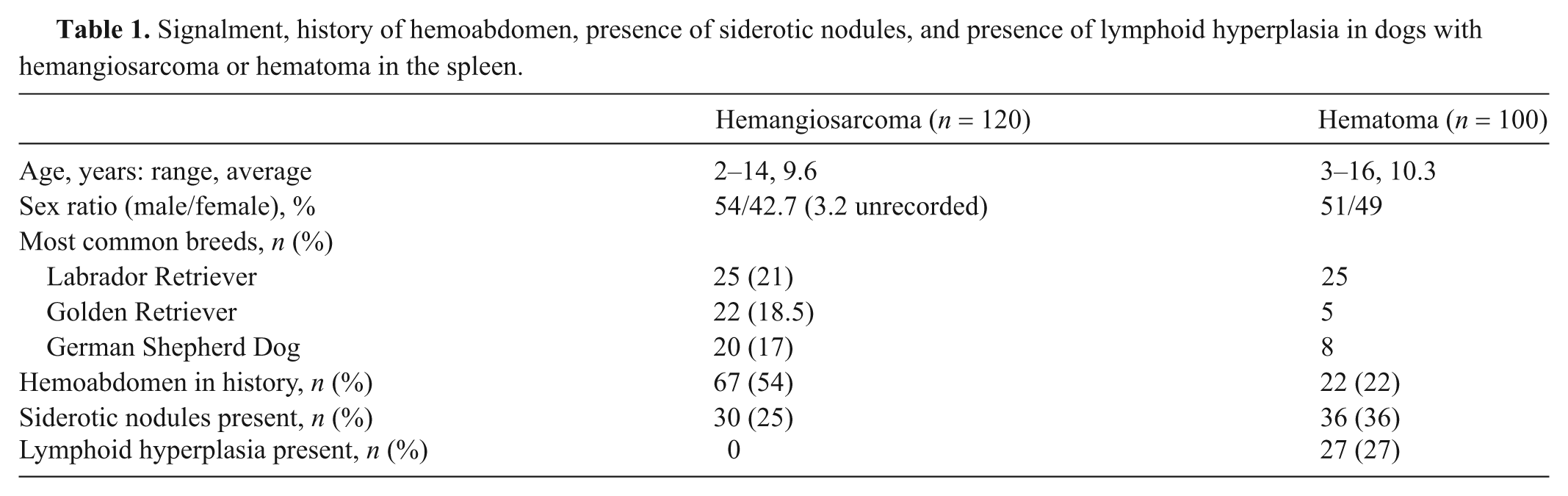
Information about these 220 cases is shown in Table 1. The average age and range of ages are similar for hemangiosarcoma and hematoma cases. Males are more numerous in hemangiosarcoma cases, but there was no sex difference in hematoma cases. The 3 most common breeds of dogs were Labrador Retriever, Golden Retriever, and German Shepherd Dog, representing 105 of the 220 dogs. In addition, dogs with hemangiosarcoma included 6 (4.8%) Boxers and 5 (4.0%) Australian Shepherds. The 104 other dogs were animals from a wide variety of pure or mixed breeds. The clinical histories include hemoabdomen in 67 (54%) cases of hemangiosarcoma and in 22 (22%) cases of hematoma. In the remaining cases, a splenic mass was discovered during a physical examination conducted for evaluation of a nonspecific illness, such as lethargy or anemia, or as part of a wellness examination. Siderotic nodules were more common in hematoma cases (36%) than in hemangiosarcoma cases (24%). Lymphoid hyperplasia was present in 27% of the 100 cases of splenic hematoma and in none of the 120 cases of hemangiosarcoma.
Signalment, history of hemoabdomen, presence of siderotic nodules, and presence of lymphoid hyperplasia in dogs with hemangiosarcoma or hematoma in the spleen.
Siderotic nodules are thought to be areas of previous hemorrhage; therefore, it is possible that their incidence would increase with increased age of the dog, reflecting accumulated damage within the spleen. However, in dogs with hemangiosarcoma or hematoma, siderotic nodules were present in roughly equal proportion in dogs younger or older than the average age (2–10 or 11–14 years; Table 2). It appears that their presence is not strictly an age-related phenomenon. Similarly, lymphoid hyperplasia might be expected to increase with age, but it was found at similar levels in dogs ≤10 years of age (15 cases) as in dogs ≥11 years (12 cases).
Numbers of dogs either younger or older than the average age of 10 years with siderotic nodules in the spleen and their association with either hemangiosarcoma or hematoma.
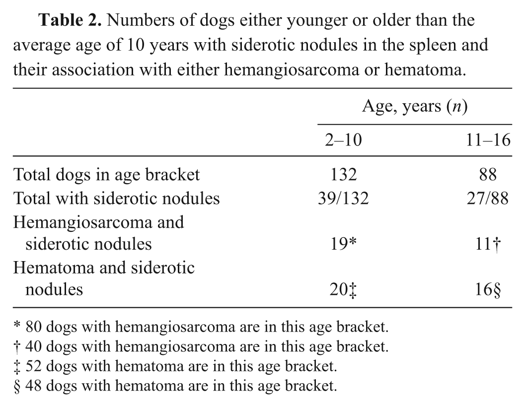
80 dogs with hemangiosarcoma are in this age bracket.
40 dogs with hemangiosarcoma are in this age bracket.
52 dogs with hematoma are in this age bracket.
48 dogs with hematoma are in this age bracket.
Splenic hemangiosarcoma was more common than splenic hematoma during the period from which these cases were selected. Previous reports have shown hemangiosarcoma to be more common than hematoma, 4 less common, 3 or the same. 2 Age ranges and averages were similar for these cases of hemangiosarcoma and hematoma and similar to those reported previously.1–4 In the current study, more males than females were diagnosed with hemangiosarcoma, but equal numbers of the sexes were diagnosed with hematoma. Previous reviews of hemangiosarcoma cases have reported higher numbers of males, 1 higher numbers of females, 3 or no difference.2,4 Also, previous reviews of hematoma cases reported more males 3 or no difference.2,4
In the current study, Labrador Retrievers were the most common breed affected with hemangiosarcoma and hematoma. Golden Retrievers were second for hemangiosarcoma and third for hematoma. This could be a reflection of the total numbers of dogs of these breeds rather than evidence of breed predilection for development of hemorrhagic splenic masses. Labrador Retrievers and Golden Retrievers or their crosses were the first- and second-most-common breeds of all canine submissions to the CSUVDL during this period (15.1% and 9.3%, respectively). However, German Shepherd Dogs may be overrepresented for these conditions because they constituted only 5% of total canine submissions to CSUVDL but represented 17% and 8%, respectively, of the hemangiosarcoma and hematoma cases. Also, Golden Retrievers may be underrepresented for hematoma development, as they represent 5% of hematoma cases but 9.3% of the total canine submissions. Previous reports list German Shepherd Dogs as the most common breed with splenic hemangiosarcoma,1–4 but these previous reports do not state what breeds were seen in the total canine case submissions in the periods and geographic regions that were assessed. The popularity of different breeds has changed since these studies were published between 15 and 25 years ago and may have varied by geographic areas, so it is not possible to draw any conclusions about differences in breed numbers compared to the current study.
A history of hemoabdomen was more than twice as common in dogs with hemangiosarcoma than in dogs with hematoma. Previous studies have also reported hemoabdomen to be more common with hemangiosarcoma.3,4 But hemoabdomen also occurs with enough frequency in hematoma cases that its presence does not appear to be useful for predicting whether a mass is hemangiosarcoma or hematoma.
Siderotic nodules were more common in cases of hematoma than hemangiosarcoma. But these foci are also so common in hemangiosarcoma cases that finding these foci would not increase confidence in the diagnosis of hematoma.
In the present series of cases, lymphoid hyperplasia was commonly associated with splenic hematoma but was not seen in hemangiosarcoma cases. This suggests that the presence of lymphoid hyperplasia is supportive of a diagnosis of hematoma, although not diagnostic in itself. Of the factors reviewed, only the presence of lymphoid hyperplasia appeared to be useful for increasing confidence that a lesion is truly a hematoma and that hemangiosarcoma had not been overlooked.
Footnotes
Declaration of conflicting interests
The author(s) declared no potential conflicts of interest with respect to the research, authorship, and/or publication of this article.
Funding
The author(s) received no financial support for the research, authorship, and/or publication of this article.