
Abstract
Splenic epithelial cysts are rare in humans and have not been reported in animals, to our knowledge. During a routine medical examination of a 12-y-old castrated male Maltese dog, a splenic mass was found and subsequently removed via splenectomy. Histologically, a well-defined multilocular cyst in the spleen was lined mostly by simple cuboidal, multifocally by stratified cuboidal, or occasionally by stratified squamous epithelium. Immunohistochemically, the lining cells were positive for cytokeratin and negative for vimentin, CD31, and Wilms tumor protein 1. The case was diagnosed as a primary splenic epidermoid cyst.
Keywords
Cystic lesions of the spleen are rare in humans. 14 Splenic cysts can be classified as either parasitic or non-parasitic. Non-parasitic cysts are divided into primary (true) cysts and secondary cysts (pseudocysts). 8 Primary cysts are lined by epithelial cells, and are further subdivided into epidermoid, dermoid, and mesothelial types according to the type of lining epithelium: stratified squamous epithelium, stratified squamous epithelium with skin appendages, and mesothelium, respectively. 2 In addition, endothelial cell–derived hemangioma or lymphangioma can occur as cystic lesions in the spleen. 11 In humans, non-parasitic splenic cysts comprise 0.07% of splenic conditions; among the non-parasitic cysts, epidermoid cysts only account for ~10%. 14
Small splenic cysts usually cause no clinical problems and are found incidentally. Large splenic cysts can cause abdominal pain and discomfort. 6 Given that cysts often grow slowly and surreptitiously, enlarged cysts can compress the renal artery, causing systemic hypertension and rupture of nearby organs. 1
Given that the routine use of ultrasonography and CT makes detection of splenic cysts easier, the incidence of splenic cysts is expected to increase. 16 Primary non-parasitic epithelial cysts have not been reported in any of 3 retrospective studies of splenic biopsies in dogs.5,7,9 Here we describe a splenic epidermoid cyst in a dog.
A 12-y-old castrated male Maltese dog was presented to a local referral hospital for splenectomy because of a splenic mass identified via ultrasound examination during a comprehensive medical examination at another hospital. There were no reported clinical signs related to the splenic mass. Additional ultrasound findings included severe gallbladder sludge. Serum chemistry revealed increased activity of liver enzymes. The dog had unilateral nephrectomy performed previously as treatment for nephrolithiasis and hydronephrosis. In addition, the thyroid hormone (T4) concentration was consistently low, and thyroid medication had been administered. Total splenectomy and cholecystectomy were performed. The dog recovered well after splenectomy. The spleen was submitted for microscopic evaluation.
Grossly, the spleen had a 2-cm multilocular mass with variably sized cystic spaces in the body (Fig. 1A–C). The splenic mass was fixed in 10% neutral-buffered formalin, processed routinely, and slides were stained with H&E. To reveal the origin of the lining cells, immunohistochemistry (IHC) was performed for cytokeratin (clone AE1/AE3, 1:200; Dako), Wilms tumor protein 1 (WT1; ab15249, 1:100, Abcam), and CD31 (clone JC70A, 1:100; Dako).
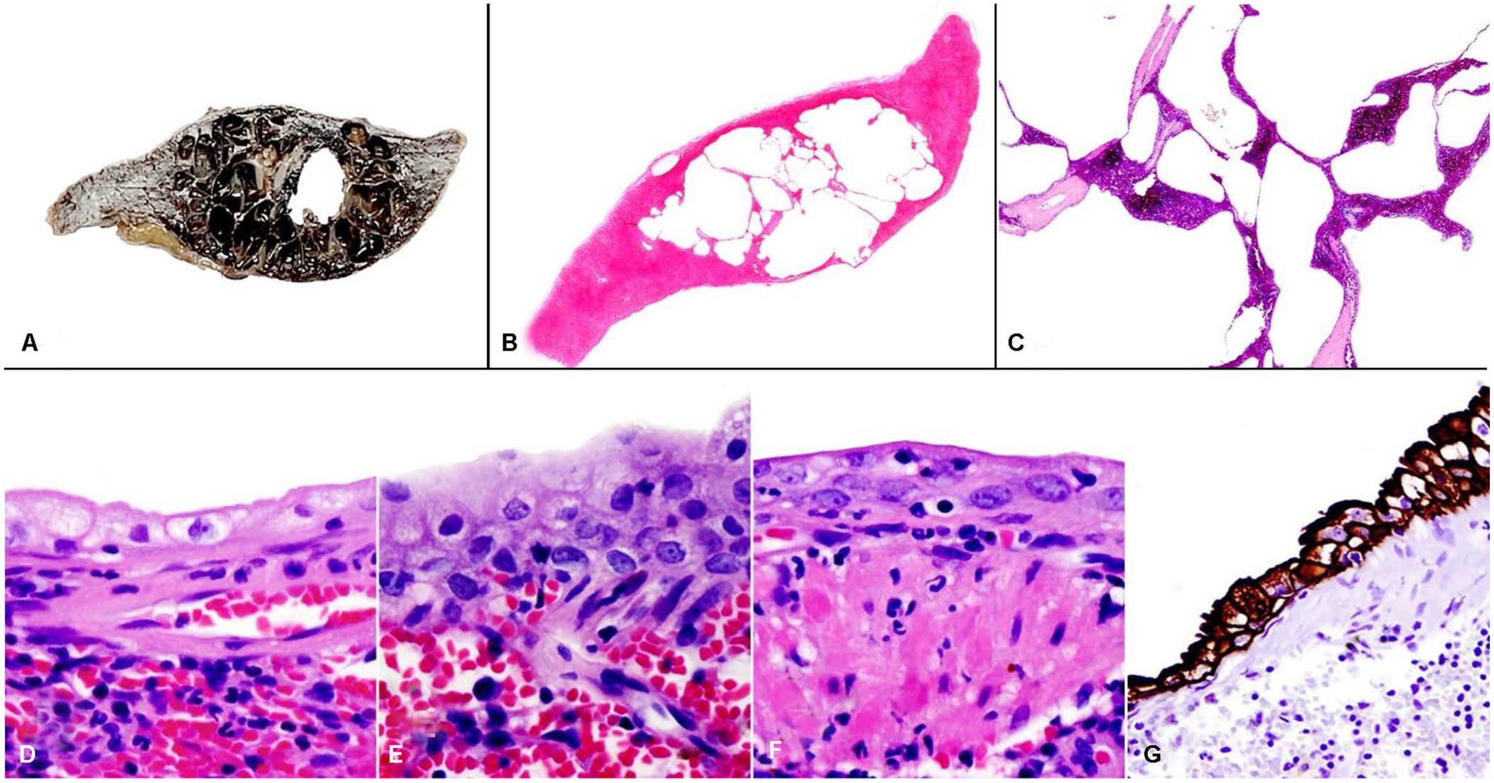
Splenic epidermoid cyst in a dog. Figures 1B–F are H&E sections.
Histologically, cystic spaces were multichambered, lined with simple or stratified cuboidal epithelium and segmentally with squamous epithelium, and supported by thin fibrous connective tissue and splenic parenchyma (Fig. 1D–F). The cystic spaces were compressive and clearly demarcated from the surrounding splenic parenchyma. No evidence of neoplasia was noted. Immunohistochemically, the lining cells were positive for cytokeratin (Fig. 1G) but were negative for vimentin, WT1, and CD31. The IHC results were highly suggestive of a primary epithelial cyst, more specifically an epidermoid cyst. No evidence of neoplasia was noted.
To our knowledge, a non-neoplastic and non-parasitic primary splenic epidermoid cyst has not been reported previously in a domestic animal. The splenic cysts found in animals are commonly neoplasms including hematoma and hemangiosarcoma 15 (rarely lymphangioma) 13 or parasitic cysts such as Echinococcus granulosus.3,15 It is not clear how primary epithelial cysts develop. One proposed mechanism is the mesothelial invagination theory, which states that the mesothelium from the splenic capsule was included into the parenchyma of the spleen during development. 10
The epithelium lining the splenic cysts could easily be mistaken for endothelium, leading to a misdiagnosis of lymphangioma or hemangioma. However, primary epithelial cysts are positive for cytokeratin and negative for factor Ⅷ–related antigen and CD31. 12 In our case, cyst-lining cells were CD31-negative and cytokeratin-positive, ruling out an origin from vascular or lymphatic endothelium. Dermoid cyst was also ruled out given that no skin appendages, including hair follicles, were observed histologically.
The cells lining epithelial cysts may be squamous, transitional, or mesothelial cells. 11 These 3 epithelia may appear at different sites, even within the same cyst. Therefore, IHC may be required to identify the lining cells. Mesothelial cysts are immunoreactive to both cytokeratin and WT1; epidermoid cysts are positive for cytokeratin and negative for WT1. 4 In our case, immunostaining was positive only for the cytokeratin marker; therefore, we confirmed that the splenic cyst was an epidermoid cyst.
If the demand for annual medical checkups of companion animals increases, and ultrasonography and CT are used routinely, the probability of finding a splenic cyst also increases. Epithelial cysts should be added to the differential diagnoses of splenic cystic lesions in dogs.
Footnotes
Declaration of conflicting interests
The authors declared no potential conflicts of interest with respect to the research, authorship, or publication of this article.
Funding
This study was supported by BK21 FOUR Future Veterinary Medicine Leading Education and Research Center.