
Abstract
The broad spectrum of heat-related injury (HRI) and its associated lesions is well described in the human literature, with rare reports of similar findings in farm animals. In the current case series, lesions from 4 of 8 lambs that presented with clinical signs of heat stress are reported. Gross lesions at necropsy consisted of acute renal swelling and pallor in 2 of 4 lambs, muscle pallor in 2 of 4 lambs, and chronic bronchointerstitial pneumonia in each of the 4 lambs. Histological lesions considered heat-related included acute renal tubular necrosis, pigment casts, tubular epithelial regeneration, multifocal myocyte degeneration, necrosis, and dropout with histiocytic influx and regeneration. Chronic, bronchointerstitial pneumonia, present in each lamb, was considered a condition predisposing to HRI. Compatibility between observed lesions and those reported in human beings with injury secondary to elevated body temperatures established a diagnosis of HRI in these animals. Diagnostic pathologists should consider HRI in lambs with histological evidence of renal tubular necrosis and/or rhabdomyolysis and even in cases where the clinical picture is strongly suggestive but lesions are not demonstrable.
Heat-related injuries (HRIs) include a spectrum of progressively more severe clinical diseases, which, in human beings, are commonly termed heat stress, heat exhaustion, and heat stroke. 10 Heat-related injury occurs when thermoregulatory mechanisms fail to maintain an appropriate core body temperature after either environmental acquisition or endogenous production of heat exceeds the body’s ability to dissipate heat adequately. Excess heat can result from exposure to high environmental temperature (classic heat stroke) or exercise-associated generation of heat (exertional heat stroke). 2 Compounding factors such as high humidity, low environmental air movement, and heavy insulation (hair coat) additionally reduce the body’s ability to disperse heat. Development of HRI may be enhanced in patients with poor thermoregulatory ability, such as neonates and geriatric patients, or by conditions such as underlying disease or lack of acclimation. 9
During prolonged hyperthermia, direct cytolytic effects of heat on tissues, secondary endotoxemia, release of inflammatory cytokines, injury to the vascular endothelium, and activation of the coagulation cascade are well-recognized processes that, when untreated, can lead to multiple-organ dysfunction (MOD), disseminated intravascular coagulation (DIC), and death. 2 A wide spectrum of clinical conditions including cardiac, renal, hepatic, and respiratory failure as well as rhabdomyolysis, seizures, and DIC are associated with progressively severe HRI in human beings. 2 A comparable report has not been made in the veterinary literature, which has focused on the clinical diagnosis and lesions of only the most severe HRI in dogs4,5 while largely overlooking other nonhuman species. Contemporary literature is limited largely to reproductive and lactational effects of heat stress on dairy cattle, increased susceptibility to disease and decreased weight gain in feedlot cattle, and prevention of exertional heat-induced injury in horses.1,7 Clinical manifestations and gross and histopathological changes attributable to HRI have not been described in farm animals. Studies of experimentally induced HRI in sheep report aberrations in physiological and biochemical parameters but contain no mention of gross or histopathological changes.6,8
Diagnosis of HRI in veterinary species, as in human beings, relies heavily on clinical signs. 4 When pathologists are presented with cases that lack a clinical history, diagnosis becomes more difficult. The current report describes an aggregate of postmortem lesions in 3 of 4 affected lambs whose clinical features led to a strong presumptive diagnosis of HRI.
Fourteen Hampshire-cross and Suffolk-cross lambs aged 4–6 months, 12 breeding-age ewes, and 2 adult rams resided on a small farm in central Oklahoma. Eight lambs were born and raised on the premises, whereas 6 were purchased from breeders in adjacent states. The lambs occupied three 4.5 m × 9 m dirt pens essentially devoid of vegetation and partially shaded by a large oak tree. Sheep had continuously occupied the pens for several years. All animals had free access to feed (native grass hay and custom-formulated sheep concentrate; 0.7 kg per head per day), water, and covered shelter ventilated by fans. Despite measures to avoid heat stress, 8 of the 14 lambs developed clinical signs of HRI over a period of approximately 4 weeks, during which daytime high temperatures were rarely less than 37.8°C and frequently as high as 44.4°C; concurrent daily lows failed to drop below 27.8°C.
On July 17, 2011, a 4-month-old castrated male lamb (lamb 1) was presented to the Center for Veterinary Health Sciences (CVHS) at Oklahoma State University (Stillwater, Oklahoma) after a 1-day history of lethargy, muscle fasciculation of the hind limbs, an arched back, respiratory difficulty, and dribbling urine. On initial examination, the lamb was hyperthermic (41.7°C; reference range, 38.3–39.4°C), was recumbent, and exhibited open-mouth breathing. Immediate cooling successfully normalized body temperature; however, tachycardia (120 bpm; reference range, 80–100 bpm) and tachypnea (44 bpm; reference range, 12–30 bpm) persisted. When assisted to a standing position, the lamb had a dorsally arched back with hind limbs tucked cranially, dribbled urine, and within 15–20 sec developed severe tremors of the hind limbs and fell down. Additional clinical findings included bilateral serous nasal discharge, increased bronchovesicular lung sounds, and cranioventral pulmonary consolidation, confirmed via transthoracic ultrasonography. When amputation of the urethral process failed to alleviate urinary dribbling, and radiographs of the pelvis and caudal spine revealed no abnormalities, a clinical determination of decreasing neurological function was made. The lamb was euthanized with intravenous pentobarbital and underwent necropsy approximately 4 hr later.
Gross findings were limited to bilaterally, moderately enlarged, moist, pale kidneys; cranioventral pneumonia; multifocal urethral hemorrhage (secondary to catheterization); and a single 3-mm-diameter granular bladder calculus. Histological examination revealed asymmetrically severe, acute renal tubular necrosis; tubular epithelial regeneration; hyaline and cellular casts (Fig. 1); and bronchointerstitial pneumonia with bronchiolitis obliterans. Significant lesions were not observed in the brain or spinal cord. In this initial case (lamb 1), the renal lesion was considered most significant; however, its cause and pathogenesis were obscure. Heat-related injury was included in the differential diagnosis due to the clinical history, initial physical examination findings, and ongoing severe climatic conditions.
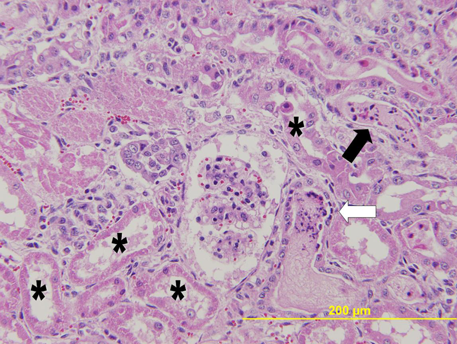
Lamb 1. Acute tubular necrosis (asterisks), marked tubular epithelial attenuation with tubular necrotic cellular and karyorrhectic debris, neutrophil influx (black arrow), and tubular regeneration (white arrow) evidenced by flattened, tubular epithelium, and cellular basophilia. Hematoxylin and eosin. Bar = 200 µm.
During the following 3 weeks, 7 additional lambs became similarly affected, initially presenting with arched backs (Fig. 2), urine dribbling, tremors of the hind limbs, respiratory difficulty, and marked hyperthermia. Four animals (lambs 5–8) recovered after treatment by the referring veterinarian with a combination of antibiotics, vitamin E and selenium, vitamin B12, fluids, and attempts at cooling. The 14 cohabitant adult sheep remained unaffected. Serum from lamb 5, as well as feed and water samples, was submitted to the Oklahoma Animal Disease Diagnostic Laboratory (Stillwater, OK). Serum selenium and copper levels were adequate, blood lead and Leptospira species titers were negative, and feed sulfur levels were within normal limits. Water levels of total dissolved solids, nitrites, nitrates, and sulfates were acceptable for livestock.
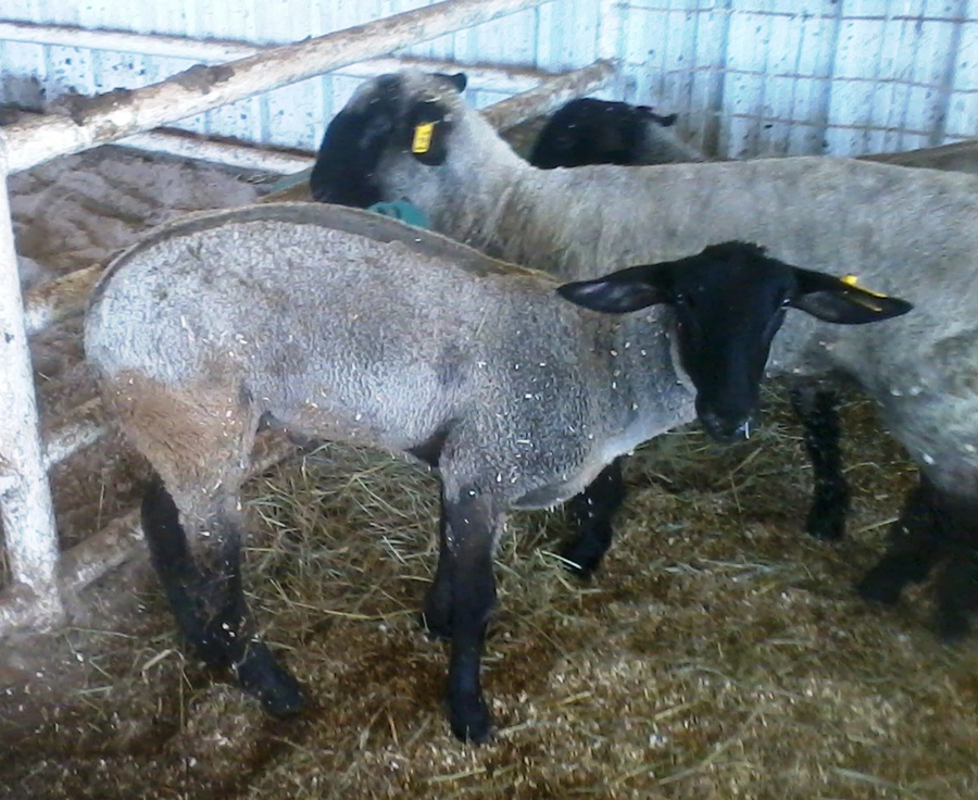
An initial presenting sign of heat-related injury in lambs. Note the arched back, tucked-up hind limbs, and nasal discharge.
On August 5, 2011, 20 days after submission of lamb 1, 2 live lambs (lambs 2 and 3) with particularly severe and progressive clinical signs, together with tissue from a lamb necropsied on the farm (lamb 4), were submitted to the CVHS for clinical examination and necropsy. Initial physical findings for both lambs 2 and 3 were similar to lamb 1 and included severe respiratory distress, marked hyperthermia, tachycardia, and tachypnea, with the addition of hypersalivation, marked dehydration, crackles and wheezes on lung auscultation, and progression to total recumbency (lamb 3) and stupor (lamb 2). Each lamb’s body temperature was ≥42.2°C. Significant findings on complete blood cell count and chemistry for both lambs included markedly elevated muscle enzymes and moderately to markedly increased blood urea nitrogen and creatinine (Table 1). Hyperfibrinogenemia, present in both lambs, tended to rule out overt disseminated intravascular coagulation but was considered a relatively specific marker for an ongoing, severe inflammatory process.
Salient clinical signs and clinicopathological data of lambs in the current study.*
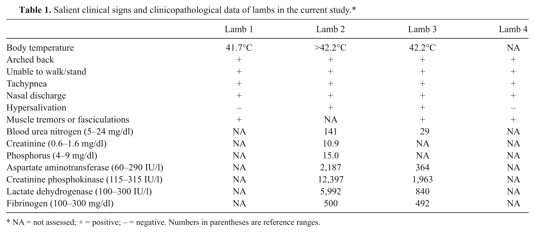
NA = not assessed; + = positive; − = negative. Numbers in parentheses are reference ranges.
Postmortem examinations of lambs 2 and 3 were initiated within 30 min of euthanasia. Gross renal changes were found only in lamb 2 and included bilaterally, markedly swollen, pale, and excessively moist kidneys. Muscle pallor, more prominent in lamb 2, was present in axial and hind limb muscles of both lambs 2 and 3. Mesenteric and sublumbar lymph nodes of lamb 3 were moderately enlarged and edematous. Both lambs had cranioventral pneumonia. Gross evaluation of tissues submitted from lamb 4 revealed chronic bilateral pneumonia; portions of both kidneys, liver, and the sacral section of the spinal column were grossly normal. Histologically, lesions in lamb 2 consisted of acute, severe, renal tubular necrosis with adjacent infiltrates of neutrophils; frequent single cell necrosis; numerous pigment and cellular casts; and marked dilation of cortical tubules with flattened epithelium. Periodic acid–Schiff staining revealed prominent loss of the ciliated brush border of proximal convoluted tubules. Skeletal muscle lesions were similar in lambs 2 and 3 and consisted of multifocal myocyte degeneration, necrosis, and dropout with variable histiocytic influx and moderate regeneration with satellite cell activation (Fig. 3, Table 2).
Histopathological findings of lambs in the current study.*

E = considered equivocal; NA = not assessed; + = mild; ++ = moderate to severe; − = negative.
The most striking histological lesion in both lambs 1 and 2 was the renal tubular necrosis. Causes considered reasonable for the renal lesions included any of a number of nephrotoxins, shock, hypovolemia, sepsis, hemolysis, trauma, and ischemic injury. The probability of exposure to nephrotoxic agents was considered unlikely given the controlled environment, unremarkable results of feed and water analysis, the extended time gap between onset in affected animals, the lack of recognizable renal lesions in lambs 3 and 4, and the unequal severity of the renal changes in lamb 1. Although specific morphological evidence of vascular change leading to an ischemic event was not identified in any of the kidney sections examined, the single cell necrosis and loss of the renal proximal tubular ciliated brush border are findings reported in ischemic, heat-induced injuries in human beings 3 and thus were considered heat-related ischemic injuries in these lambs.
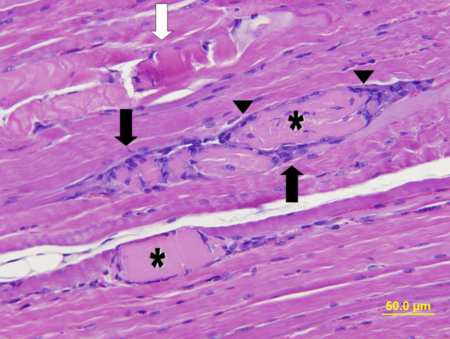
Lamb 2. Myocyte degeneration and necrosis characterized by fractured, hypereosinophilic (white arrow), and swollen, homogenously eosinophilic (asterisks) fibers. Note the histiocytic influx (black arrows) and numerous, prominent, activated satellite cells with small, deeply basophilic nuclei (arrowheads). Hematoxylin and eosin. Bar = 50 µm.
Of interest is the regenerative response evident in the kidneys of lamb 1 and absent from subsequent lambs. Although in human beings and event horses, HRI can often be directly associated with a single exertional or environmental event, the animals in the present case series were farm animals exposed to recurrent episodes of high environmental temperature (daily highs). Given these conditions, the regenerative response and concurrent acute necrosis are considered either the result of a single illness for a longer period of time than reported by the owner or the result of an unrecognized earlier HRI, resulting in a previous episode of acute tubular necrosis subsequently followed by a later episode. Because recognition of illness in lambs 2–4 occurred at least 2 weeks after illness in lamb 1, it is postulated that critical conditions for development of HRI in these lambs were met at a different time to lamb 1. Although individual tolerance may have allowed subsequent animals to survive the initiating events in lamb 1, the concurrent muscle and kidney damage and rapid clinical decompensation suggest that these animals faced a more severe single bout of heat exposure leading to severe and widespread damage. As in people, susceptibility of each animal is presumably based on its individual level of fitness, concurrent disease (pneumonia), and environmental exposure.
Rhabdomyolysis is a common HRI in both human beings and domestic animals and is believed to be the direct effect of heat on myocytes. 11 Additional factors such as vascular compromise may add an ischemic element to the damage.5,10 Causes of primary myopathies are numerous, but histological changes in these animals were consistent with heat-induced as well as nutritional and toxic myopathies. The apparent unavailability of myotoxic plants in the lambs’ environment justified downgrading the likelihood of a toxic myopathy. Nutritional (vitamin E and/or selenium) deficiency myopathy was considered, but confirmation of adequate serum selenium levels, and the rarity of confirmed vitamin E and/or selenium deficiency myopathy in the region, minimized nutritional deficiency as a likely primary cause.
Rhabdomyolysis and pigment casts in renal tubules of lamb 2 are not directly associated with renal damage; these 2 histological findings in lamb 2 are independent and unrelated lesions of HRI. Chronic bronchointerstitial pneumonia was present in each of the 4 lambs. Attempts at identification of a primary infectious agent via viral isolation, fluorescent antibody test, and polymerase chain reaction failed to identify a specific viral cause. Regardless of cause, pneumonia was considered an underlying condition that predisposed these animals to heat injury.
In the present case series, animals presented in various stages of collapse, including an inability to stand unsupported (lamb 1), prostrate but able to eat (lamb 3), and nearly comatose (lamb 2). Clinically, these signs were interpreted as expressions of neurological dysfunction; however, attribution of many clinical signs specifically to the central nervous system is challenging in nonhuman animal species. In the absence of histological lesions to account for the neurological signs in these lambs, it is not known whether the signs were attendant to primary neurological dysfunction, a requirement for confirmatory diagnosis of the most severe stage of HRI (i.e., heat stroke),2,10 or rather were secondary to pain, weakness, overt exhaustion, or other factors. Because the designation of heat stroke requires clinical signs specifically attributable to central nervous system dysfunction, the syndrome reported in these lambs is classified within the broader continuum of HRI.
The owner was initially skeptical of the preliminary diagnosis of HRI, such that circumstance resulted in submittal of additional animals and tissues that provided the opportunity to observe multiple, repeated gross and histological lesions in animals whose clinical signs supported a strong presumptive diagnosis of HRI. Elucidation of this spectrum of lesions is of value to the pathologist examining animals without the advantage of a collaborative clinical history.
Footnotes
Declaration of conflicting interests
The author(s) declared no potential conflicts of interest with respect to the research, authorship, and/or publication of this article.
Funding
The author(s) received no financial support for the research, authorship, and/or publication of this article.