
Abstract
Two 2- and 3-month-old beef calves from 2 separate herds, locations, and times were found dead and were submitted to the veterinary diagnostic laboratory for diagnostic work-up. In both cases, no premonitory signs were seen by the owners. Histopathology revealed acute panlobular hepatic necrosis in both calves. In addition, calf A had copper and selenium deficiency, and calf B had oxalate nephrosis, and selenium and zinc deficiencies. Alpha-amanitin was detected in the urine from calf A, and in the liver and rumen contents from calf B using liquid chromatography–mass spectrometry. The cause of panlobular hepatic necrosis and death of both calves was determined to be amanitin toxicosis from ingestion of amanitin-containing mushrooms based on microscopic changes and toxicological analysis of tissues. In cases of sudden death in cows with histopathological findings of panlobular hepatic necrosis, toxicological analysis for amanitin is needed for a definitive diagnosis of poisoning by amanitin-containing mushrooms.
Keywords
There are approximately 100 poisonous mushrooms in North America. Most toxic mushroom fatalities in animals and human beings are caused by cyclopeptides in the mushrooms. Three genera, Amanita, Galerina, and Lepiota, are known to contain hepatotoxic cyclopeptides (amanitins) with Amanita phalloides and Amanita ocreata commonly found along the West Coast.2,15,18 Such mushrooms contain several amanitins, but α-amanitin and β-amanitin are found in the highest concentration. Exposure to amanitin-containing mushrooms can occur easily under grazing conditions. The 2 cases in the present report illustrate that analysis of postmortem specimens for amanitin can be used to confirm amanitin poisoning in cattle.
In case 1, a 2-month-old beef calf (calf A) from Napa County, California, was submitted to the Davis branch of the California Animal Health and Food Safety Laboratory System (CAHFS) for necropsy in April 2008. The calf was found dead with no premonitory signs of illness. No other calves had died or were showing clinical signs, but 2 cows in an adjacent pasture had died in the previous 2 months. Gross examination revealed a calf in good flesh with dark pink, wet lungs. The liver was friable and had an enhanced lobular pattern. Portions of brain, sciatic nerve, heart, liver, kidneys, lungs, spleen, thymus, forestomachs, abomasum, and small and large intestines were immersed in 10% buffered neutral formalin and embedded in paraffin. Four-micron sections were cut, stained with hematoxylin and eosin, and examined by light microscopy. Histopathological examination revealed severe, acute, diffuse, centrilobular to panlobular hepatic necrosis (Fig. 1) with light nonsuppurative inflammatory cell infiltrates in the portal tracts.
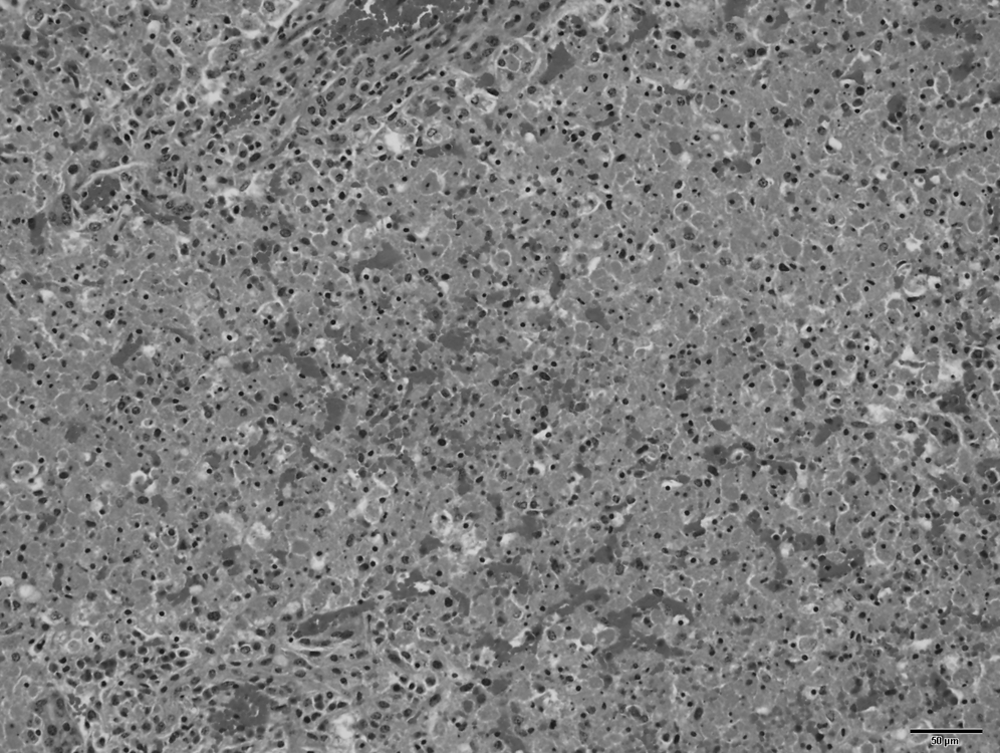
Photomicrograph of liver from calf A with acute massive hepatocellular necrosis and lobular collapse. Portal tracts are at the top and bottom left. Bar = 50 μm.
In case 2, in early December 2009, a 3-month-old Hereford steer (calf B), weighing approximately 84 kg, was submitted to CAHFS, Davis for necropsy. The calf came from a herd pastured in Sonoma County, California and had been found dead. The calf had not shown signs of illness prior to death, and no other animals in the herd were affected. Gross examination revealed a moderately fleshed male with a heavy tick burden. The cranial ventral lungs were mottled dark pink–red and wet, and there were petechial hemorrhages on the epicardium and thymus. There was focal fibrous adhesion of the pericardial sac to the right cranial lung. The liver was swollen. No other gross lesions were observed. Portions of the brain, heart, liver, kidneys, lungs, spleen, thymus, small and large intestines, reticulum, rumen, omasum, abomasum, and tongue were fixed in 10% buffered neutral formalin, processed as previously described, and examined by light microscopy. The most significant histopathologic change was acute panlobular coagulation necrosis and hemorrhage in the liver. In scattered lobules, a single layer of hepatocytes in the periportal regions were spared. Hepatocellular necrosis was characterized by dissociated hepatocytes with intense eosinophilic cytoplasm and pyknotic nuclei. Additionally, refractile crystals were dispersed throughout the kidney cortex within all types of tubules, many associated with tubular necrosis. Special stain (Pizzato stain) demonstrated that the renal tubular crystals were calcium oxalate. Miscellaneous findings included multifocal hemorrhages in the thymus associated with discrete lymphocellular necrosis, and patchy filling of alveoli with proteinic edema fluid and multifocal interstitial and pleural fibrosis in the lungs.
In both cases 1 and 2, a full diagnostic work-up was performed including aerobic cultures of the liver and lung on MacConkey and blood agar plates, Salmonella culture of feces, flotation of the feces for parasite ova, and reverse transcription polymerase chain reaction on spleen for Bovine viral diarrhea virus, all of which were negative. Initial toxicological analyses included analyses for nitrate and nitrite in ocular aqueous humor of calves A and B, and heavy metal (As, Cd, Cu, Fe, Hg, Mn, Mo, Pb, Zn) and selenium analyses of the liver of calf A and of the liver and kidney of calf B. Nitrates or nitrites were not detected in aqueous humor in either case at a reporting limit of 1 mg/l. The liver of calf A contained below optimal concentrations of copper (18 mg/kg wet weight [WW], reference [ref.] range: 25–100 mg/kg WW) and selenium (0.17 mg/kg WW, ref. range: 0.25–0.5 mg/kg WW). The liver of calf B contained a normal concentration of copper, a below optimal selenium concentration (0.092 mg/kg WW), and a slightly low zinc concentration (17 mg/kg WW, ref. range: 35–100 mg/kg WW). All other metals were within acceptable reference ranges in the liver. The kidney contained normal levels of all metals listed.
Based on the histopathologic findings and the nontoxic tissue copper levels, the rumen contents from calf A were tested for carboxyatractyloside, the hepatotoxin of Xanthium sp. (Elizabeth Torr, personal communication, 2010), which was not detected. Tissue samples from both calves were tested for amanitin (hepatotoxin in Amanita spp.) in 2009 concurrent with the diagnostic work-up of calf B. A validated test for amanitin was not yet available in 2008 during the diagnostic testing of calf A. Urine (frozen since April 2008) from calf A (rumen contents, liver, and kidney no longer available), and fresh liver and rumen contents from calf B were analyzed for α-amanitin using liquid chromatography–mass spectrometry (LC/MS), as previously described. 10 In brief, α-amanitin was extracted from all 3 samples with acetonitrile in 0.1 M phosphate buffer. The acetonitrile was removed from the aqueous phase by extraction with methylene chloride, and the resulting extracts were purified by solid-phase extraction using a modified version of a previously reported purification procedure. 6 High-pressure liquid chromatography (HPLC) separation was performed using a commercial HPLC system a equipped with a 150 mm × 4.6 mm column. b The HPLC system was interfaced with a linear ion trap mass spectrometer, c and α-amanitin was detected in MS/MS/MS (MS 3 ) mode. A method detection limit study of 7 replicate analyses of liver fortified with 1 μg/kg of α-amanitin gave an average recovery of 98% and a calculated method detection limit of 0.5 μg/kg. Analysis of unfortified negative control fresh and fixed liver, and rumen contents, showed no interferences at the α-amanitin retention time. Alpha-amanitin was positively identified in the urine from calf A and in fresh liver and rumen contents from calf B based on comparison of the retention time and MS 3 spectrum obtained from analysis of purified α-amanitin standard material. d Detection of α-amanitin was considered diagnostic to confirm exposure to amanitin-containing hepatotoxic mushrooms in both calves A and B. Liver from calf B that had been fixed in buffered neutral formalin for 1 month was tested similarly to see if amanitin could be detected in formalin-fixed tissue. Despite 2 testing attempts as described, no α-amanitin was detected in formalin-fixed liver.
Based on the history of acute death and pathological and toxicological findings, a diagnosis of amanitin toxicosis was made in both cases. Mushroom poisonings have traditionally been confirmed through indirect methods such as positive identification of amanitin-containing mushrooms with a history of exposure or ingestion, clinical presentation, and postmortem findings with histopathology. Typically, the suspect mushrooms are not submitted for analysis. In case 2, 1 month after the calf’s death, the producer brought in mushrooms found growing where the calf was pastured; LC/MS analysis did not detect any α-amanitin in these mushrooms. The delay in collection of mushrooms likely resulted in missing hepatotoxic mushrooms that were present in the pasture at the time of death.
Differential diagnoses for acute centrilobular–panlobular hepatic necrosis include anemia, heart failure, copper toxicosis, and ingestion of hepatotoxins such as amanitin-containing mushrooms, cocklebur (Xanthium spp.), and hepatotoxic blue-green algae (microcystins). In both cases, anemia and heart failure were ruled out based on gross and microscopic examinations. Although both calves had selenium deficiency, neither had gross or microscopic lesions in the heart compatible with white muscle disease. Additionally, neither had centrilobular congestion in the liver that would suggest changes due to heart failure. The acute panlobular necrosis was not suggestive of heart failure. Copper toxicosis was ruled out by heavy metal analysis of liver (calves A and B) and kidney (calf A). Blue-green algae–contaminated ponds or troughs were not found on inspection by the veterinarians, and cocklebur was not found in the pasture. Additionally, the toxic metabolite of cocklebur, carboxyatractyloside, was not detected in the rumen contents of calf A. Cases of cocklebur toxicosis and blue-green algae toxicosis usually occur in the summer and early fall in California and were considered unlikely in both cases.
Amanitin-containing mushrooms are abundant in California. Two species that are common in California are A. phalloides and A. ocreata. Amanita phalloides is particularly common in the San Francisco Bay area, and along the Pacific Coast, and is most abundant in warm, wet years. 19 Amanita phalloides is commonly found in association with oaks, birch, and pine, and has also been found in open pastures in the Central Valley of California. The large fruiting bodies appear in the late summer and fall and have several characteristics: a smooth, yellowish-green to yellowish-brown cap, white gills, a white ring around the upper part of the stem (veil), and a white cup-like structure (volva) around the base of the stem. Amanita ocreata is commonly known as Western American destroying angel, and the mushroom grows from Baja California, Mexico, along the Pacific Coast to Washington. Amanita ocreata grows well in sandy soils under oak or pine, and the mushroom has been commonly found in the foothills and valley of the Central Valley of California. The fruiting bodies are usually found in late winter and spring. Amanita ocreata has a white or cream-colored cap; white, short gills; a white stem with a white, thin, broken, partial veil (annulus); and a white, thin volva. 1
Amanitins are extremely toxic, and poisonings have been confirmed in dogs 20 and horses. 3 Reported intravenous median lethal dose of α-amanitin in dogs is 0.1 mg /kg body weight.7,8 Considering the average concentration of amanitins per mushroom, 1 A. phalloides has the potential to kill an animal as large as an adult horse or cow. 19
There is only limited data on the toxicokinetics of amanitins. Alpha-amanitin is taken up by cells in the gastrointestinal tract where the first damaging effects are seen. Following systemic absorption, α-amanitin is taken up by hepatocytes via OATP1B3, an organic anion-transporting polypeptide. 14 Following intravenous administration in dogs, it was shown that plasma half-life of amanitins is short, ranging from 25 to 50 min, and that amanitins are not detectable in plasma after 4–6 hr. 7 There is no known metabolism, or plasma protein binding of the α-amanitin. Between 80% and 90% of the administered dose of amanitins are eliminated in urine and up to 7% are eliminated in bile. 9 After oral ingestion of A. phalloides in human beings, α- and β-amanitins were detected in plasma up to 36 hr after ingestion, and in urine up to 72 hr post-exposure. 12 Plasma and urine amanitin concentrations do not seem to correlate with the clinical severity or outcome. In human beings that died from A. phalloides intoxication, amanitin was detected in kidneys up to 22 days post-ingestion. 5 The kidneys appear to contain higher concentrations than liver, indicating that toxins are bound to renal tissue. There is no known toxicokinetic data available for ruminants, but based on the confirmed cases in the present report, rumen contents, urine, and liver are useful diagnostic specimens in suspected amanitin cases involving ruminants. Unfortunately, amanitin was not detected in 1 formalin-fixed liver, which tested positive prior to fixation. Thus, fresh samples are likely to be necessary for detection of the toxin.
Amanitins inhibit nuclear RNA polymerase II, which leads to decreased messenger RNA and protein synthesis.16,21 Cells with a high metabolic rate, such as hepatocytes, crypt cells, and proximal convoluted tubules of the kidneys are most commonly affected. While this mechanism is well established, additional cellular effects contribute to the pathogenesis. In mice and cultured dog hepatocytes, apoptosis contributed to amanitin-induced liver failure.13,17
Long-term consequences of amatoxin poisoning are best documented in human beings. Approximately 20% of people who “recover” from A. phalloides ingestion will develop immune complex–mediated chronic active hepatitis and anti–smooth muscle antibody activity. The pathogenesis of the sustained chronic liver disease is thought to arise from irreversible alteration of hepatocytes during the acute toxicity, rendering hepatocytes unrecognizable to, and as a result, a target of the host’s immune system. 4 Previous nonfatal exposure to amanitins may be one etiology for the pathologist to consider in livestock with lesions of chronic hepatitis of unknown cause.
Acute proximal convoluted tubular necrosis seen in the kidneys of amanitin-poisoned human beings is likely a result of reabsorption of amanitins by renal tubules after glomerular filtration. 11 Acute necrosis of only the proximal convoluted tubules of the kidneys was not evident in either calf described in the present report and may be related to dose and rapid clinical course. Intestinal crypts are also targeted by amanitin in human beings. Intestinal hemorrhage and crypt necrosis were not seen in either calf on gross or microscopic examination.
Calcium oxalate nephrosis seen in calf B was prominent. Crystals were numerous and were associated with tubular necrosis. As previously mentioned, amanitin causes acute renal tubular necrosis of the proximal tubules, but calcium oxalates have not been noted to occur associated with nephrosis caused by amanitin. Foci of necrosis associated with oxalate crystals in calf B were randomly distributed in the renal cortex and were not confined to the proximal tubules and therefore not likely related to the toxic effect of the amanitin. Sources of calcium oxalates include ethylene glycol and soluble oxalate-containing plants such as sorrels or docks (Rumex spp.), halogeton (Halogeton glomeratus), greasewood (Sarcobatus vermiculatus), garden rhubarb (Rheum rhaponticum), Bermuda buttercup (Oxalis pes-caprae), and other Oxalis spp. Additionally, fungi such as Aspergillus niger and Aspergillus flavus can produce large quantities of oxalates on feedstuffs. The source of the oxalates in calf B was not identified. Halogeton and greasewood are not found in western California. Curly dock and Bermuda buttercup are abundant along the coastal regions of California and thus the most likely sources.
The present report documents amanitin toxicosis in cattle. Over the past 10 years, the CAHFS laboratory has identified cases of centrilobular to panlobular hepatic necrosis of unknown cause in cattle. Mushroom poisonings were suspected due to season or mention of transient presence of mushrooms in the pasture, but the detection method for amanitin was not previously available. Amanitin toxicosis should be considered in these cases and in cases of sudden death in cattle on pasture. Microscopic findings of acute panlobular hepatic necrosis will help narrow potential diagnoses to include hepatotoxic mushroom poisoning. Because pathological findings in cases of amanitin toxicosis are not specific, toxicological analysis must be performed for a definitive diagnosis of mushroom poisoning. Rumen contents, liver, or urine are the tissues of choice for toxicologic analysis. In any suspicious case and whenever possible, collection of mushrooms found in the environment of the animal is also recommended for positive identification and possible amanitin testing.
Footnotes
Acknowledgements
The authors wish to thank Mike Filigenzi, Elizabeth Torr, and Sam Stump for their technical assistance.
a.
1100 HPLC system, Agilent Technologies Inc., Santa Clara, CA.
b.
Polar-RP, Phenomenex Inc., Torrance, CA.
c.
Thermo Fisher Scientific Inc., Wilmington, DE.
d.
Axxora LLC, San Diego, CA.
The author(s) declared no potential conflicts of interest with respect to the research, authorship, and/or publication of this article.
The author(s) received no financial support for the research, authorship, and/or publication of this article.