
Abstract
A 7.5-kg female European badger (Meles meles) was submitted under the United Kingdom Government’s Wildlife Incident Investigation Scheme to the Thirsk Regional Laboratory of the Animal Health and Veterinary Laboratories Agency. Postmortem examination revealed a large, poorly encapsulated retropharyngeal abscess full of necrotic debris and pus. Arcanobacterium haemolyticum was isolated from the lymph node and confirmed by phenotypic profiling and 16S ribosomal RNA DNA sequencing. Workers should be aware of the potential for badgers to harbor zoonoses other than those more traditionally associated with the species.
Arcanobacterium haemolyticum, formerly designated Corynebacterium haemolyticum, has been isolated from human beings with pharyngitis and systemic disease2,4,8 and more recently associated with pyothorax 5 and Lemierre syndrome. 1 Although human beings are considered to be the reservoir of infection, 4 reports of sporadic isolations from other animals, including horses, 3 sheep, 7 cattle, 6 and a pet rabbit 9 have been published.
In October 2009, a 7.5-kg female European badger (Meles meles) was submitted under the Wildlife Incident Investigation Scheme, funded by the United Kingdom Department for Environment, Food and Rural Affairs, to the Thirsk Regional Laboratory of the Animal Health and Veterinary Laboratories Agency (Thirsk, North Yorkshire, United Kingdom). The carcass was in reasonable body condition, although it had undergone mild autolysis and harbored a severe Trichodectes melis louse infestation. There were no external signs of trauma. Postmortem examination revealed a large, poorly encapsulated retropharyngeal abscess full of necrotic debris and pus. Other significant gross pathology included multifocal beige lesions up to 3 mm in diameter throughout the liver parenchyma, diffuse purple pulmonary discoloration with edema of cut surface, and polycystic kidneys. Histopathological examination demonstrated nonsignificant acute focal myocarditis and widespread severe pulmonary edema. To rule out malicious poisoning, samples were submitted for toxicological analysis by the Food and Environmental Research Agency (York, United Kingdom), with negative results for a standard suite of tests comprising alpha-chloralose, paraquat, diquat, strychnine, and a range of anticoagulant rodenticides and carbamate pesticides.
Ziehl–Neelsen-stained smears of the retropharyngeal abscess revealed no acid–alcohol-fast bacteria, and no mycobacterial species were isolated. A swab from the retropharyngeal lymph node cultured onto 5% sheep blood agar and incubated aerobically at 37°C yielded a mixed growth including Pasteurella multocida, a Streptococcus sp., and tiny β-hemolytic colonies consisting of Gram-positive branching rods. Catalase and urease tests on the latter were negative, and the organisms were positive for the reverse Christie, Atkins, Munch-Peterson (CAMP) reaction with Staphylococcus aureus β-hemolysin.
The tiny β-hemolytic isolate was provisionally identified as A. haemolyticum using a manual microorganism identification test kit a (profile 0530361) and confirmed using extended phenotypic profiling b and 16S ribosomal (r)RNA DNA sequencing. The 16S rRNA gene was amplified using primer pair 5’-AGTTTGATCCTGGCTCAG-3’ and 5’-ACCTTGTTA CGACTT-3’. Sequencing of this product generated a contig of 1,281-bp showing >99% similarity with the 16S rRNA sequence of the A. haemolyticum type strain entry by BLAST analysis (http://www.ncbi.nlm.nih.gov/blast/Blast.cgi; GenBank accession no. FR715327). A phylogenetic tree, constructed with type strains of all members of the genus Arcanobacterium using MEGA4, confirmed the identification of the isolate as A. haemolyticum (Fig. 1).
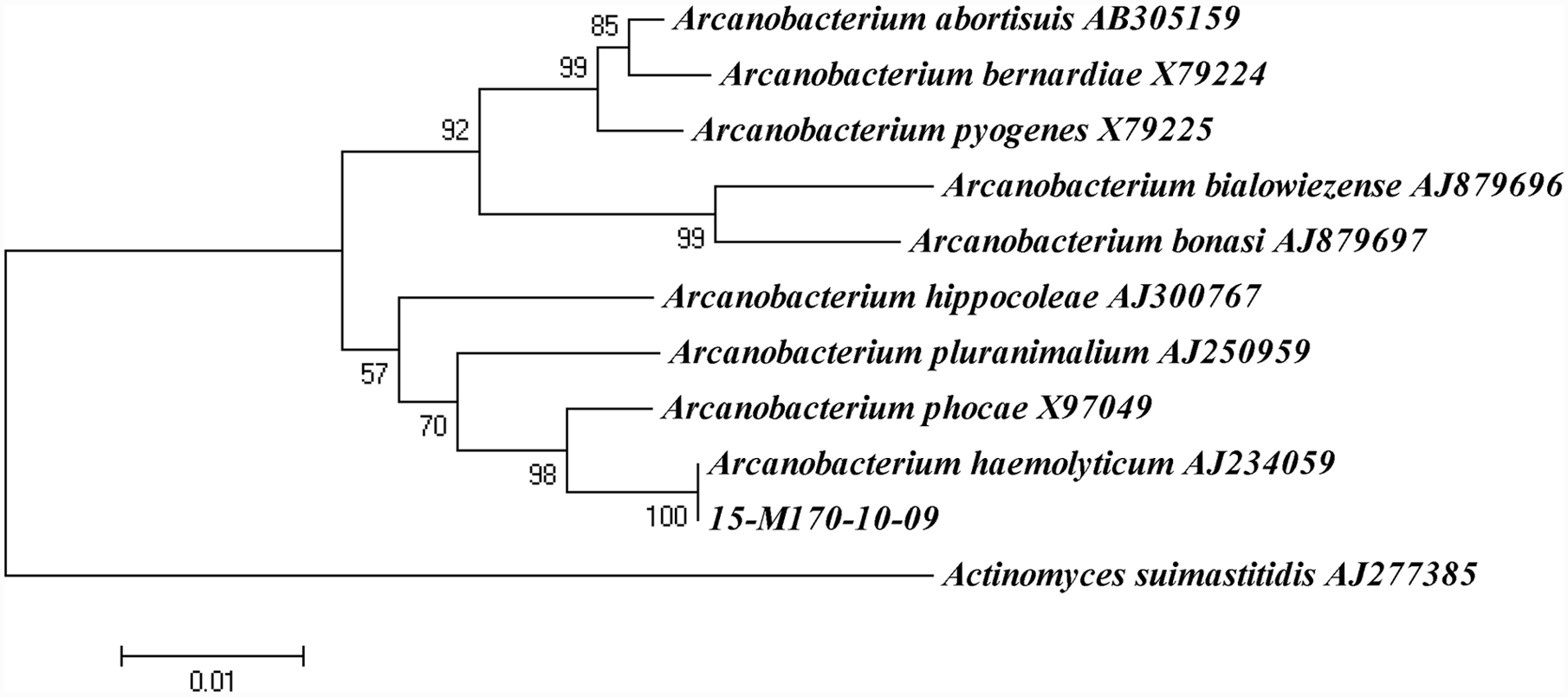
Phylogenetic analysis based on 16S ribosomal RNA sequences of type strains of Arcanobacterium species and isolate 15-M170-10-09. The tree was constructed using the neighbor-joining approach following CLUSTAL alignment of sequences trimmed to a 1,164-bp consensus contig. The sequence of Actinomyces suimastitidis was used as an outgroup. Bootstrap confidence analysis was performed with 500 replicates to determine the reliability of the tree topology. Bar = 0.01 substitutions per nucleotide position.
Previous reports of isolations of A. haemolyticum from animals have involved domesticated3,9 and farmed animal hosts6,7 for which human contact is a possible source of infection. The current study reports the isolation of A. haemolyticum from a wildlife host for which the potential for direct human contact is minimal. The likely role of A. haemolyticum in this single case is uncertain, and further study is necessary to determine how widespread this potentially zoonotic organism is among badgers. Nonetheless, those required to work in close association with badgers, including rehabilitation workers and wildlife veterinarians, should include A. haemolyticum in the list of known zoonotic agents associated with this species.
Footnotes
Acknowledgements
The authors wish to thank colleagues at the Food and Environmental Research Agency for toxicological data.
Notes
The author(s) declared no potential conflicts of interest with respect to the research, authorship, and/or publication of this article.
The authors wish to thank the United Kingdom Department for Environment, Food and Rural Affairs for its funding of the Wildlife Incident Investigation Scheme (project OM0047). SAC Veterinary Services receives financial support from the Scottish Government as part of its Public Good Veterinary and Advisory Service.