
Abstract
A 4-year 10-month-old, intact female dromedary camel had progressive left carpal joint swelling and lameness for 7 months. Radiographs showed multifocal lytic lesions in the carpal and proximal metacarpal bones. Surgical biopsy of the synovial capsule and carpal bones suggested neoplasia, and the camel was subsequently euthanized. At necropsy, a white to pale pink, firm, multilobulated, soft tissue mass was located on the palmar aspect of the left carpal joint. Two smaller masses were present on the dorsal aspect of the carpal joint. The masses infiltrated all the carpal bones and the proximal region of the metacarpal bone. The joint capsule was diffusely thickened. The articular surfaces of the carpal bones and the metacarpal bone were multifocally eroded. The lungs contained multiple, firm, raised, gray, randomly distributed nodules. The neoplastic cells stained positive for vimentin and S-100. Chondrosarcoma arising from around the carpal joint with infiltration of carpal and metacarpal bones, and pulmonary metastasis, was diagnosed based on the histopathological and immunohistochemical evaluation.
Primary tumors of bone are common, and chondrosarcoma represents 10% of the primary bone neoplasms in dogs. 13 Among other animal species, chondrosarcoma is reported to be common in sheep. 7,13 A few reports exist in cows and goats. 3,9,12 It is rare in all other animal species including human beings. 8,13 Further, there is only a single report of chondrosarcoma involving the mandible of a Bactrian camel (Camelus bactrianus), 11 and there are no reports of chondrosarcoma in dromedary camels. A chondrosarcoma in a dromedary camel (Camelus dromedarius) is reported in the current study.
A 4-year 10-month-old, intact female dromedary camel had left carpal joint swelling and non–weight bearing lameness that had become progressively more severe during the previous 7 months. The camel was treated at times with varying combinations of support bandaging, dexamethasone, and antibiotics. The owner reported marked worsening of the condition over the several weeks prior to admission. Radiographs of the left carpal joint showed multifocal radiolucent, smoothly marginated lesions in the carpal bones and proximal metacarpi (Figs. 1, 2). Open arthrotomy was performed to retrieve biopsy specimens for culture and histopathology, and a full-limb cast was placed on the limb. Based on the initial histopathological evaluation with a diagnosis of chondrosarcoma, the camel was euthanized and presented for necropsy.
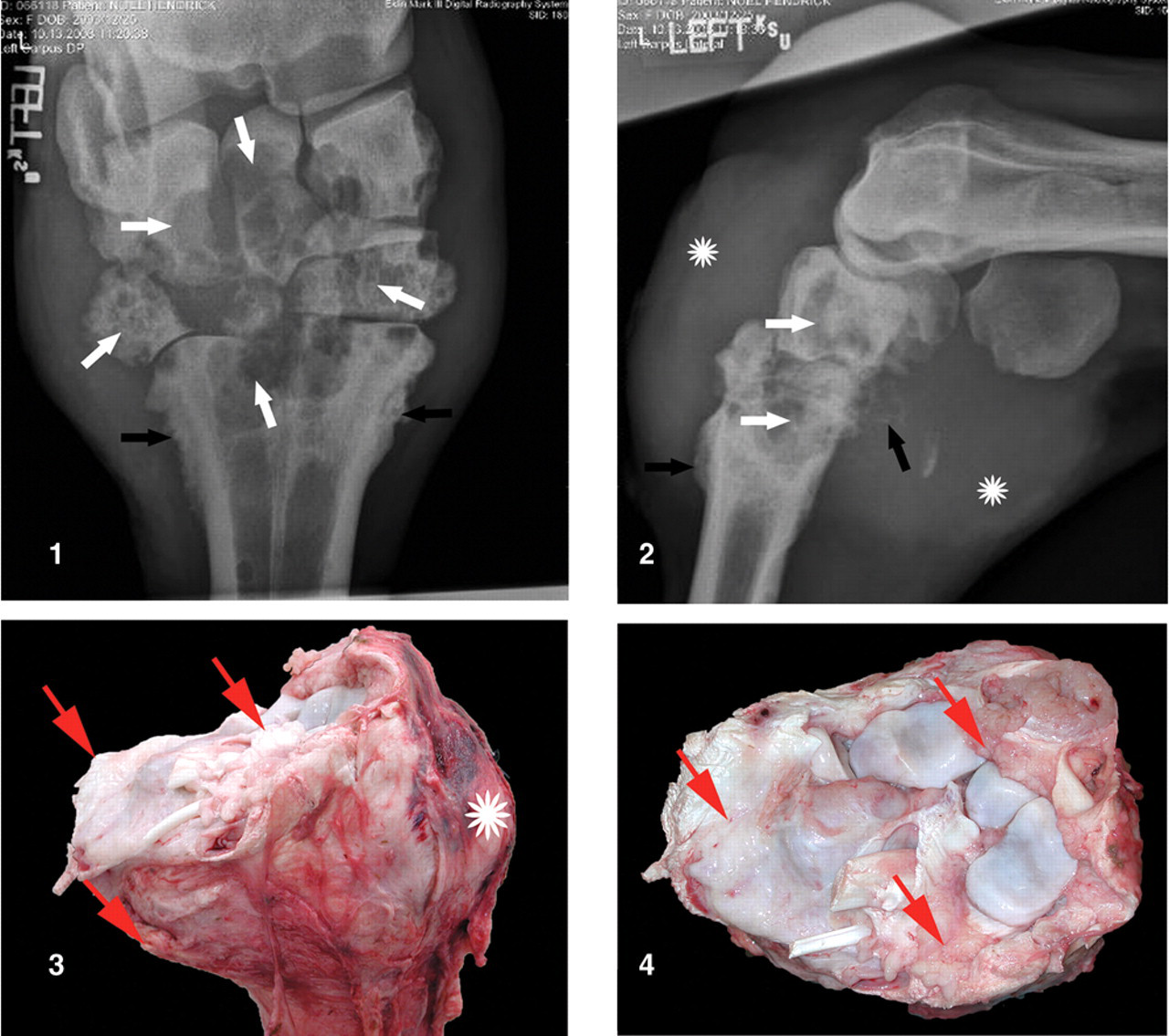
Chondrosarcoma; dromedary camel (Camelus dromedarius). Cranial to caudal projection radiograph of the carpus. The bones of the proximal and distal carpal rows are abnormal as noted by multifocal, multiform radiolucent areas (white arrows). Note the presence of proliferative new bone formation (black arrows) and minimal disruption of articular surfaces. Figure 2. Chondrosarcoma; dromedary camel (Camelus dromedarius). Lateral to medial projection radiograph of the carpus. The bones of the proximal and distal carpal rows are abnormal as noted by multifocal, multiform radiolucent areas (white arrows). Note proliferative new bone formation (black arrows) and extensive soft tissue enlargement centered on the middle carpal joint and carpometacarpal joint (asterisks). Figure 3. Left carpal joint, chondrosarcoma, lateral view; dromedary camel (Camelus dromedarius). A mass is located on the palmar aspect of the left carpal joint and within the left carpal joint (arrows). Figure 4. Also note the masses involving the dorsal proximal and dorsal distal aspect of the carpal metacarpal joint (asterisk). Left carpal joint, chondrosarcoma, dorsal view; dromedary camel (Camelus dromedarius). A multilobulated mass surrounds and invades into the left carpal joint (arrows).
The camel was in good body condition. The left carpal joint and the surrounding soft tissues were markedly swollen. A 12 cm × 10 cm × 7 cm, white to pale pink, firm mass was located on the palmar aspect of the left carpal joint, as well as within the left carpal joint, infiltrating all carpal bones and the proximal region of the metacarpal bone (Figs. 3, 4). Two similar masses (4 cm × 3 cm and 2 cm × 3 cm, respectively) were present on the dorsal proximal and dorsal distal aspect of the carpal metacarpal joint, respectively. The masses were multilobulated on both external and cut surface. The joint capsule and the synovium were diffusely thickened (Figs. 3, 4). The articular surfaces of the carpal bones and the metacarpal bone were multifocally eroded. The lungs contained approximately 50, firm, raised, gray, 3–5 mm in diameter, randomly distributed nodules (Fig. 7). The uterus contained a fetus that was in the third trimester of gestation. There were no other significant findings in any of the other organs.
Tissue samples for histopathology were routinely processed and stained with hematoxylin and eosin. In the carpal bones, metacarpal bone, and surrounding soft tissue, there was an unencapsulated, poorly delineated, invasive neoplasm composed of islands of chondrocytes suspended in variable amounts of cartilage matrix (Fig. 5). The islands of chondrocytes were separated by dense fibrous connective tissue, which was multifocally infiltrated by lymphocytes, plasma cells, and macrophages. Individual neoplastic chondrocytes had variably distinct cellular borders, abundant eosinophilic cytoplasm, and a round eccentrically placed nucleus containing coarsely stippled chromatin. Occasional mitotic figures were present (Fig. 5). In a few places, the chondrocytes contained 2–9 nuclei, and there was marked anisocytosis and anisokaryosis. The neoplasm invaded the carpal bones (Fig. 6) and metacarpal bone resulting in lysis of the bone. There was no osteoid present in any of the sections. The lung contained multifocal neoplastic nodules with similar cell features as described above (Fig. 8). Based on the cellular atypia, multinucleation, presence of mitotic figures, and absence of osteoid production, a diagnosis of chondrosarcoma with infiltration of carpal and metacarpal bones, and pulmonary metastasis, was made. The diagnosis of chondrosarcoma was further supported by immunohistochemistry using vimentin and S-100 antibodies. For vimentin, sections were incubated with prediluted mouse monoclonal anti-vimentin antibody a and an enhanced alkaline phosphatase red detection kit. b For S-100, the sections were incubated with polyclonal rabbit anti–S-100 antibody c (1:400) and goat anti-rabbit biotinylated antibody d (1:200) followed by an enzyme detection kit. e The antigen–antibody complex was visualized using 3,3’-diaminobenzidine peroxidase substrate kit. f Diffuse cytoplasmic staining for vimentin and S-100 was observed in the neoplastic cells (Fig. 5, insets). Similar staining has been reported in chondrosarcomas. 10
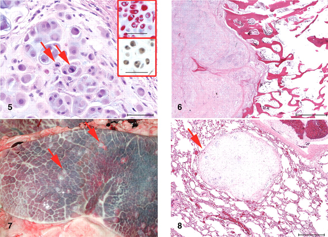
Chondrosarcoma; dromedary camel (Camelus dromedarius). The neoplasm is composed of nests of chondrocytes suspended in the basophilic cartilage matrix. Two mitotic figures are present in the neoplastic chondrocytes (arrows). Hematoxylin and eosin. Bar = 50 µm. In the insets, diffuse cytoplasmic staining for vimentin (red) and S-100 (brown) in the neoplastic chondrocytes is shown. Bar = 50 µm. Figure 6. Chondrosarcoma; dromedary camel (Camelus dromedarius). The chondrosarcoma is invaded into the carpal bone replacing the trabecular bone. Hematoxylin and eosin. Bar = 2 mm. Figure 7. Lung; dromedary camel (Camelus dromedarius). The lungs contain multiple, firm, raised, gray, 3–5 mm diameter, randomly distributed nodules of metastatic chondrosarcoma (arrows). Figure 8. Lung; dromedary camel. The lung contains a well-delineated nodule of metastatic chondrosarcoma (arrow). Hematoxylin and eosin. Bar = 500 µm.
According to the World Health Organization’s classification, chondrosarcoma in human beings is classified into primary and secondary based on the origin of the neoplasm; primary chondrosarcoma arises from a previously normal bone while a secondary chondrosarcoma is a progression from a benign lesion such as osteochondroma. 8 A similar classification scheme is followed in animals. 13 Primary chondrosarcoma is further classified into central (arising within the bone) and periosteal chondrosarcoma. 13 Chondrosarcomas in other locations are referred to as extraskeletal chondrosarcomas. In the current case, the masses were present adjacent to the carpal joint and within the joint. Therefore, it is difficult to speculate and classify the neoplasm as primary or secondary, or as extraskeletal. In dogs, the most common locations of chondrosarcoma include rib, appendicular skeleton, and nasal cavity. 6 In sheep, chondrosarcoma has been reported to most commonly involve cartilage in the sternocostal junction. 12 In cows, chondrosarcomas more frequently involve flat bones. 9 In a report of chondrosarcoma in a goat, the neoplasm involved the proximal third of the humerus. 12 Mandibular chondrosarcoma has been described in a single Bactrian camel. 11 In the present case, the neoplasm was present around the left carpal joint, which is not a very common location for chondrosarcoma. According to a recent review of tumors of the hand in human beings, based on the tumors registered in the Basel Bone Tumor Reference Center from1972–2008, the malignant neoplasms involving the hand are extremely rare. 1 But, among the 37 malignant neoplasms reported, 28 were chondrosarcomas. However, most involved metacarpal bones or phalanges, and none involved carpal joint. Only one report in a horse, which had chondrosarcoma of distal radius with infiltration into the carpal joint and carpal bones, was found. 2
The histological features considered while classifying a cartilage neoplasm into benign or malignant include cells with large nucleus, large cells with multiple nuclei, and presence of even a single mitotic figure. 5,13 In the present case, the neoplasm contained all these features and hence was classified as a malignant neoplasm. Human chondrosarcomas are graded as 1, 2, and 3 based on the cellularity, nuclear features, mitotic figures, degree of differentiation, matrix, and necrosis. 4,8 The histological grade is the most important indicator of local recurrence and metastasis. 4,8 However, in animals, the histologic grading of chondrosarcomas is not used routinely. Grading is used in occasional studies and appears to be a useful prognostic indicator. 4,14 Based on the histological criteria, the current neoplasm can be classified as a grade 2 chondrosarcoma. Although metastasis is not very common in chondrosarcomas, it does occur in approximately 25% of the cases in dogs 14 and mainly involves the lungs. 13 In other species, although chondrosarcoma is rare, metastasis to lung has been reported in a cow, a Bactrian camel, and a goat. 9,11,12 Similarly, metastasis to the lung was observed in the present case. Based on the clinical history and gross lesions, metastasis probably occurred later in the clinical course in the current case.
Chondrosarcoma in camels may be underreported because of the scarcity of camels examined at postmortem. However, the incidence is low in other domestic species with the exception of dogs. 13 Clinically, chondrosarcoma and/or osteosarcoma was not suspected in the present case because of the frequency of septic arthritis and trauma as opposed to joint neoplasia, which has not been reported for the species. Although the current case was not a diagnostic challenge, it is an important report from the clinician’s perspective. Based on the review of existing literature, this is the first report of a chondrosarcoma in a dromedary camel.
Footnotes
Acknowledgements
The authors thank Ms. Jennifer Hill in the histology laboratory for her help with the immunohistochemistry.
a.
Dako North America Inc., Carpinteria, CA.
b.
Ventana Medical Systems Inc., Tucson, AZ.
c.
Dako North America Inc., Carpinteria, CA.
d.
Vector Laboratories Inc., Burlingame, CA.
e.
Vectastain Elite ABC kit, Vector Laboratories Inc., Burlingame, CA.
f.
Vector Laboratories Inc., Burlingame, CA.
The authors declared that they had no conflicts of interest with respect to their authorship or the publication of this article.
The authors declared that they received no financial support for their research and/or authorship of this article.