
Abstract
Following unilateral enucleation, 4 Domestic Shorthair cats with an average age of 12.5 years (range: 9–16 years) were histologically diagnosed with a presumed primary intraocular chondrosarcoma at the Comparative Ocular Pathology Laboratory of Wisconsin (Madison, Wisconsin). Medical records and follow-up were available for 3 of the 4 cats. Clinically, only 1 eye was affected in each cat; a mass lesion was noted in 2 cats, and a neoplasm was suspected in the other 2 cats. Grossly, 3 tumors presented as coalescing, poorly demarcated, white, friable masses filling the vitreous and intraocular chambers; 1 tumor presented as a solitary, well-demarcated, tan mass involving the iris and ciliary body. Histologically, all 4 neoplasms were composed of haphazardly arranged plump neoplastic spindle cells surrounded by irregular islands and thick trabeculae of abundant, variably basophilic, and Alcian blue–positive chondromatous matrix. None of the cats presented histologically or clinically with signs suggestive of feline posttraumatic ocular sarcoma. Two cats are still alive and healthy 6 months and 3 years following enucleation. One cat died 6 months following enucleation; however, this cat suffered from poorly controlled diabetes mellitus, and the cause of death is undetermined. No other tumors or skeletal lesions were identified that could suggest a metastatic tumor to the eye. The origin of primary intraocular chondrosarcoma is unclear, but is presumed to be ocular multipotent mesenchymal stem cells. Four cases of intraocular chondrosarcoma in cats not associated with the posttraumatic sarcoma complex of intraocular tumors are described.
Keywords
Chondrosarcomas are uncommon malignant neoplasms with predominating cartilaginous matrix 4 that account for the second most common primary skeletal tumor after osteosarcoma in both veterinary and human medicine.7,11 The average age of cats affected by chondrosarcomas is 9.6 years, and there is a slight male predilection for the development of the tumors. 4 Extraskeletal chondrosarcomas, and visceral ones in particular, are rare tumors in dogs, 8 cats, 4 and people. 11 In a case series of 67 cats with chondrosarcomas, 47 were tumors of skeletal origin and 20 presented in subcutaneous sites, several of which were thought to be vaccine-site sarcomas. 4 Intraocular chondrosarcoma is reported in 2 dogs as a primary 9 or metastatic 10 neoplasm. Metastatic intraocular chondrosarcoma was also reported in 1 human patient. 5 In 1959, a single account was published describing an intraocular chondrosarcoma in a cat presenting with multiple intraocular lesions. 1 To the authors’ knowledge, there are no other reports of feline intraocular chondrosarcomas in the literature.
The Comparative Ocular Pathology Laboratory of Wisconsin (COPLOW; Madison, Wisconsin) is a referral laboratory focused on the diagnosis of ocular pathology in animal patients. The laboratory maintains more than 40,000 histological samples from submitted ocular tissues, all filed in a searchable database. Following a diagnosis of primary intraocular chondrosarcoma in a feline globe, the COPLOW database was searched for feline patients diagnosed with intraocular cartilaginous tumors from 1998 to 2014. Within the database, 9,000 submitted specimens were from feline ocular tissues, and 4,800 of those were neoplasms. Sixteen feline globes submitted and diagnosed with intraocular chondrosarcomas were identified. Twelve cats presented with either a history of ocular trauma or histological features supportive of a traumatic ocular event (e.g., lens capsule rupture, corneal or scleral perforation) and were diagnosed with feline posttraumatic ocular chondrosarcoma. Four globes presented with a purely cartilaginous neoplasm and no medical history or findings supportive of a metastatic tumor or traumatic event. These 4 cases were presumed to be primary intraocular chondrosarcomas.
Cats 1–4 had available submission forms including medical background and signalment. All 4 cats had been subjected to a complete ophthalmic examination by veterinary ophthalmologists certified by the American College of Veterinary Ophthalmologists by the time of referral. Complete medical records and follow-up information were available for cats 1–3. The 4 cats were Domestic Shorthair cats; cats 1, 2, and 4 were spayed females; and cat 3 was a neutered male. The cats had an average age of 12.5 years (range: 9–16 years). All 4 cats presented with unilateral ocular disease. Clinically, a mass lesion was seen in cats 1 and 3 (Fig. 1), and neoplasia was suspected in cats 2 and 4 where flocculent white material was seen in the anterior chamber or vitreous. Cats 2–4 were systemically healthy, and cat 1 suffered from poorly controlled diabetes mellitus. The duration of intraocular disease from observation to enucleation averaged 6 weeks (range: 3–9 weeks). No other tumors or skeletal lesions were identified.
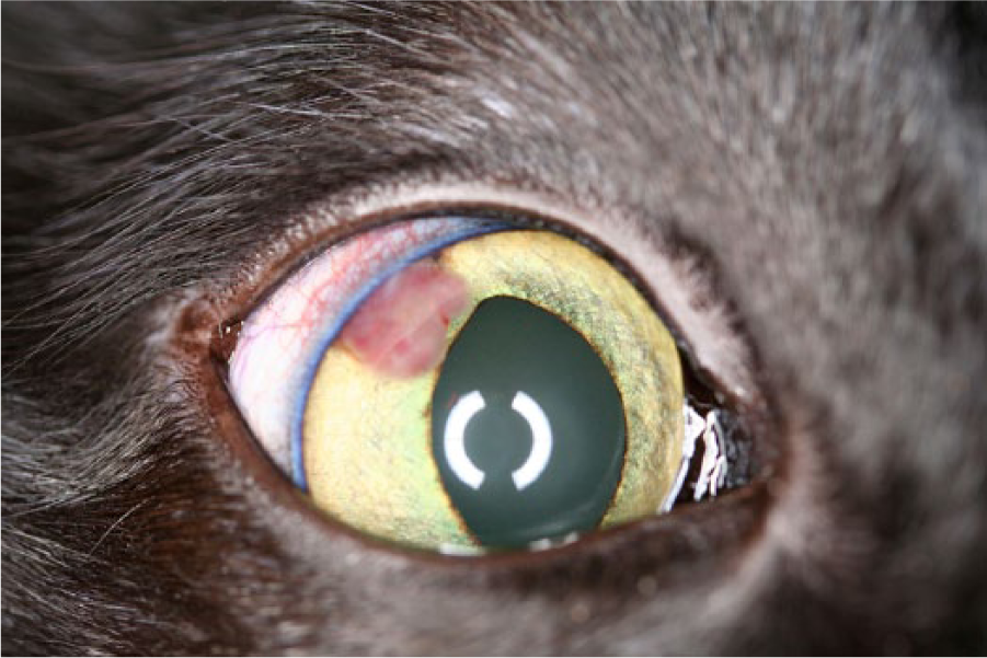
Clinical image of the right eye of cat 3 with presumed primary intraocular chondrosarcoma, which presented clinically and histologically as a solitary mass lesion. The mass is seen at the dorsolateral aspect of the iris, extending from the 10 to 12 o’clock position. It is tan and moderately vascular, and extends into the anterior chamber. The remainder of the eye appears within normal limits. (Courtesy Dr. Eric Smith.)
Grossly, tumors from cats 1, 2, and 4 presented as poorly demarcated, white, friable masses that partially effaced the vitreous and anterior and posterior chambers (Fig. 2A, 2B); the tumor from cat 3 presented as a well-demarcated, white mass involving the iris and ciliary body. Histology was available for the 4 cats, and cytology was available for cat 3. Histological and cytological evaluations were all performed by veterinary pathologists certified by the American College of Veterinary Pathologists.
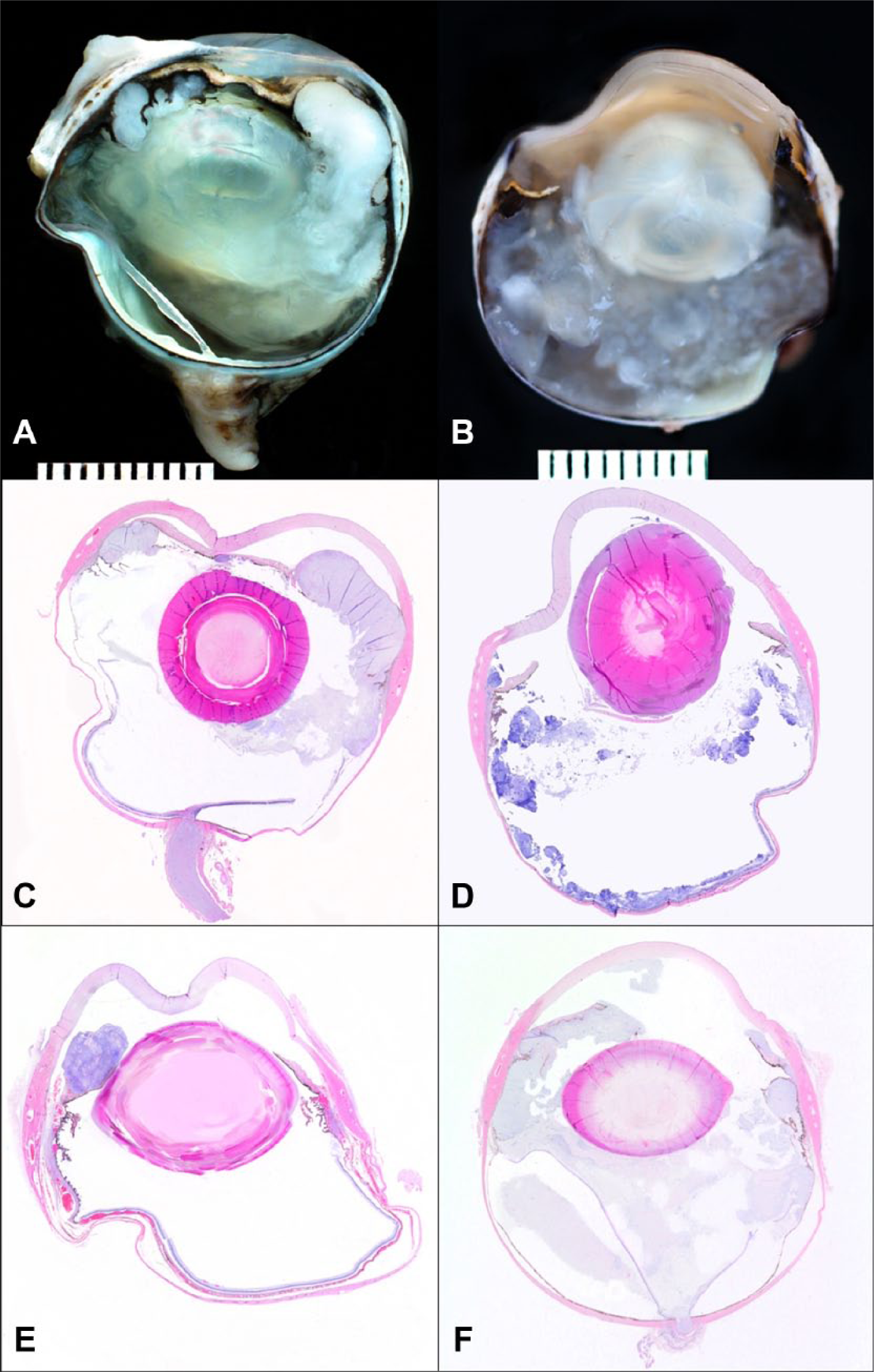
Gross and subgross images of the globes of cat 1 (
A transcorneal fine-needle aspiration was taken from the mass of cat 3 that presented as a solitary lesion. Cytology revealed moderate numbers of monotypic, pleomorphic neoplastic cells and lightly eosinophilic extracellular matrix. Cells were round to oval or spindle-shaped with variable amounts of basophilic cytoplasm. Nuclei were round to oval with coarse to clumped chromatin and 1–2 prominent nucleoli. Anisokaryosis, binucleated, and multinucleated cells were seen. Cytological differential diagnoses were amelanotic melanoma or anaplastic sarcoma, and histology was recommended.
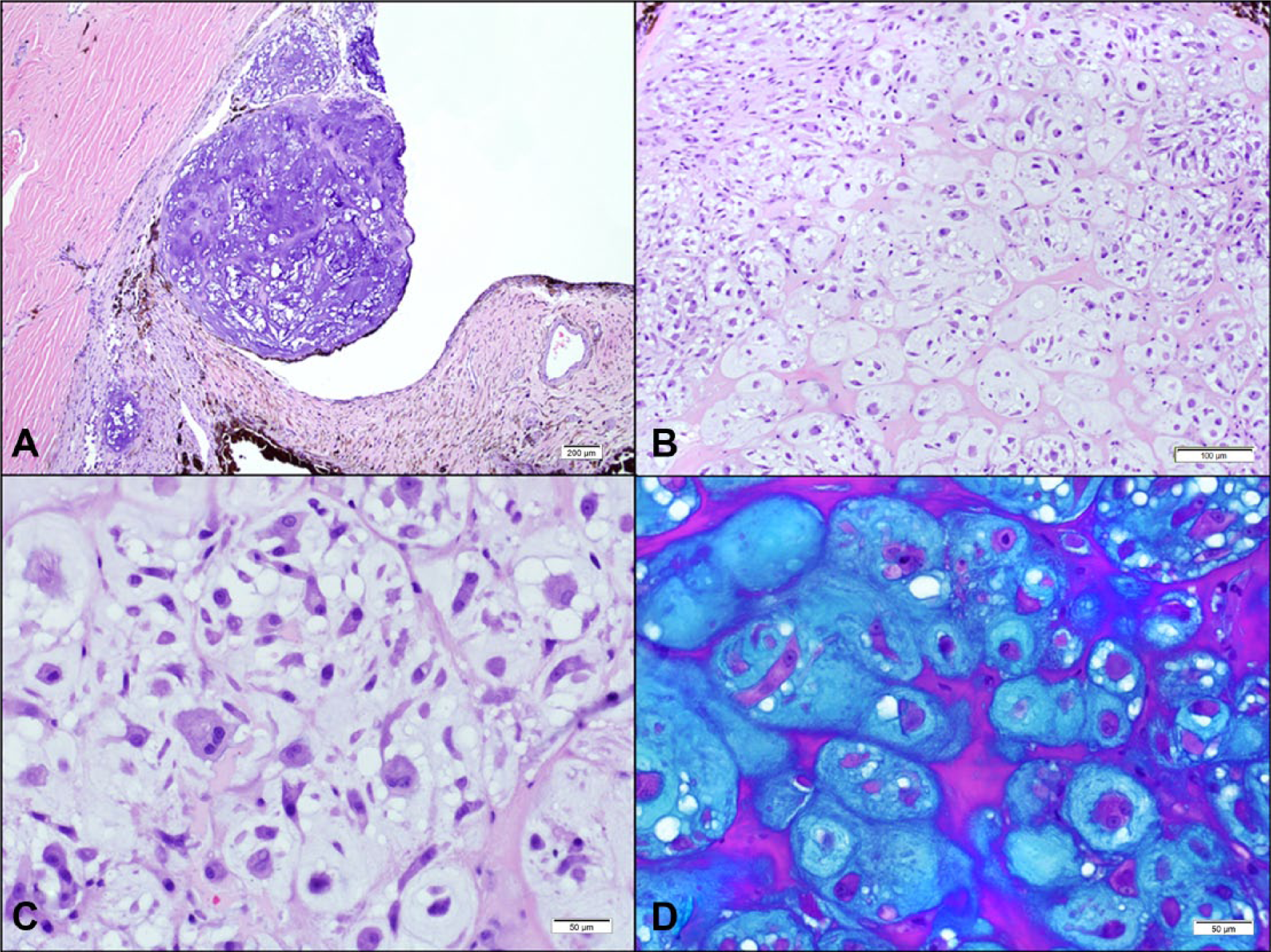
Following routine paraffin embedding, standard 5-μm tissue sections from each globe were stained with hematoxylin and eosin and Alcian blue periodic acid–Schiff. Histologically, the globe from cat 3 (Fig. 1) presented with a solitary, well-demarcated, nonencapsulated mass (Fig. 2E). The mass was highly cellular and composed of round to polygonal neoplastic cells haphazardly embedded in an abundant lightly basophilic and Alcian blue–positive amorphous extracellular material interpreted as cartilaginous matrix (Fig. 3B–3D). Neoplastic cells presented with a finely stippled chromatin and often 1 single prominent nucleolus. Cellular pleomorphism was marked with multiple cells presenting karyomegaly and prominent nuclear pseudoinclusions. Three mitotic figures were seen in 10 high-power fields. The remaining 3 globes presented with a different neoplastic distribution but similar cellular morphology. The neoplastic tissue carpeted the intraocular surfaces and variably effaced the intraocular chambers, causing compression and atrophy of the adjacent uveal and retinal tissues (Fig. 2C, 2D, 2F). Neoplastic cells also multifocally infiltrated the trabecular meshwork, iris and ciliary body stroma, retina, and optic nerve (Fig. 3A). These 3 cases presented abundant Alcian blue–positive cartilaginous extracellular matrix and focally extensive areas of tissue necrosis ranging from 10% to 30% of the neoplastic tissues.
Histological images showing neoplastic cells within the anterior chamber, and ciliary cleft (
All 4 globes presented with glaucoma either clinically or histologically. The globes from cats 1, 2, and 4 had histological features consistent with glaucoma, including diminished numbers of ganglion cells in the inner retina, optic nerve atrophy, and optic nerve cupping. Preiridal fibrovascular membranes were present in all 4 globes, 2 of which (cats 2 and 4) also presented with a peripheral anterior synechia. The globes from cats 1 and 2 presented with a cortical cataract interpreted by the presence of Morgagnian globules and liquefaction of lens fibers within the lens cortex, as well as posterior migration of the lens epithelium. No evidence of lens capsule rupture was observed in the reported cases. Hyphema was present in the globe from cat 3.The globe from cat 4 presented with complete retinal detachment and a mild to moderate lymphoplasmacytic anterior uveitis. None of the cases had clinical presentations that supported a diagnosis of feline posttraumatic ocular sarcoma nor was the histology compatible with such a diagnosis.
Six months and 3 years following enucleation, cats 2 and 3, respectively, are still alive and healthy. Cat 1 died 6 months following enucleation; however, this cat suffered from poorly controlled diabetes mellitus, and the cause of death is undetermined. Cat 4 was lost to follow-up.
Following careful examination of the case reported in 1959, 1 it is likely that it represented a case of feline posttraumatic ocular sarcoma prior to the recognition of this condition in 1990 3 rather than a primary tumor. In the 1959 case, the tumor was reported to fill the globe, invade the cornea, destroy the lens and iris, and only lens capsule remnants were seen. The neoplasm was also reported to exhibit ossification, and tumor cells were seen within choroidal and scleral vessels. The cat died 2 weeks following enucleation, and a necropsy was not performed. This description is compatible with feline posttraumatic ocular chondrosarcoma with regards to invasiveness, lens capsule rupture, ossification, and perhaps poor prognosis, although the cause of death is undetermined. Feline posttraumatic ocular neoplasms can appear with different morphological features, but invariably involve ocular trauma. Osseous metaplasia is reported in the literature describing feline posttraumatic ocular sarcoma. 3 Chondromatous metaplasia is yet to be reported in the literature in association with feline posttraumatic ocular sarcoma; however, it is recognized at the COPLOW as a sole entity or in conjunction with osseous metaplasia. The cases presented in the current study are reports of intraocular chondrosarcoma in cats not associated with the posttraumatic ocular sarcoma complex.
Cats, similar to all mammals, do not have cartilage or bone within their globes and, therefore, the tissue of origin for these tumors is unclear. One case of primary intraocular chondrosarcoma is reported in a blue discus (Symphisodon aequifasciatus), 6 which could have arisen from naturally occurring cartilage in fish eyes. In mammals, it is likely that primary intraocular chondrosarcomas arise from ocular multipotent mesenchymal stem cells or cancer stem cells. 13 In human eyes, multipotent mesenchymal stem cells have been derived from the trabecular meshwork. 12 The trabecular meshwork could be a likely origin of the tumors seen in the current cases, all of which showed extensive involvement in the vicinity of the iris, ciliary body, and ciliary cleft (Fig. 3C). These trabecular stem cells were differentiated in vitro to chondrocytes, as well as adipocytes and osteocytes. 12 Vascular pericytes derived from the bovine retina were also stipulated to have the potential to give rise to osteoblasts and chondrocytes, 2 offering another possible origin for the tumors described herein.
In summary, primary intraocular chondrosarcomas in cats are rare, comprising 4 out of 4,800 feline ocular neoplasms within the COPLOW collection. Primary intraocular chondrosarcomas may appear as well-demarcated, solitary masses, or as flocculent coalescing lesions. All of the cases in the present study were associated with glaucoma and preiridal fibrovascular membrane formation. Although only a small number of cases were available and only 3 had a complete follow-up, it appears that surgical excision by enucleation may provide favorable survival rates. The favorable outcome could be explained by the fact that the tumors were well contained within the globe in all of these cases. In all 4 globes, the chondrosarcomas primarily compressed, rather than infiltrated, ocular tissues and did not extend beyond the globe. Primary intraocular chondrosarcomas likely arise from mesenchymal stem cells within the globe; however, this mechanism has not been elucidated.
Footnotes
Declaration of conflicting interests
The author(s) declared no potential conflicts of interest with respect to the research, authorship, and/or publication of this article.
Funding
The author(s) declared that they received no financial support for their research and/or authorship of this article.