
Abstract
An outbreak of an acute disease in buffalo (Bubalus bubalis) caused by the ingestion of Baccharis megapotamica var. weirii occurred in the southern region of Brazil. Ten out of 50 buffalo died 24–48 hr after being introduced into a pasture containing abundant amounts of the plant. Factors influencing the ingestion of the plant and consequent toxicosis included hunger, stress caused by shipment, and unfamiliarity with the plant. Clinical signs included serous ocular discharge, incoordination, mild bloat, and muscle trembling. One buffalo was necropsied. Gross findings included dehydration, abundant liquid in the rumen, reddening of the mucosa of forestomachs, abomasum, and intestine, and edema of the wall of the rumen. The main histologic lesions were superficial to full thickness degeneration and necrosis of the stratified epithelium lining the forestomachs, necrosis of the intestinal mucosa, and widespread lymphoid necrosis. A calf (Bos taurus) was fed a single dose of 5g/kg/body weight of B. megapotamica var. weirii harvested from the same site where the buffalo died. Twenty hours after the administration of the plant this calf died with clinical signs and lesions similar to those observed in the naturally poisoned buffalo.
The Baccharis genus (Asteraceae: tribe Asteraceae) includes nearly 500 species. All are found in the New World with the exception of Baccharis halimifolia, which was introduced into Australia from the United States. 12 This species is suspected of poisoning cattle in both countries 9 and proved toxic when administered experimentally to chicks. 8 Baccharis glomerulifolia, another North American species, was toxic to mice and chicks 8 under experimental conditions, and Baccharis pteronioides has been associated with cattle poisoning in the southwestern United States. 18,26 Baccharis pteronioides toxicosis was produced in hamsters dosed with 100–200 mg of the plant. 26 Baccharis artemisioides causes disease in cattle in a restricted zone of Argentina northwest of Buenos Aires and southeast of Cordoba. 22
Nearly 120 species of Baccharis have been recorded in Brazil; of those, only Baccharis coridifolia 4,27 and Baccharis megapotamica 7,19,28 have been proven to be toxic to livestock. Both B. megapotamica and B. coridifolia are found in southern Brazil, but they occupy different habitats; B. megapotamica is found in marshy areas 28 whereas B. coridifolia grows in pastureland. 4 Two varieties of B. megapotamica with essentially the same distribution and toxic effects on livestock are known, namely B. megapotamica var. megapotamica and B. megapotamica var. weirii. 28
Baccharis coridifolia and the 2 varieties of B. megapotamica cause a severe acute poisoning in livestock characterized by degeneration and necrosis of the epithelial lining of gastrointestinal tract and necrosis of lymphocytes in lymph nodes, spleen, tonsils, and several lymphoid aggregates. 4,27,28,30,31 Baccharis megapotamica, 15 B. coridifolia ,6,10 and B. artemisioides 22 contain a series of potent cytotoxic agents belonging to the macrocyclic trichothecene complex of antibiotics previously believed to be produced only by fungi. These cytotoxic substances were demonstrated to be the toxic principles in these plants. 13,31 In the case of B. megapotamica, the macrocyclic trichothecenes accumulate in the plant as baccharinoids, which are named B1, B2, B3, B4, etc. B4 is the most abundant baccharinoid, but there are also large concentrations of B1–B8.13 To date, no macrocyclic trichothecenes have been demonstrated in B. halimifolia, B. pteronioides, or B. glomerulifolia.
Spontaneous poisoning by B. coridifolia occurs frequently in cattle, 21 occasionally in sheep, 24 and rarely in horses. 2 Spontaneous outbreaks involving B. megapotamica var. weirii have been reported only once in cattle, 7 and once in sheep. 19 There are also some anecdotal accounts of spontaneous toxicosis by B. megapotamica var. weirii in cattle. Typically, the toxicosis in livestock occurs when naïve animals raised in areas free of Baccharis spp. are transferred to pastures infested by the plant. The risk of toxicosis increases considerably if the animals are subjected to such stress factors as fatigue, hunger, or thirst. 4 Interestingly, cattle that are raised in pastures where Baccharis spp. exist will graze it very rarely if ever, 4 although in the case of B. megapotamica var. weirii there are anecdotal accounts that particularly hungry cattle familiar with the plant, may, on occasion, ingest it and get poisoned. The present report describes a spontaneous outbreak of B. megapotamica var. weirii in water buffalo (Bubalus bubalis).
Fifty 3- to 4-year-old water buffalo of both sexes were purchased from a farm (farm 1) in southern Brazil and shipped by truck to another farm (farm 2) 300 km away. The buffalo were kept in a holding pen on farm 1 for 12 hr without food or water, immediately prior to being transported for 8 hr to farm 2. As a result, the animals had no food or water for a total of 20 hours. Upon arrival at farm 2, the animals were released into a 200-hectare pasture that already held 200 cattle (Bos taurus) that had been there for several months. Ten buffalo died within 24–48 hr of being placed in the pasture. Most of the affected buffalo were found dead, but the owner was able to observe one moribund buffalo that had serous ocular discharge, incoordination, mild bloat, and muscle trembling. This buffalo was necropsied 2 hr after death. Gross findings included a dehydrated cadaver with a markedly distended rumen with abundant liquid content. There was mild to moderate edema of the wall of the rumen, particularly in the pillars, along with diffuse reddening of the mucosa of the forestomachs (Fig. 1), abomasum, and intestine (Fig. 2).
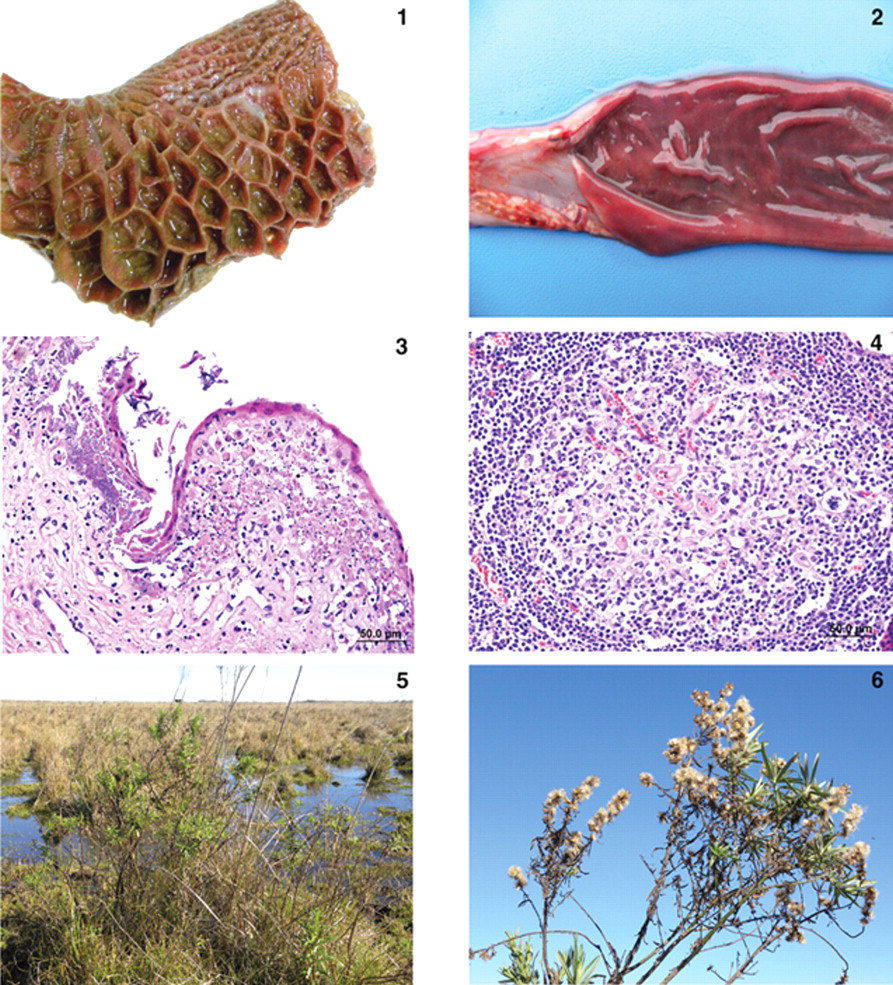
Diffuse reddening of the mucosa of the reticulum of a water buffalo (Bubalus bubalis) naturally poisoned by Baccharis megapotamica var. weirii. Figure 2 . Diffuse reddening of the intestinal mucosa and hemorrhagic intestinal content in a water buffalo (Bubalus bubalis) naturally poisoned by Baccharis megapotamica var. weirii. Figure 3 . Photomicrograph of the rumen of a water buffalo (Bubalus bubalis) naturally poisoned by Baccharis megapotamica var. weirii. Notice the extensive necrosis affecting the entire thickness of the stratified epithelium. Basophilic aggregates (bacteria) can be observed at left attached to the necrotic epithelial lining. Hematoxylin and eosin. Bar = 50 µm. Figure 4 . Photomicrograph of a mesenteric lymph node of a water buffalo (Bubalus bubalis) naturally poisoned by Baccharis megapotamica var. weirii. Notice the extensive lymphoid necrosis in secondary germinal centers. Hematoxylin and eosin. Bar = 50 µm. Figure 5 . A specimen of Baccharis megapotamica var. weirii is observed in the first plane of the photography. The plant was in association with marshy land in the pasture where the water buffalo were poisoned. Figure 6 . Female specimen of the plant Baccharis megapotamica var. weirii.
Significant microscopic changes were found in the forestomachs, lymph nodes, spleen, lymphoid aggregates, and liver. Epithelial cells lining the forestomachs displayed coagulative necrosis, the severity of which varied, so that in some segments the more superficial cells of the stratified epithelium were affected, sparing deeper cells. In some instances, these deeper cells had hydropic degeneration. In some other segments, coagulative necrosis affected the entire thickness of the stratified epithelium. Myriads of bacterial aggregates could be observed attached to segments of necrotic epithelial lining (Fig. 3). Necrosis of the intestinal mucosa was also observed. Mesenteric lymph nodes had necrosis of lymphocytes in secondary germinal centers (Fig. 4). The subcapsular, trabecular, and medullary sinuses were filled with macrophages, and erythrophagocytosis was conspicuous. Lymphoid necrosis was also observed in the white pulp of the spleen. Gut-associated lymphoid tissue and other lymphoid aggregates were not available for histology. Hepatic lesions consisted of intense hepatocellular vacuolization with eosinophilic globules (Councilman bodies), and marked dilatation of lymphatics in portal triads. The lumina of these lymphatics were filled by faint eosinophilic homogenous material.
The pasture in farm 2 where the 10 water buffalo died was inspected carefully, and several spots of marshy land were observed (Fig. 5). These areas contained large amounts of a plant later identified as B. megapotamica var. weirii (Fig. 6), and there was evidence that livestock had been consuming these plants. Baccharis megapotamica was absent from farm 1.
The plant was harvested on-site, and the fresh green leaves were fed to an 8-month-old, 120-kg Bos taurus calf as a single dose of 5 g/kg/body weight (bw). The calf died 20 hr after the plant was fed, after displaying clinical signs similar to those seen in the water buffalo. Necrotic lesions similar to those seen in the water buffalo were present in the lining epithelium of forestomachs and in the lymphoid tissue including mesenteric, hepatic, and gastric lymph nodes, tonsils, and ileal mucosal-associated lymphoid tissue.
The diagnosis of B. megapotamica toxicosis in the buffalo was made based on the characteristic acute clinical disease, the presence of the plant in large amounts in its characteristic habitat, and the experimental reproduction of the disease by feeding the plant present in the pasture to a calf. Baccharis megapotamica var. megapotamica and var. weirii had been previously experimentally fed to calves, 28 and lethal doses were determined to be between 3 and 4 g/kg/bw for var. megapotamica and 1 g/kg/bw for var. weirii. Fatal poisoning was acute with both varieties. The most important postmortem findings were edema of the ruminal wall along with congestion of the rumen, abomasum, small intestine, cecum, and colon. Histologically, the rumen showed necrosis characterized by pyknosis and karyorrhexis of epithelial cells, mainly of the stratum spinosum. Lymphoid tissue (spleen, lymph nodes, Peyer patches) showed necrosis characterized by pyknosis and karyorrhexis of the lymphoid cells. Thus, the disease observed in the spontaneous outbreak in buffalo was essentially the same as described previously in cattle 7,28 and sheep. 19
Livestock will not usually ingest either variety of B. megapotamica 28 but a combination of hunger, dehydration, and lack of familiarity with the plant most likely led to the lethal ingestion of B. megapotamica var. weirii. In the southern region of Brazil there are reports of the spontaneous poisoning by 4 plants that induce similar disturbances in the gastrointestinal tract and should be included in the differential diagnosis, namely B. coridifolia, B. megapotamica var. weirii, Baccharidastrum triplinervium, and Eupatorium tremulum. The toxic principle of the latter 2 plants is as yet undetermined. Poisoning caused by the first 2 plants is virtually clinically and pathologically indistinguishable; however, the habitats of B. megapotamica var. weirii (marshy areas) and B. coridifolia (dry pastureland) differ 4,28 and this could help to differentiate between the 2 intoxications. Additionally, lymphoid necrosis is reportedly less severe in cases of B. coridifolia poisoning 28 and E. tremulum, 17 and does not occur in the poisoning caused by B. triplinervium. 16 Lesions in the forestomachs are much less severe in B. triplinervium poisoning 16 than in the poisonings caused by the other 3 plants. Larger amounts (20–30 g/kg/bw) of E. tremulum 17 and B. triplinervium 16 must be consumed to induce disease and death in cattle; this is translated in much lower mortality ratios for these 2 plants when compared to Baccharis spp. toxicosis. To confirm the diagnosis of poisoning by any of these plants, it is important to find evidence of plant consumption.
From the standpoint of the morphological aspects of the lesions in the forestomachs, B. megapotamica var. weirii poisoning closely resembles ruminal acidosis. However, ruminal acidosis usually follows the ingestion of excess carbohydrate in the form of grain or other fermentable foodstuffs, and is associated mainly with intensive beef and dairy production 5 and not with cattle at pasture.
The cause of death in cases of B. megapotamica poisoning is unknown. However, since the disease is virtually identical to B. coridifolia poisoning, comparisons can be made. Baccharis-induced death is believed to be caused by dehydration and acid-base imbalance resulting from fluid loss into the ruminal compartment in a similar manner to what happens in ruminal acidosis. 3,21 The finding of myriads of bacteria attached to the necrotic ruminal mucosa in cases of B. coridifolia and B. megapotamica poisoning, even in animals freshly dead, suggests the possibility that bacteremia could play a role in the mechanism of death. 21
Trichothecenes are terpenoids, which can be divided into 2 groups: the simple trichothecenes (e.g., deoxynivalenol [DON], diacetoxyscirpenol [DAS], and T-2 toxin) and the macrocyclic trichothecenes (e.g., baccharinoids, roridins, and verrucarins). They exhibit a wide range of biological activity, which includes dermatonecrosis, gastroenteritis, feed refusal, coagulopathy, and immunosuppression. 12,15,20 Trichothecenes are also potent phytotoxins, and the macrocyclics are particularly toxic to plants. 12 T-2 toxin and DAS are highly toxic, causing necrosis of mucous membranes (mouth, pharynx, esophagus, rumen, stomach) on contact, 14 similar to lesions produced by plant-associated macrocyclic trichothecene poisoning. In this regard, the lymphoid necrosis associated with baccharinoid-induced poisoning has a close morphological resemblance with the lymphoid necrosis induced by T-2 toxin, and extracts of B. megapotamica were used in treatment trials of B-cell leukemia in rats. 15 The cytotoxicity of trichothecenes is attributed to ribosomal binding and subsequent inhibition of protein synthesis in actively dividing cells of lymph nodes, spleen, bone marrow, and thymus. 23 Induction of apoptosis in these cells by the trichothecenes is likely to contribute to lesion expression. 11,25,29 Changes in cell membrane structure, with resultant lipid peroxidation due to amphophilic trichothecene molecules, inhibition of RNA and DNA synthesis, and inhibition of mitosis are additionally recognized deleterious effects of T-2 toxin on cells. 23 Although the mode of action of macrocyclic trichothecenes in B. megapotamica on subcellular levels is not completely determined, macrocyclic trichothecenes are believed to act by compromising protein synthesis by inhibiting the peptide bond formation step, 1 and it is fair to assume that mechanisms associated with all types of trichothecene toxicoses are similar.
Interesting results that could shed some light in the pathogenesis of Baccharis-induced toxicosis stemmed from experimental B. pteronioides poisoning in hamsters. 26 Hamsters in the highest dosed group (200 mg) developed multiple hemorrhagic infarcts in the liver and kidney, with severe hemorrhagic enteritis and severe necrotizing vasculitis with vascular thrombosis of hepatic and renal vessels associated with fibrin thrombi in glomerular capillaries. The authors of the hamster study 26 compared their findings to those of bacterial endotoxin–produced vasculitis and infarction. However, neither vasculitis nor fibrin thrombi were found in the cases reported herein; however, studies to clarify the exact pathogenesis of B. megapotamica–induced toxicity are ongoing.
Footnotes
The authors declared that they had no conflicts of interest with respect to their authorship or the publication of this article.
This work was financially supported by the Brazilian agency National Council of Scientific and Technologic Development (CNPq), grant no. 473493/2010-1.