
Abstract
A challenge faced by veterinary diagnosticians in serologic analysis for exposure to pathogens is the need for a protein conjugate capable of antibody attachment in many animal species. The advent of protein conjugates that are less specific in nature allows diagnosis across many species with little or no modification of technique. Toxoplasma gondii is an organism of veterinary interest that has been demonstrated to infect a plethora of warm-blooded animals. However, the serologic tests available for simultaneous diagnosis in this broad range are limited in number. The current study examined the use of an immunoglobulin G enzyme-linked immunosorbent assay (ELISA) modified by the use of non-species-specific protein conjugates in domestic animal species commonly submitted to diagnostic laboratories for evaluation of Toxoplasma exposure status. Comparison with results from an established indirect hemagglutination technique revealed very good agreement between the 2 test methods. This modification of the ELISA provides a useful method for veterinary diagnosticians to perform rapid and accurate evaluation of multiple animal species for Toxoplasma exposure using a single test.
Introduction
Toxoplasma gondii is a protozoal organism that can cause clinical disease in both definitive and intermediate hosts and is believed to be one of the most common parasitic infections in the world. 14 Toxoplasmosis has been described in human beings and numerous wild and domestic animals, including birds, canids, felids, rodents, and ruminants. 15 The risk of congenital transmission with potentially severe consequences for the fetus in acutely infected pregnant women has generated great public interest in this organism, as has emerging recognition of its importance in causing disease in immunocompromised patients. 16 In France, concern about congenital toxoplasmosis is such that pregnant women are tested monthly to detect the presence of recent infection. 18 Felids have been demonstrated to be the definitive host for the organism, while a great diversity of vertebrates, including human beings and many domestic and wild animals, have been found to be intermediate hosts.
Infection of the definitive felid host can be acquired through predation or scavenging of meat from an infected intermediate host via tissue cysts, by ingestion of oocysts shed by an infected felid, or by congenital transmission from an infected queen. Infection is established in the intestinal tract where asexual and sexual reproduction takes place, leading to fecal shedding of oocysts, potentially accompanied by more generalized infection. Intermediate hosts can similarly be infected by either ingestion of oocysts or tissue cysts. In intermediate and dead-end hosts, sporozoites from ingested oocysts or bradyzoites from ingested tissue cysts enter the wall of the small intestine. Tachyzoite development leads to systemic spread and invasion of numerous organs where bradyzoite-containing tissue cyst development proceeds. 7,11,12,17,19 The complexity of the life cycle of Toxoplasma and the relatively short period of oocyst shedding from the definitive host have led to the development of numerous serologic techniques to diagnose infection. The ideal serologic test for diagnosis in the veterinary setting is objective, has high throughput potential, and demonstrates both high sensitivity and specificity in numerous species.
Clinical signs in definitive and intermediate hosts due to infection with Toxoplasma vary in severity from negligible to terminal disease. A great deal of concern in both human and animal health involves the risk of abortions and fetal malformations in the presence of acute infection. Among domestic animals, abortion in sheep and goats is widely noted, while significance in cattle is less well defined, likely due to the similarity to manifestation of Neospora caninum infection. 10 Most cats are believed to remain asymptomatic upon initial infection, though some display nonspecific signs such as fever, dyspnea, and abdominal pain. 8 Space-occupying lesions created by enlarging tissue cysts can produce disease, related to their site of localization in the vertebrate intermediate host, notably causing neurologic and/or ocular deficits.
The relatively brief period of oocyst shedding in the definitive felid host, combined with the persistent immunoglobulin G (IgG) antibody response in both definitive and intermediate host infections, make serologic evaluation the method of choice for detecting exposure to Toxoplasma in all potential host species. 3-7,9 Techniques that have been commonly used include the Sabin–Feldman dye test, latex agglutination, hemagglutination assays, fluorescent antibody tests (FATs), and enzyme-linked immunosorbent assays (ELISAs). Some of these are hampered in the veterinary diagnostic environment by the use of live organism (Sabin–Feldman) or by the requirement for species-specific conjugate (FAT and ELISA). In the current study, the results of a commercially produced indirect hemagglutination assay were compared with those of a commercially available IgG ELISA modified to detect IgG antibody in multiple animal species.
Material and methods
Animals and samples
Blood samples were collected into clot tubes from animals by referring veterinarians. The extracted serum samples were submitted to the Animal Health Diagnostic Center at Cornell University (Ithaca, New York) from 2001 to 2008 for detection of Toxoplasma exposure by indirect hemagglutination. After storage at 5°C, positive samples with a sufficient amount of serum remaining and a sampling of negative samples were reassessed using the modified IgG ELISA technique in 2008–2009. The sera tested included samples from 46 alpacas, 26 cats, 53 dogs, 39 goats, 21 horses, and 42 sheep.
Indirect hemagglutination assay
Seroreactivity to Toxoplasma antigens was initially evaluated using an indirect hemagglutination assay (IHA). a The commercial preparation of the IHA had perfect qualitative agreement with the Center for Disease Control and Prevention (CDC) Toxoplasma indirect FAT in their evaluation of human serum samples. 23 Serum containing anti-Toxoplasma IgG antibody causes agglutination of the sheep red blood cells coated with antigen as part of the test procedure. 2,13 The test procedure distinguishes these reactions from nonspecific agglutination through the use of both coated and noncoated red blood cells. Interpretation is based on observation of presence or absence of a red blood cell mat covering the bottom of the test well on completion of incubation. The test is equally effective for human and animal diagnostic work, as no species-specific conjugate is required for antibody detection. 22 This procedure was used as a reference test to evaluate the performance of the modified IgG ELISA.
The IHA was prepared by adding 25 μl of sample diluent (buffered saline containing 0.1% sodium azide and 1% normal rabbit serum) to the bottom of as many U-shaped wells on a 96-well microdilution test tray as required and adding an equal volume of the liquid to be tested. Two rows were used to test the reactivity of the negative control (normal human serum) and 2 were used to test the reactivity of the positive control (anti-Toxoplasma human serum or globulin). Two rows were reserved for each sample to be tested. Sample diluent was tested for agglutination. The negative control was tested for agglutination at ratios of 1:32 and 1:64. The positive control was tested at ratios of 1:32 to 1:4,096 dilutions. Samples were initially screened from 1:32 to 1:256 and were tested at higher dilutions if positive at all of these dilutions. Sheep red blood cells sensitized with a Toxoplasma extract were added to 1 row of each control and sample group. Unsensitized sheep red blood cells were added to the other row. The plate was shaken on a vibrating shaker briefly and then allowed to incubate at room temperature for 3 hr. A positive result was indicated by the formation of a smooth mat of cells covering the bottom of the well, with no such reaction noted in the unsensitized row. The latter reaction indicates the presence of nonspecific agglutinins and invalidates the test. A negative result produces a collection of red blood cells in a small group in the center of the bottom of the well. Serum samples that produced agglutination in the 1:64 well were considered positive for anti-Toxoplasma IgG antibody.
Enzyme-linked immunosorbent assay
A commercial IgG ELISA b was modified by use of protein A c or protein G conjugate d to replace the conjugated goat anti-human IgG included in the kit. A similar method was used to create an ELISA for detection of anti-Toxoplasma antibody in sheep samples using protein G. 21 Samples, controls, and a calibrator were evaluated after dilution 1:21 with kit-supplied diluent (containing Tween-20, bovine serum albumin, and phosphate buffered saline). These were added to kit wells that had been sensitized by passive absorption with T. gondii antigen. Any antigen-specific antibody contained in the sample binds to the immobilized antigen. Protein A or G conjugate, depending on the vertebrate species from which the sample was collected, was added to each well to attach to any immobilized IgG antibody. Protein A conjugate was used for canine and feline samples, whereas protein G conjugate was used for the remaining species. Following washing, microwells were then incubated with peroxidase substrate solution, which changes color when hydrolyzed by the presence of peroxidase. Color intensity of the sample indicated antibody concentration in the microwell. This was evaluated using a microplate reader with light at a wavelength of 450 nm. Comparison to kit-provided calibrators guided the identification of positive versus negative serum, with a ratio of sample color intensity to kit calibrator color intensity of greater than or equal to 1.1 indicating a positive reaction for anti–T. gondii IgG antibody as described by the kit insert. A ratio between 0.9 and 1.1 (noninclusive) was considered an indeterminate result and less than or equal to 0.9, a negative result.
Protein A and protein G conjugate
Protein A and protein G horseradish peroxidase conjugates were used in the IgG ELISA assays instead of the kit conjugate to allow testing of multiple animal species. Protein A conjugates are reported to provide strong binding to antibody of cats, dogs, mice, pigs, and rabbits, whereas protein G conjugates are reported to provide strong binding to IgG antibody of cows, goats, guinea pigs, horses, mice, pigs, rabbits, and sheep. d Protein G conjugate also showed strong binding to alpaca antibody on testing with this procedure (unpublished data). Both proteins were received as lyophilized samples and were diluted 1:20,000 with phosphate buffered saline (pH 7.2) prior to use, with the dilution determined by testing against kit controls to satisfy quality control parameters. Similar methods have been used with these proteins and a chimera of the 2 proteins in previous studies. 1,20,24
Data analysis
The significance of association between the results of the IHA and the ELISA was evaluated using the McNemar chi-square test. The degree of agreement between the results from the 2 tests was quantified using κ statistics. The accuracy of the ELISA test in detecting exposure to Toxoplasma was evaluated in comparison to the IHA and measured using the relative sensitivity and specificity. The expected performance of the ELISA at different cut-off points was examined using the receiver operating characteristic (ROC) curves for all data and additionally separated by animal species.
Results
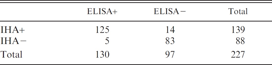
Serum samples were evaluated from multiple animal species using both the IHA and the IgG ELISA as described. The protein G conjugate replacement was used to analyze alpaca, caprine, equine, and ovine sera. Protein A conjugate was used to analyze canine and feline sera. The sera tested included samples from 46 alpacas, 26 cats, 53 dogs, 39 goats, 21 horses, and 42 sheep. There was no significant difference detected between positive and negative results when comparing IHA and ELISA results with McNemar chi-square (P = 0.07). The associated κ value using the kit standard cut-off parameters was 0.83 (95% confidence interval [CI]: 0.75–0.90 calculated using GraphPad e ), indicating very good agreement between the 2 test results. The combined sensitivity of the protein A/G–modified ELISA kit was found to be 90% (95% CI: 83–94%) and specificity 95% (95% CI: 87–98%) when the IHA results were utilized as a gold standard, and positive versus negative ELISA results were determined using the manufacturer's guidelines. It is noted that adjusting the range of indeterminate results to 0.8–1.0 ratios improves sensitivity based on this data to 92% from 90%, while specificity remains at 95% (Figs. 1, 2), and should be considered for animal samples. The prevalence of positives in the samples tested was 59% (Table 1); at a prevalence of 10% (the approximate fraction of positive samples submitted to the Animal Health Diagnostic Center based on IHA results), the calculated positive predictive value would be 67% with a negative predictive value of 99%. Table 2 documents the range of test performances by species using the kit-recommended parameters for distinguishing positive from negative results. In addition, ROC curves are provided for all results collectively and by species to evaluate the ELISA's performance when compared with the IHA (Figs. 1–3).
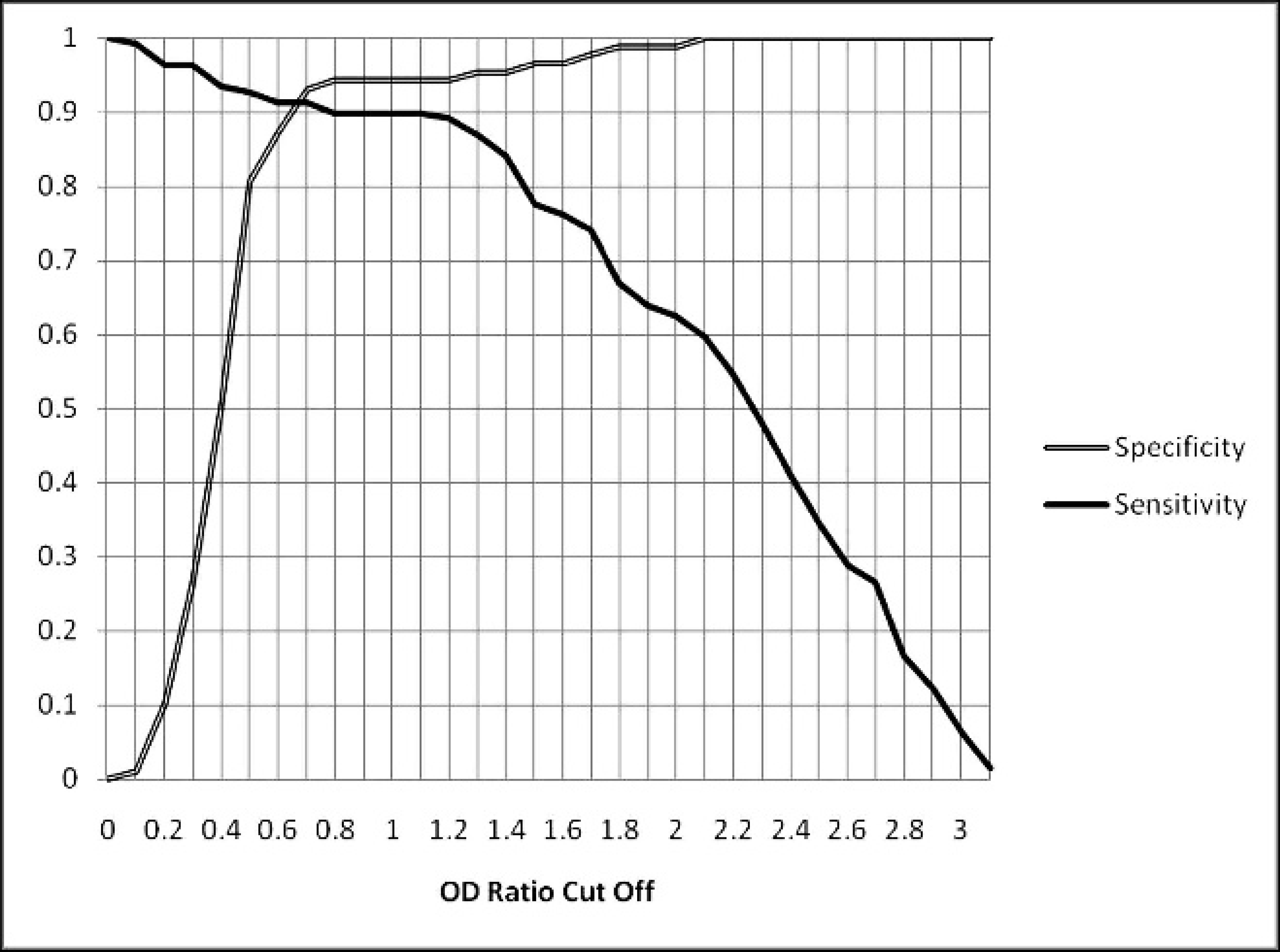
Effect of enzyme-linked immunosorbent assay optical density (OD) ratio cut-off value on relative sensitivity and specificity.
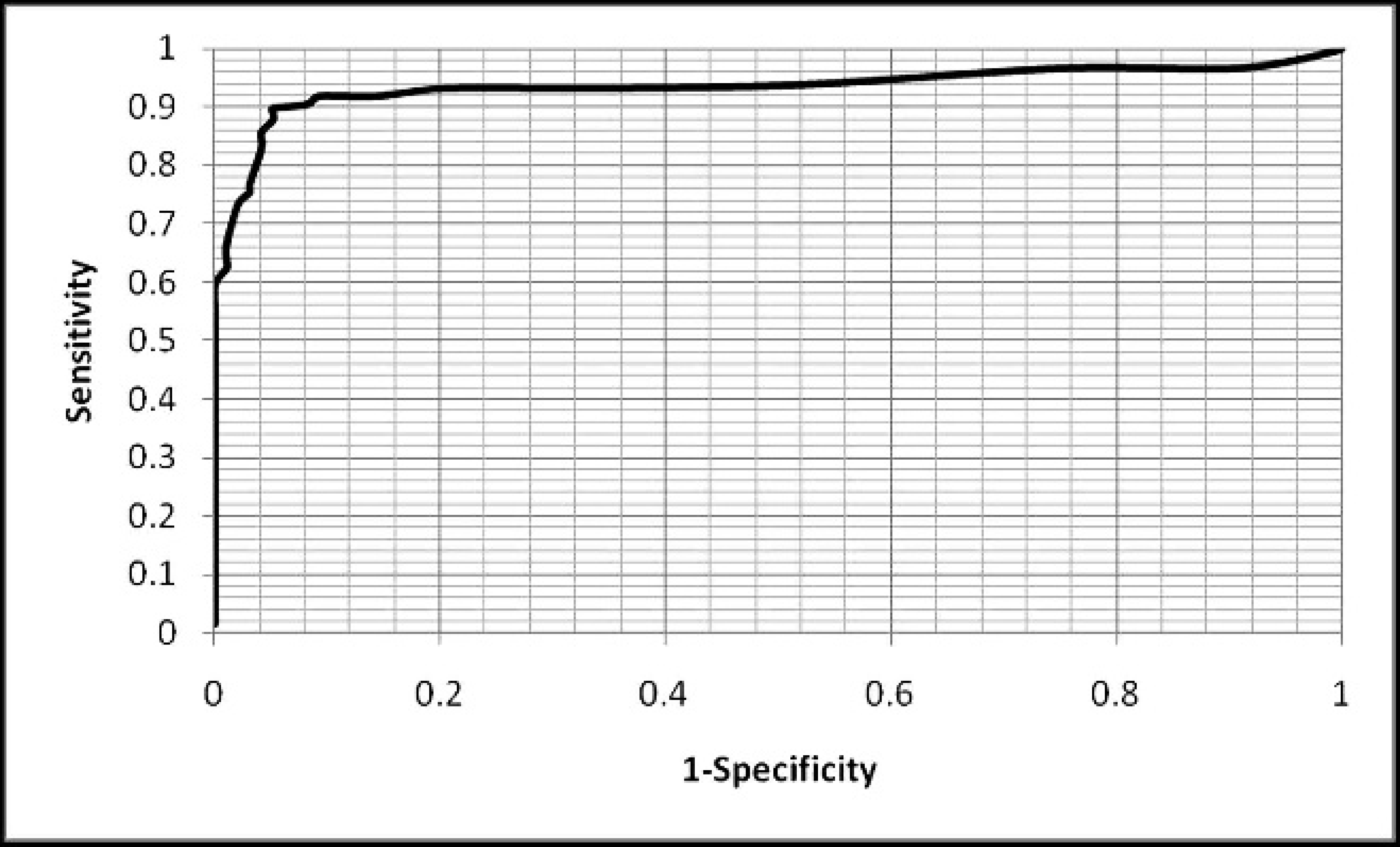
Receiver operating characteristic calculation for modified immunoglobulin G enzyme-linked immunosorbent assay.
Two by two comparison of indirect hemagglutination assay (IHA) to modified immunoglobulin G enzyme-linked immunosorbent assay (ELISA) testing.
Discussion
Antemortem diagnosis of toxoplasmosis presents a challenge owing to the nonspecific nature or lack of clinical signs. Serologic evaluation provides a useful tool by the ease of access to the requisite sample and its high sensitivity to detection of recent and chronic infection. However, care must be used in linking an animal's serologic status to active illness because of this same ability to detect historic infection. The cessation of production of a commercially available IHA kit in the United States has led to a critical gap in diagnostic options for serologic diagnosis of Toxoplasma infection across a wide range of animal species, notably intermediate hosts like goats and sheep in which toxoplasmosis can cause significant economic losses. The use of ELISAs for similar detection has been limited by the requirement for species-specific anti-IgG conjugate to produce valid results. The successful testing of an ELISA that uses conjugates effectively across many of the animal species of interest provides an additional tool for veterinary diagnostic laboratories to use in determining an animal's serologic status.
Both the previously available IHA and the currently available IgG ELISA allow detection of IgG in serum samples, and both provide an antigen substrate to which antibody present in the serum will attach. Beyond that, the 2 procedures utilize quite different technology to quantify antibody in a given sample. The IHA uses antigen-coated red blood cells to which antibody attaches for detection, while the ELISA uses U-shaped wells coated with antigen. The IHA results depend on the subjective determination by the operator of the extent of agglutination in an individual reaction to establish a maximum positive dilution. The interpretation of ELISA results do not depend on subjective evaluation by the operator, as the optical density in the reaction well is quantified by a microtiter plate reader.
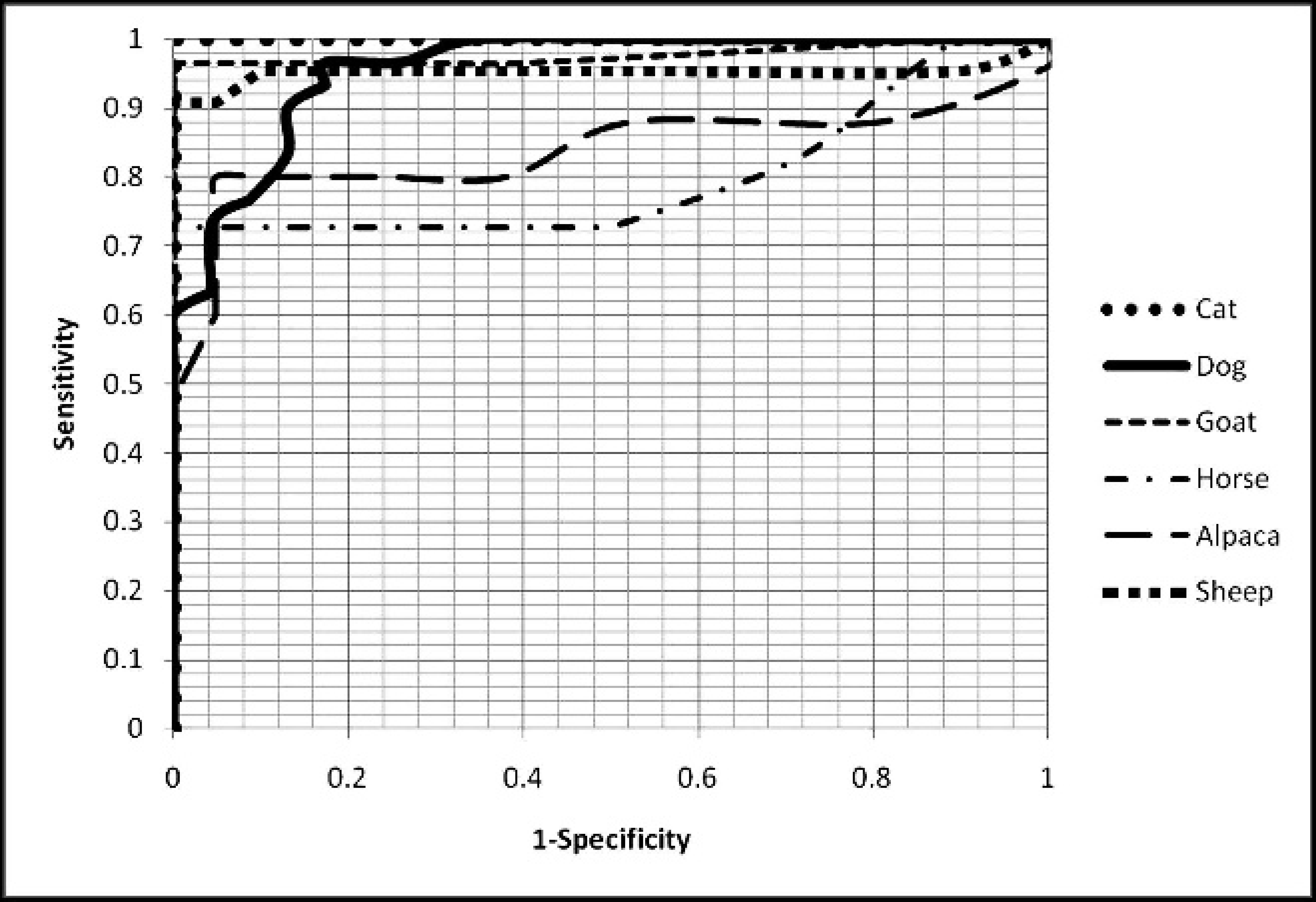
Receiver operating characteristic calculation by species.
In the present experiment, 227 serologic samples from diverse animal species were evaluated pairwise for the presence of anti-Toxoplasma IgG antibody both by the IHA and the IgG ELISA modified with protein A or G techniques. Results indicated very good agreement between the 2 tests in determining positive versus negative samples, indicated by a κ value of 0.83. While good agreement is also noted as indicated by κ values for results in each species, evaluation of horse and alpaca samples showed lower sensitivities than those of other species (0.72 and 0.76, respectively), which should be noted in future use of this method. The modified ELISA described herein provides a strong alternative for veterinary diagnostic laboratories in detecting exposure to Toxoplasma by serologic evaluation across a range of commonly tested animal species.
Acknowledgements
The authors thank Linda Baitman (Wampole Laboratories) for assistance in securing the requisite materials for the study; and Dr. Bettina Wagner and Aziza Solomon for providing feline serum samples. This research was made possible by the United States Department of Agriculture federal formula fund grant NYC-478430.
κ value calculation by species.*
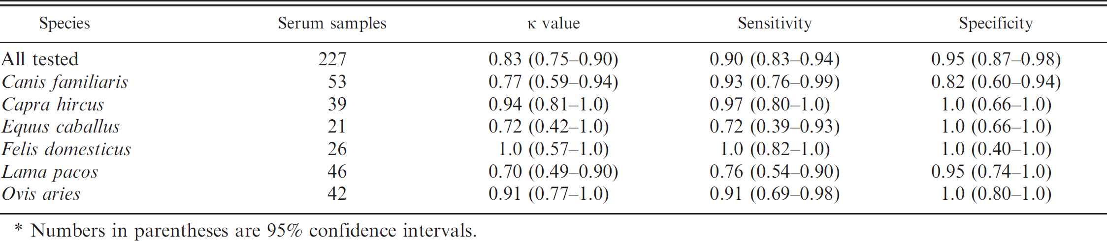
Numbers in parentheses are 95% confidence intervals.
Footnotes
a.
Toxoplasmosis TPM-Test indirect hemagglutination kit, Wampole Laboratories, Princeton, NJ.
b.
Toxo IgG II ELISA kit, Wampole Laboratories, Princeton, NJ.
c.
Protein A peroxidase from Staphylococcus aureus/horseradish, Sigma-Aldrich, St. Louis, MO.
d.
Protein G horseradish peroxidase conjugate, Molecular Probes, Invitrogen Corp., Carlsbad, CA.
e.
GraphPad Software Inc., La Jolla, CA.