
Abstract
Since 2004, there have been several reports of Influenza A virus (FLUAV) infection in dogs. Dogs have been infected with equine influenza H3N8, avian influenza H3N2 and H5N1, and the pandemic H1N1 virus. Because ofrecent avian and equine influenza outbreaksin Italy, the objectives of the present study were to estimate the level of exposure of Italian dogs to influenza A viruses and to assess a diagnostic algorithm for detection of FLUAV exposure in dogs. Sera collected from 6,858 dogs from 2006 to 2008 were screened in a competitive enzyme-linked immunosorbent assay (cELISA) for antibodies to the highly conserved influenza A nucleoprotein. Samples positive in the cELISA were confirmed by testing in hemagglutination inhibition (HI) and fluorescent antibody test (FAT). Two seropositive dogs had antibodies to H3 hemagglutinin proteins, consistent with exposure torecent canine and equine subtype H3N8 viruses. Using a Bayesian model, the sensitivity and specificity of the cELISA were estimated as 93.98% (probability intervals [PI]: 81.67–99.08%) and 98.71% (PI: 98.43–98.96%), respectively. After accounting for the imperfect sensitivity and specificity ofthe cELISA, the Bayesian posterior prevalence of FLUAV exposure among tested Italian dogs was 0.5% (PI: 0.1–1.4%). The study results indicate that screening with a cELISA for influenza A nucleoprotein antibody, followed by confirmatory testing with HI and/or FAT, is a highly sensitive and highly specific approach for diagnosing FLUAV exposure in dogs.
Keywords
Introduction
Since the recognition of canine Influenza A virus (FLUAV) subtype H3N8 (CIV H3N8) in the United States, there has been renewed interest in the potential role of dogs in the ecology of influenza A viruses. In other countries, serological surveys 14,15 of dogs for evidence of exposure to CIV H3N8 have been conducted with largely negative results; however, additional transmissions of influenza A viruses from multiple species to dogs have been reported. Retrospective studies implicated equine FLUAV subtype H3N8 as the cause of isolated respiratory outbreaks in dogs in the United Kingdom in 2002 and 2003 12,20 and in Australia, 13 where dozens of dogs in contact with infected horses developed influenza-like illness during the 2007 equine influenza outbreak.
During the spread of highly pathogenic avian FLUAV subtype H5N1 throughout Asia, a dog in Thailand died after ingesting an H5N1-infected duck. 1,25 A subsequent serosurvey 5 revealed that 25% of 629 asymptomatic dogs had been exposed to the virus in Thailand. The transmission of an entire avian FLUAV subtype H3N2 to dogs in South Korea was identified in 2007, 23 and additional studies 16,24 support the sustained transmission of this avian-origin H3N2 virus between South Korean dogs. Since its appearance in April 2009, pandemic H1N1 has also been reported in dogs in both the United States and China [ProMED-mail: 2009, PRO/AH/EDR>Influenza pandemic (H1N1) 2009, animal (40): USA (NY) canine. Available at http://www.promedmail.org/pls/apex/f?p=2400:1000, archive number 20091222. 4305. Accessed July 17, 2010; ProMED-mail: 2009, PRO/AH/EDR>Influenza pandemic (H1N1) 2009, animal (30): China, canine. Available at http://www.promedmail.org/pls/apex/f?p=2400:1000, archive number 20091128.4079. Accessed July 17, 2010]. Clearly, these examples confirm the susceptibility of dogs to influenza virus infection.
Serological studies 11,15–17,21,23 of influenza A exposure in dogs have been conducted by means of enzyme-linked immunosorbent assay (ELISA) and/or hemagglutination inhibition (HI) assay as screening and confirmatory tests, respectively. The sensitivity (Se) and specificity (Sp) of these assays under unknown field conditions have not been reported for canine sera. Several ELISA kits for influenza A nucleoprotein have recently been developed. Most commercially available kits are indirect ELISAs, which have been validated for select species. Others are based on a competitive ELISA (cELISA) format and could theoretically be used for detection of serum antibodies to avian or mammalian influenza A viruses in any species. No ELISA kits, however, have been developed, validated, or authorized for use with canine sera. The HI assay is universally considered the gold standard for serological diagnosis of influenza A viruses in animals, and it generates indirect evidence of the viral hemagglutinin subtype to which an animal has been exposed. However, its reliability is dependent on a variety of factors, including the receptor affinity of the virus (α-2,3 or α-2,6); the quality, concentration, and origin of the erythrocyte suspension used in the assay; and the homology of antigen used with the circulating virus. 6,19 In addition, the HI assay is a time-consuming and labor-intensive test that has the major diagnostic characteristic of being subtype specific and, thus, is not suitable for a serosurvey aimed at detection of multiple virus subtypes.
Since 2000, several FLUAV outbreaks have occurred in poultry (H7N1, H7N3, H5N2) and horses (H3N8) in Italy, 7,8,18 providing ample opportunities for dogs to be exposed to these viruses by direct or indirect contact. A recent study, 22 which investigated the potential exposure of 637 dogs and cats to influenza A viruses in northeastern Italy using an in-house ELISA, did not identify any seropositive animals. To date, however, there have not been any studies that have comprehensively investigated the potential exposure of dogs to influenza A viruses in Italy. The objectives of the present study were to estimate the level of exposure of Italian dogs to influenza A viruses and to assess a diagnostic algorithm for detection of FLUAV exposure in dogs using a combination of serological tests, including cELISA, HI, and fluorescent antibody test (FAT). A Bayesian analysis was used to determine the Se and Sp of the cELISA for identifying FLUAV exposure in dogs with unknown potential for contact with infected animals.
Materials and methods
Estimating the level of exposure of Italian dogs to influenza A viruses
Samples. Serum samples collected from pet dogs in Italy from 2006 to 2008 were originally submitted to the Istituto Zooprofilattico Sperimentale delle Venezie (IZSVe; Legnaro, Padova, Italy) for assessment of rabies vaccine efficacy. Serum samples from 6,858 dogs that were at least 1 year of age and represented all 20 regions of Italy were selected for testing for antibodies to influenza A viral proteins (Fig. 1). Serum samples had been stored at −20°C prior to testing. The selected samples included 2,041 dogs from 2006; 2,038 dogs from 2007; and 2,779 dogs from 2008. The total sample size is equivalent to accepting an error in the point estimates of 0.15%, assuming an expected prevalence of 0.4%, a 95% confidence level, and a population size that approximates to infinity. For example, if the true prevalence was 0.4% and the diagnostic test was perfect, this sample size would provide detection of a prevalence value between 0.25% and 0.55% with a 95% level of confidence.
A panel of 97 convalescent serum samples collected from racing greyhounds naturally infected with A/canine/FL/04 (H3N8) served as reference sera in the study. These samples were previously confirmed 11 as seropositive for antibodies to the H3 protein in HI and virus neutralization assays. The reference sera served as positive controls in all assays. Sera collected from specific-pathogen-free dogs housed in a barrier research facility served as negative controls.
Diagnostic tests. Testing for antibody to influenza A nucleoprotein was performed using a commercially available cELISA. a In addition to its high throughput format, this assay was selected as the initial screening test because it detects exposure to any FLUAV strain, regardless of the subtype. The test was performed according to the manufacturer's instructions. Samples were diluted at a 1:10 ratio in the provided dilution buffer. Positive and negative control sera provided in the kit as well as a reference positive and negative canine serum sample were included in each test plate.
All Italian dog sera that had positive or doubtful results in the cELISA (competition percentage greater than 45%) were tested in a FAT, as an influenza type A–specific confirmatory test. An in-house FAT was developed to detect antibodies to influenza A viral proteins in infected cells. Briefly, Madin–Darby canine kidney cells were cultured in Eagle minimum essential medium with 10% fetal calf serum and 1% L-glutamine. b A 50-μl volume of cell culture suspension (1.5 × 10 4 cells/well) was dispensed into 96-well plates c and incubated for 24 hr at 37°C (±2°C) in the presence of 5% CO2. Plates were washed with Earle balanced salt solution d to remove residual calf serum. The wells were inoculated with 1 multiplicity of infection of A/canine/FL/04 (H3N8) diluted in Eagle minimum essential medium containing 1% L-glutamine and 0.01% trypsin. d When the beginning of cytopathic effects appeared at 24 hr postinfection, the cells were fixed with 80% acetone (v/v in sterile distilled water). As a result of the small amount of available sera, samples were tested in singlet. A reference positive and negative canine serum sample was tested on each plate. Positive and negative reference sera were also tested in wells containing uninfected cells to control for nonspecific reactions. Starting at a dilution of 1:8, 10 serial log2 dilutions of each canine serum sample were prepared in phosphate buffered saline (PBS). Briefly, 50 μl of each serum dilution was added to the wells and the plates were incubated for 30 min at 37°C and then washed 3 times with PBS. Specific anti–influenza A antibodies in tested sera were detected with fluorescein isothiocyanate (FITC)–conjugated rabbit anti-dog immunoglobulin G b diluted 1:5,000 in Evan Blue solution as the contrast dye. A 50-μl volume of the FITC-conjugated detection antibody was added to each well and incubated for 30 min at 37°C, followed by 3 washes with PBS. Using a fluorescent microscope, serum samples were considered positive for influenza A antibodies based on the presence of green foci in the cellular membrane and cytoplasm on a brown–red background. The endpoint FAT titer was defined as the reciprocal of the last dilution of serum that produced fluorescent foci.
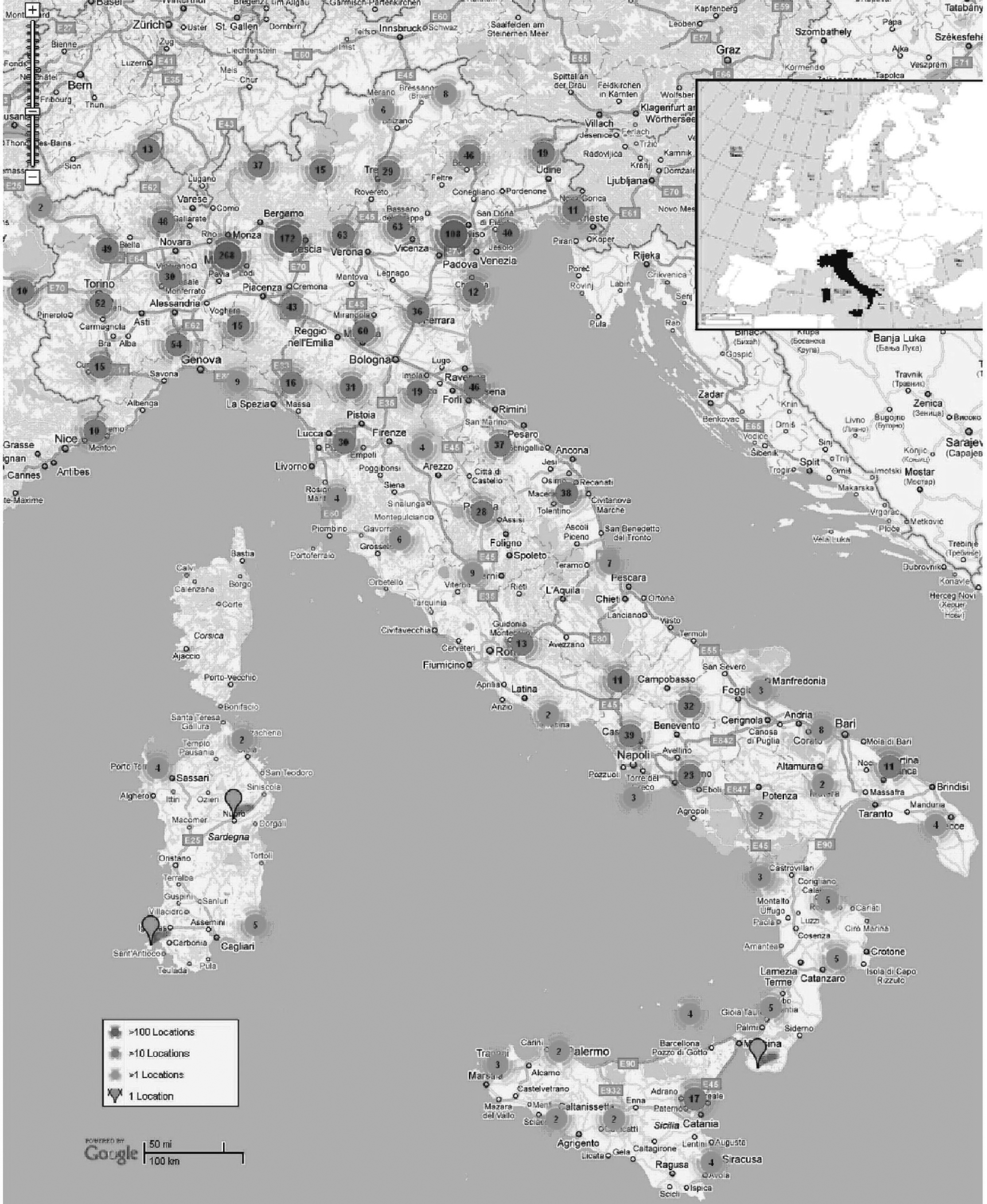
Map showing the distribution and number of serum samples collected from dogs in Italy from 2006 to 2008. Purple dots: >100 samples; light blue dots: 10–100 samples; green dots: <10 samples. The actual number of samples is indicated for each dot.
All Italian dog sera that had positive or doubtful results in the cELISA (competition percentage greater than 45%) were also tested in subtype-specific HI assays. The HI assays were performed using 9 different influenza A viruses as antigens: H1N1 (A/swine/Italy/711/06); H2N3 (A/duck/Germany/1215/73); H3N8 (A/canine/Florida/04, A/equine/NM/99, and A/passerine/Italy/6000/00); H5N3 (A/duck/Italy/775/04); H7N3 (A/turkey/Italy/2962/V03); H9N2 (A/mallard/Italy/3817-34/05); and H10N1 (A/ostrich/South Africa/01). These subtypes were selected as prototypes of viruses likely circulating in poultry and mammals during the period of canine serum collection. Each virus was propagated in 10-day-old embryonated chicken eggs. The viruses in the harvested allantoic fluids were inactivated with 0.0005% β-propiolactone e and stored at −80°C or were lyophilized and stored at −20°C.
Prior to testing, the Italian canine serum samples and control sera were incubated with receptor destroying enzyme (RDE b ; 1 part serum to 3 parts RDE) for 16 hr at 37°C, followed by heat-inactivation at 56°C for 30 min. The HI assays were performed using 4 hemagglutinating units/25 μl of virus, 8 log2 serial dilutions of serum samples in PBS starting at 1:8 ratio, and 0.5% (mammalian-origin virus) or 1% (avian-origin virus) suspensions of chicken erythrocytes in PBS. The 2 different erythrocyte suspensions were used based on World Organization for Animal Health 28–30 standard procedures established for use of avian- and mammalian-origin viruses in HI assays. The HI titers were estimated visually after incubating at room temperature for 45 min with 0.5% erythrocytes or for 30 min with 1% erythrocytes. The endpoint HI titer was defined as the reciprocal of the last dilution of serum that completely inhibited hemagglutination. The cutoff titer for seropositivity was 32, based on previous testing of sera from specific-pathogen-free dogs and dogs experimentally infected with canine FLUAV subtype H3N8. 11
The agar gel immunodiffusion (AGID) assay is a standard serological test for diagnosis of avian FLUAV infection based on detection of antibodies to the highly conserved viral matrix protein and nucleoprotein in the nucleocapsid. 2 An AGID assay using A/canine/FL/04 as the antigen was performed on the known positive reference sera using standard procedures. 2,28–30 However, as a result of poor performance (43/97 sera detected as positive), further AGID testing was not pursued with the Italian dog sera.
Sensitivity and specificity of the cELISA
A Bayesian approach was used to estimate the Se and Sp of the cELISA. The Se and Sp of the HI and FAT could not be estimated because of the low number of positive results in the cELISA and the negligible discordant results between the HI and FAT. The methodology for application of Bayesian theory to inference of diagnostic test Se and Sp has been described elsewhere. 3,4 Briefly, uncertainty regarding the true value of the Se and Sp of the test and the disease prevalence (p) was modeled using probability distributions referred to as prior distributions. Prior values for the cELISA, based on the uncertainty for the Se and Sp values, were modeled using data reported in peer-reviewed literature 9–11,15,27 and at professional conferences (Rigoni M, Viale E, Mancin M, et al.: 2009, Development of a competitive ELISA system for the detection of influenza A virus nucleoprotein antibodies and validation in five species of poultry. In: Proceedings of the World Association of Veterinary Laboratory Diagnosticians 14th International Symposium, p. 174. Madrid, Spain). The prior distribution for the value of p was estimated based on an author's (Capua) expert opinion. Alternatively, the prior distribution of p was parameterized using a beta (1,1) distribution to assess the robustness of the results to the prior uncertainty on the true value of p. The prior beta distributions of these test parameters were fitted using the betabuster software, available at the Bayesian Epidemiologic Screening Techniques webpage (http://www.epi.ucdavis.edu/diagnostictests/betabuster.html). Data collected from the field were then used to modify the prior distributions to produce new distributions, referred to as posterior distributions. Posterior distributions represent the uncertainty for the Se, Sp, and p values for the cELISA given the researchers' prior uncertainty for these values and the test results. The Se and Sp of the cELISA were estimated using the 6,858 canine sera, assuming that samples were obtained from 1 single population (i.e., dogs in Italy; 1 test, 1 population). 3,4 All calculations were performed using the WinBUGS software, 26 and results were computed for 100,000 iterations of the model after the initial 1,000 were discarded. The results of the cELISA, HI, and FAT were securely stored in the FMD BioPortal database (https://fmdbioportal.ucdavis.edu/), which is a Web-based system for visualization and analysis of infectious animal diseases.
Results
Estimating the level of exposure of Italian dogs to influenza A viruses
A video displaying results from the 6,858 Italian dogs tested is available at the FMD BioPortal Web site (http://fmd.ucdavis.edu/ma/AI_Italy.avi). Of the 2,041 Italian canine serum samples from 2006, 18 (0.88%) were positive for nucleoprotein antibody by cELISA, and 2 (0.09%) samples were doubtful. For the 2,038 samples from 2007, 23 (1.12%) were positive by cELISA, and 14 (0.69%) were doubtful. For the 2,779 samples from 2008, 16 (0.58%) were positive by cELISA, and 9 (0.32%) were doubtful. All 82 cELISA-positive and doubtful samples were retested in the HI and FAT. Only 1 cELISA-positive serum sample from 2006 and 1 cELISA-positive sample from 2007 were also positive in both the HI and FAT. The 2 samples were positive for antibody to the H3 proteins of A/canine/FL/04 (H3N8) and A/equine/NM/99 (H3N8) viruses. The HI titer was 256 and the FAT titer was 1,024 for the 2006 serum sample, while the HI titer was 64 and the FAT titer was 256 for the 2007 serum sample. The remaining cELISA-positive or doubtful samples had HI titers of less than 8. The apparent prevalence of FLUAV exposure for the tested Italian dogs was, therefore, 0.03% (2/6,858 dogs).
Prior and Bayesian posterior estimates of the sensitivity and specificity of a competitive enzyme-linked immunosorbent assay for detection of antibodies to influenza A viruses in Italian dogs and of the virus prevalence in the population. *
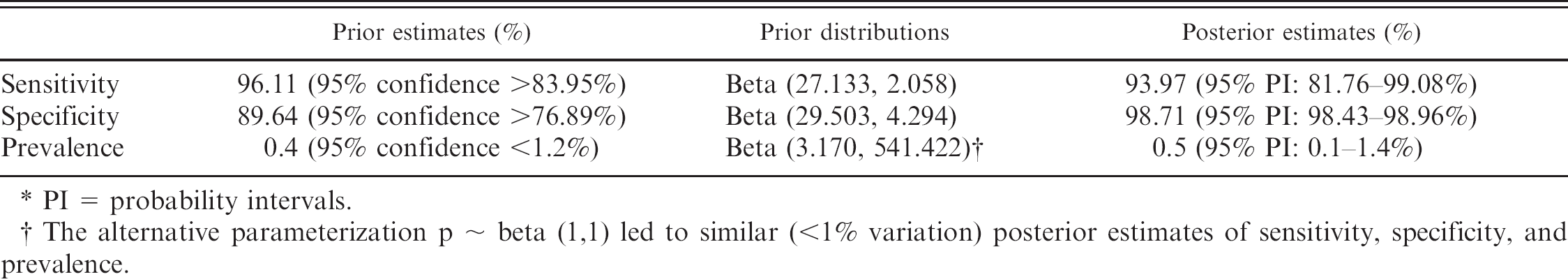
PI = probability intervals.
The alternative parameterization p ∼ beta (1,1) led to similar (<1% variation) posterior estimates of sensitivity, specificity, and prevalence.
For the reference serum panel from dogs naturally infected with A/canine/FL/04 (H3N8) virus, 92 (95%) tested positive in the cELISA, and 95 (98%) tested positive in the FAT. None of the serum samples from uninfected specific-pathogen-free dogs tested positive in any of the assays.
Sensitivity and specificity of the cELISA
Eighty-two samples were considered positive or doubtful in the cELISA and were therefore classified as cELISA-positive samples for purposes of the analysis. Two of these 82 samples were also positive in both the HI and FAT, 79 were negative in both the HI and FAT, 1 was HI negative and FAT positive, and none were HI positive and FAT negative. Sensitivity and Sp of the HI and FAT could not be computed using Bayesian methods because of the low proportion of cELISA-positive samples, along with the negligible proportion of discordant results for the HI and FAT. The Bayesian posterior Se and Sp estimates for the cELISA, the Bayesian posterior distribution of p after accounting for the imperfect Se and Sp of the test, and their respective 95% probability intervals are listed in Table 1.
Discussion
The present study identified 2 Italian dogs that had serum antibodies to highly conserved influenza A proteins and to H3 proteins from contemporary canine and equine H3N8 viruses using cELISA, HI, and FAT. Although evaluation of disease transmission was beyond the scope of this study, the potential source of FLUAV exposure for the 2 seropositive dogs was investigated. The dog that tested positive from 2006 was a 5-year-old mixed-breed canine that had lived in Miami, Florida, for 3 years before moving to Sardinia, Italy, in 2006. The dog was bled 3 weeks after arrival in preparation for travel to the United Kingdom and was likely infected by canine FLUAV subtype H3N8 while in Florida. The dog that tested positive from 2007 was a 4-year-old mixed-breed canine from Lombardy, Italy. The dog had never traveled outside of continental Europe; however, the owner confirmed that the dog was raised in close contact with horses, including during 2003, when there was an equine influenza outbreak in Italy. 18 The owner did not report whether the dog had been exposed to sick horses during the outbreak, and the owners of both dogs never observed any influenza-like illness in their dogs.
The current study evaluated a large-scale diagnostic approach for detection of FLUAV antibodies in canine sera. The proposed diagnostic algorithm consists of an initial screening with a cELISA for influenza A nucleoprotein antibody, followed by confirmatory testing of positive samples with a FAT for influenza A nucleocapsid antibodies and/or HI assays for hemagglutinin subtype–specific antibodies. This algorithm was supported by the Bayesian analysis results for the cELISA and the generally recognized advantages of the confirmatory HI and FAT. The commercially available cELISA demonstrated good Se (93.98%) and Sp (98.71%). In addition, the high-throughput microtiter plate format of the cELISA is ideal for screening large numbers of canine sera. A limitation, however, is that the Bayesian model used herein is not identifiable because there are more parameters than degrees of freedom. For that reason, prior information about the model parameters is required (Table 1). Because no prior information was available regarding the disease prevalence in the assessed population, best-guess (based on expert opinion) and noninformative parameterizations of p were alternatively assumed. Results were robust for alternative parameterization of p and were consistent with the presented results.
Although the Se and Sp of the HI and FAT could not be estimated using Bayesian analysis in the current study, cELISA-positive and doubtful samples should be confirmed in a highly sensitive and specific assay. The Se and Sp of the HI assay are generally high when the test antigen is homologous to the circulating virus. In the current study the HI assay was conducted with 7 hemagglutinin subtypes, including 3 H3N8 viruses representing canine, equine, and avian isolates. These viruses were selected as prototypes of those likely circulating in poultry and mammals during the period of canine serum collection. The 80 HI-negative samples had titers of <8, indicating that the Se of the assay was not negatively affected by the defined cutoff titer for seropositivity of 32. However, the authors acknowledge that exposure to the remaining virus subtypes was not definitively ruled out since sera were not tested against all 16 influenza A hemagglutinin subtypes. The FAT allows type-specific detection of influenza virus exposure, and the assay is highly sensitive and specific; however, major disadvantages of the FAT include the necessary in-house development of the test and its admittedly subjective interpretation.
Although the low seroprevalence (0.5% after accounting for the imperfect Se and Sp of the cELISA) in the present study indicates that dogs do not currently play a significant role in FLUAV ecology in Italy, the identified seropositive dogs highlight 2 potential sources of FLUAV exposure for dogs in Italy and abroad: the potential spread of canine-specific influenza A viruses by international movement of infected dogs and exposure of dogs to other species infected by FLUAV. Based on international observations, monitoring dogs during future influenza A outbreaks in other species is recommended. A diagnostic algorithm utilizing cELISA as well as HI and/or FAT is a highly sensitive and specific approach to assessing exposure to any influenza A subtype in dogs, especially in those with unknown exposure histories at the time of testing.
Footnotes
a.
ID Screen® Influenza A Antibody Competition Assay, ID Vet, Montpellier, France.
b.
Sigma-Aldrich, St. Louis, MO.
c.
BD Bioscience, San Jose, CA.
d.
Gibco®, Invitrogen Corp., San Diego, CA.
e.
Ferak Berlin GmbH, Berlin, Germany.