
Abstract
The Staphylococcus intermedius bacterial group (SIG) includes 3 distinct genetically heterogenous species: S. intermedius, S. pseudintermedius, and S. delphini. This pathogen group is associated with many opportunistic skin and ear infections in companion animals. Human infections with S. intermedius and S. pseudintermedius isolates and the emergence of methicillin-resistant isolates have been recently reported, which emphasizes the importance of nationwide identification of SIG isolate prevalence and antibiotic resistance in veterinary clinics. In the present study, a total of 178 SIG isolates were obtained from veterinary staff (n = 40), companion animals (n = 115), and the local environment (n = 23) in 8 Korean veterinary hospitals. Isolates were differentiated into 167 S. pseudintermedius (93.8%) and 11 S. intermedius (6.2%) isolates; S. delphini isolates were not identified. The most effective antibiotics against these isolates included amoxicillin-clavulanic acid, amikacin, nitrofloxacin, imipenem, and vancomycin; whereas ampicillin, penicillin, tetracycline, erythromycin, and trimethoprim-sulfamethoxazole were not effective. Surprisingly, the 128 SIG isolates (71.9%) displayed multiple drug resistance (MDR) against 3 or more antibiotic classes. Out of 52 SIG isolates carrying the methicillin-resistance gene (mecA), only 34 (65.4%) were oxacillin-resistant, and 49 (94.2%) methicillin-resistant SIG were multidrug resistant. This finding suggests the presence of greater numbers of MDR phenotypes than other isolates (P < 0.05).
Introduction
Staphylococcus intermedius is a common, coagulase-positive staphylococcus associated with pyoderma and otitis externa in dogs and cats. The bacterium was originally misclassified as Staphylococcus aureus biotypes E and F and was differentiated from S. aureus based on cell wall structure and biochemical properties. 8 Recent molecular phylogenetic analysis demonstrated the presence of S. intermedius genetic diversity and allowed reclassification of species previously identified as S. intermedius into 3 distinct species (S. intermedius, S. pseudintermedius, and S. delphini), collectively designated as the S. intermedius species group (SIG). 1
The SIGs are opportunistic pathogens in many veterinary infections. Infection with S. intermedius and S. pseudintermedius isolates are common causes of pyoderma and otitis externa in dogs and cats. 1,19,31,32 Infection with S. delphini is associated with purulent skin lesions in dolphins. 29 Human infections with S. intermedius and S. pseudintermedius have also been identified in food poisoning cases, catheter-related bacteremia, ischemic cardiomyopathy, and ventricular tachycardia cases, 3,7,16,25,28 and direct transmission between animals and human beings has also been demonstrated. 15,27 Such characteristics emphasize their potential as zoonotic pathogens. Furthermore, increasing evidence has identified the emergence and spread of methicillin-resistant S. intermedius (MRSI) and S. pseudintermedius (MRSP) isolates. 5,13,27,31 Thus, transmission seems very likely in veterinary environments. 33
Recent studies reported the high frequency of multiple drug-resistant S. pseudintermedius (MDRSP) and MRSP isolates from cases of bacterial pyoderma and otitis externa in pet dogs. 14,22 Such findings suggest that the potential for SIG cross-contamination between companion animals, veterinary staff, and the veterinary hospital environments exists. In the current study, a total of 178 SIG isolates were cultivated from veterinary staff, companion animals, and the environment in 8 veterinary hospitals drawn from 4 South Korean provinces. Isolates were further differentiated to examine the prevalence and antibiogram of the SIG organisms in veterinary hospitals in a nationwide survey.
Materials and methods
Sampling and bacterial identification
Samples (n = 766) were randomly obtained from veterinary staff members (n = 219 from 107 veterinary staff), hospitalized companion animals (384 samples from 108 dogs and 6 samples from 2 cats: 13 dogs with history of skin disease and 1 dog with otitis), and the hospital environment composed of doorknobs, waiting room floor and chairs, computer keyboards, computer mouse, telephones, microscope, examination room floors and desks, and cages (n = 157). Samples were aseptically collected from 8 veterinary hospitals located in 4 South Korean provinces between November 2006 and January 2010. Hospitals included veterinary teaching hospitals (n = 4), private referral veterinary hospitals (n = 1), and private veterinary hospitals (n = 3) located in Seoul, Chungcheong-do, Gyeonggi-do, and Jeju-do. From each dog and cat, 3–6 samples were taken from the anus, horizontal ear canal, nasal mucosa, skin, wound infection, footpads, and urine. Two to 3 samples were taken from each veterinary staff's hand and nasal passages.
Samples were collected with culture swabs a and were each cultured in 10% sodium chloride containing tryptic soy broth b and Baird-Parker agar. c Suspect Staphylococcus isolates were Gram-stained and tested for coagulase, DNase production, and hemolysis, and subsequently confirmed by polymerase chain reaction (PCR) and PCR-restriction fragment length polymorphism (RFLP) analysis using pta gene-specific primers as previously described. 1,2
Isolation of staphylococcal genomic DNA and PCR amplification
Individual SIG isolates were cultured overnight on 5% sheep blood agar plates d at 37°C. Genomic DNA was extracted using a commercial tissue kit e with 50 U/ml lysostaphin f according to the manufacturer's instructions for Gram-positive bacteria. 2 The S. intermedius American Type Culture Collection (ATCC) 29663 and S. aureus ATCC 29213 isolates were used as reference strains. The PCR analysis for the detection of the methicillin-resistance gene (mecA) and the penicillin-resistance gene (blaZ) was performed with gene-specific primers as previously described. 18,34 The PCR products were visualized with 1.5% agarose gel electrophoresis after ethidium bromide staining.
Antibiotic susceptibility
The antibiogram was obtained by a standard disk diffusion test against 15 antibiotics g from 8 different antimicrobial classes (aminoglycosides, beta-lactams, fluoroquinolones, lincosamides and macrolides, nitrofurans, phenicol, potentiated sulfonamides, and tetracyclines); ampicillin, AM (10 μg); amoxicillin-clavulanic acid, AmC (30 μg; 20 μg of amoxicillin and 10 μg of clavulanate); amikacin, AN (30 μg); chloramphenicol, C (30 μg); clindamycin, CC (2 μg); cephalothin, CF (30 μg); ciprofloxacin, CIP (5 μg); cefotaxime, CTX (30 μg); erythromycin, E (15 μg); nitrofurantoin, F/M (300 μg); gentamicin, GM (10 μg); imipenem, IPM (10 μg); penicillin, P (10 units); trimethoprim-sulfamethoxazole, SXT (23.75 μg, 1.25 μg); and tetracycline, Te (30 μg). Multiple drug resistance (MDR) was defined by resistance to ≥3 antimicrobials from different classes. The minimum inhibitory concentrations (MICs) of the antimicrobials oxacillin (OXA), h vancomycin (VA), i and orbifloxacin (Orb) j were determined with a broth microdilution method. All procedures and interpretations were followed as previously described by the Clinical and Laboratory Standards Institute (CLSI; M2-A9, M31, and M100) guidelines 4 except that the new recommended OXA breakpoint of ≥0.5 mg/l was used for S. pseudintermedius (Papich MG: 2010, Proposed changes to Clinical Laboratory Standards Institute interpretive criteria for methicillin-resistant Staphylococcus pseudintermedius isolated from dogs. J Vet Diagn Invest 22:160. Letter to the editor). The S. aureus ATCC 29213 and S. intermedius ATCC 29663 organisms were used as reference strains.
Statistical analyses
The chi- square (χ 2 ) statistic and the one-tailed P value (determined by the Mantel-Haenszel chi-square test) were used to analyze significant differences in antibiotic resistance between SIG isolates from different sources, between S. pseudintermedius and S. intermedius, and between SIG isolates with or without the mecA gene. Statistical significance was defined as P values < 0.05. The Analyse-it program k was used for all analyses.
Results
Isolation and identification of SIG isolates
The SIG isolates (n = 178) were cultured from veterinary staff, companion animals, and the local environment of 8 veterinary hospitals in 4 South Korean provinces (Table 1). Staphylococcus intermedius (n = 11, 6.2%) and S. pseudintermedius (n = 167, 93.8%) isolates were both identified. Staphylococcus delphini bacteria were not identified in the present study. Only 1 different kind of isolate (S. intermedius and/or S. pseudintermedius) per SIG-positive sample was obtained. The S. pseudintermedius isolates were detected in veterinary staff members (n = 37, 22.2%), companion animals (n = 107, 64.0%), and the veterinary hospital environment (n = 23, 13.8%). As seen in Table 1, S. intermedius isolates were identified in both veterinary staff members (n = 3, 27.3%) and companion animals (n = 8, 72.7%).
Antibiotic resistance of SIG isolates
Antibiotic resistance (AR) of the 178 SIG isolates using the disk diffusion test method in the present study revealed that the greatest antimicrobial resistance was observed with AM (97.2%) and P (96.6%), followed by Te (81.5%), E (66.3%), and SXT (60.7%). The lowest antimicrobial resistance was observed with AN (1.1%), F/M (0.6%), and IPM (0.6%), followed by CF (2.2%), AmC (6.2%), CTX (17.4%), GM (24.2%), CIP (26.4%), C (33.7%), and CC (37.6%). The proportion of resistant isolates to only 3 antibiotics differed (P < 0.05) according to the source of SIG; the veterinary staff, companion animal, and veterinary hospital environment isolates were more resistant to AmC, CIP, and GM, respectively (Table 2). A majority of isolates (n = 128, 71.9%) displayed MDR against 3 or more antimicrobial classes. The MDR isolate frequency from veterinary staff, companion animals, and hospital environments was 67.5%, 73.0%, and 73.9%, respectively (Table 3). An OXA resistance was identified in 34.8% of SIG isolates. In addition, an Orb resistance (MIC ≥ 8 mg/l) was identified in 34.8% of SIG isolates. A VA resistance (MIC ≥ 16 mg/l) was not shown in the present study. The OXA-resistance rate from the environment isolates was significantly higher than the rate in companion animals (P < 0.05).
Overview of Staphylococcus intermedius group (SIG) isolates from Korean veterinary hospitals (veterinary staff, companion animals, and veterinary hospital environment).*
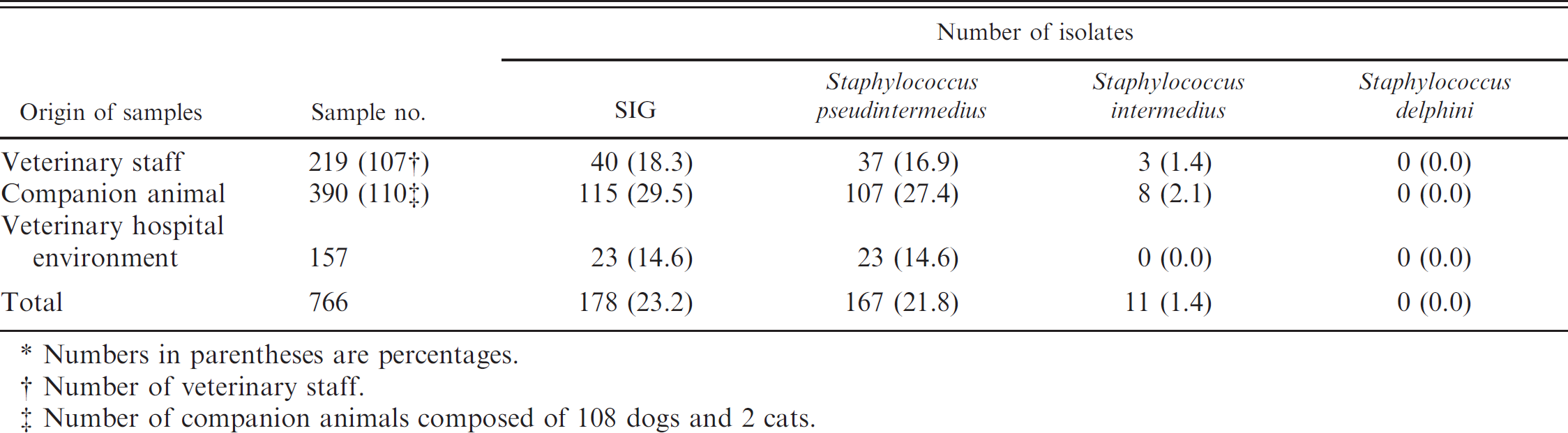
Numbers in parentheses are percentages.
Number of veterinary staff.
Number of companion animals composed of 108 dogs and 2 cats.
Comparison of antibiotic resistance rate of Staphylococcus pseudintermedius group from veterinary staff, companion animals, and veterinary hospital environment against 15 different antimicrobials from 8 different antimicrobial classes by disk diffusion method.
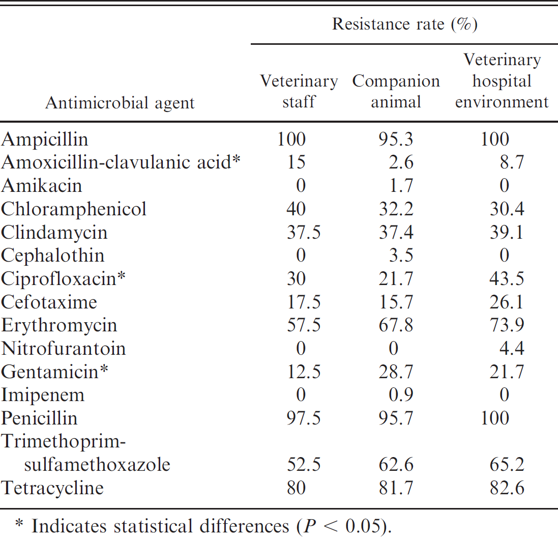
Indicates statistical differences (P < 0.05).
Prevalence of the methicillin-resistant SIG isolates
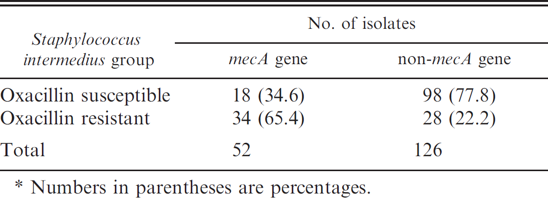
The mecA gene was detected in 52 SIG isolates by PCR (14 from veterinary staff, 29 from companion animals, and 9 from veterinary hospital environments). The AR rate of isolates with mecA gene was significantly higher for all antimicrobials (P < 0.05) except for AM, AN, C, F/M, IPM, P, and Te (Table 4). In addition, only 25 of 43 (58.1%) isolates with the mecA gene were resistant to OXA (Table 5). The regional distribution showed that the prevalence rate of methicillin-resistant S. intermedius and S. pseudintermedius isolates from Chungcheong-do was significantly higher (P < 0.01) than that which originated from Seoul or Gyeonggi-do (Table 6). Comparison of the MIC profile by broth dilution test against OXA, VA, and Orb between S. pseudintermedius and S. intermedius isolates showed a statistical difference (P < 0.05) in only the Orb-resistance rate between the 2 groups. The percentage of isolates that harbored the mecA and blaZ genes was similar in both groups (P > 0.05), as seen in Table 7. The AR rate in the 2 groups against other antibiotics determined by disk diffusion test was not significantly different except for Te (85.6% in S. pseudintermedius and 18.2% in S. intermedius).
Discussion
The current study describes the SIG distribution and antibiogram from South Korean veterinary hospitals. A total of 167 S. pseudintermedius and 11 S. intermedius isolates were identified from 756 samples in the present study; no S. delphini isolates were detected (Table 1). The results suggest that S. pseudintermedius has a wide distribution in Korean veterinary hospitals. The prevalence of SIG from animals (29.9%) in the present study was lower than that in a previous study (43.9%) but nearly 4 times (18.3% vs. 5%) higher from veterinary staff. 24 Such differences may result from the comparison of isolates from different sampling regions. Staphylococcus pseudintermedius and S. intermedius may be temporary bacterial flora on the skin and hair of companion animals. 11 Therefore, among the 3 different sources (veterinary staff, companion animals, and veterinary hospital environment), these 2 bacteria were detected mostly from companion animals (64% and 72.7%, respectively; Table 1). Antibiograms were evaluated among SIG isolates from veterinary staff, companion animals, and veterinary hospital environments; the highest resistance rates (0.01 < P < 0.05) were identified against AmC in veterinary staff, GM in companion animals, and CIP in veterinary hospital environments. Apart from those 3 antibiotics, SIG isolates from different sources did not demonstrate resistance rates that were significantly different from one another (Table 2).
Overview of multidrug-resistant Staphylococcus intermedius group isolates from Korean veterinary hospitals (veterinary staff, companion animals, and veterinary hospital environment).*
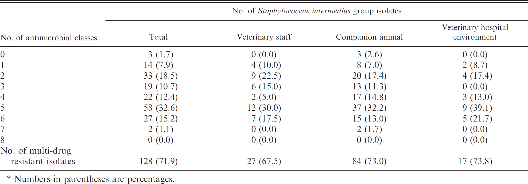
Numbers in parentheses are percentages.
Comparison of antibiotic resistance rate of Staphylococcus intermedius group with and without the mecA gene against 15 different antimicrobials from 8 different antimicrobial classes by disk diffusion method.
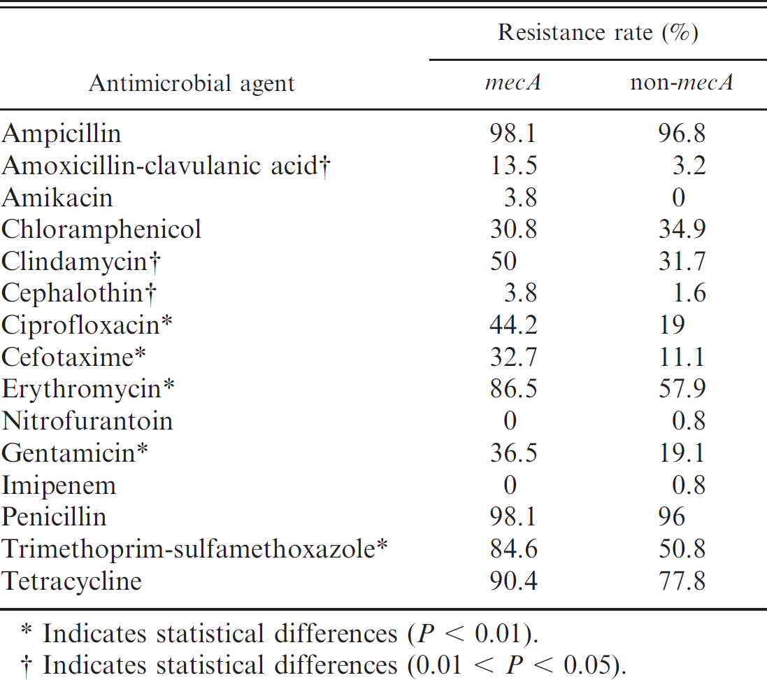
Indicates statistical differences (P < 0.01).
Indicates statistical differences (0.01 < P < 0.05).
The 9% rate of OXA-resistant SIG isolates from animals in the study was approximately 4–6 times greater than that identified in companion animals (inpatient and outpatients) in other studies 6,9,21,30 and was the same as that in the pyoderma cases in some studies. 6,14 Using a different standard of OXA breakpoint and collecting samples from either the nasal cavity or perineal region of dogs from one location, as suggested in previous studies, might lead to underestimation of AR result. 10,21 Similar to the present study, the rate of methicillin-resistant S. pseudintermedius and S. intermedius from dogs (inpatient and outpatients) at a veterinary clinic was 30% in a Japanese study 23 the rate in a German study was much lower at 6.8%. 22 Previous studies have suggested that the high methicillin resistance in East Asian countries could be due to antibiotic overuse or misuse. 24 - 23 Resistance to CIP, CTX, E, GM, SXT (P < 0.01), and AmC, CC, and CF (0.01 < P < 0.05) was more common among mecA-positive SIG isolates compared with isolates without the mecA gene (Table 2). In addition, only 65.4% (34/52; mecA–positive) and 22.2% (28/126; mecA-negative) of methicillin-resistant SIG isolates demonstrated OXA resistance (Table 5). The mecI-mediated repression of penicillin-binding protein 2 production in the regulatory region may the possible reason for OXA susceptibility of isolates with the mecA gene. 17 The 28 isolates in the current study that did not carry the mecA gene were phenotypically resistant to OXA according to the MIC test, and it seems that they were beta-lactamase hyper-producing isolates because most of them (89.3%) remained susceptible to AmC. 20 The penicillin-resistance gene blaZ was found in 174 (97.8%) SIG isolates, which reflects the resistance rate of penicillin 96.1% (171 SIG isolates) by disk diffusion test (Table 7). 34
Comparison between Staphylococcus intermedius group isolates isolated from Korean veterinary hospitals (veterinary staff, companion animals, and veterinary hospital environment) with and without the mecA gene.*
Numbers in parentheses are percentages.
Regional overview of methicillin-resistant Staphylococcus pseudintermedius (MRSP) and methicillin-resistant Staphylococcus intermedius (MRSI) isolates from Korean veterinary hospitals.*
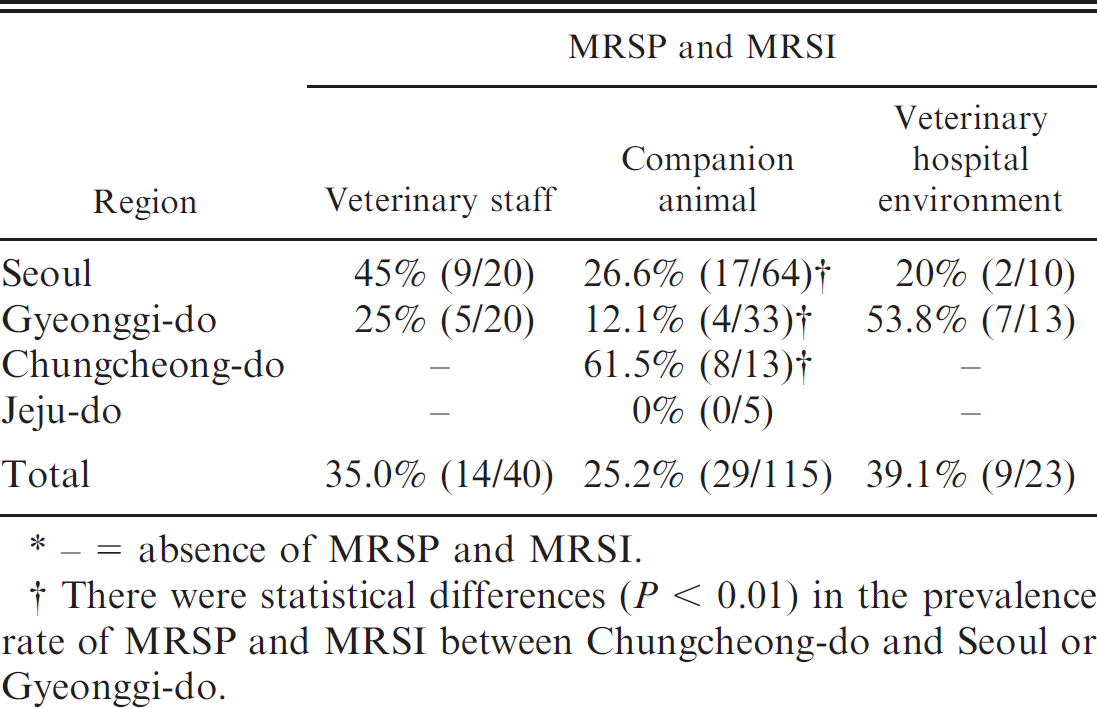
– = absence of MRSP and MRSI.
There were statistical differences (P < 0.01) in the prevalence rate of MRSP and MRSI between Chungcheong-do and Seoul or Gyeonggi-do.
Oxacillin is used in rare, severe cases as antimicrobial agents in bacterial infections when other agents have demonstrated resistance. In the current study, the S. pseudintermedius resistance rate was 25.8% with an OXA MIC breakpoint of ≥4 4,26 and 34.8% with the new recommended OXA MIC breakpoint of ≥0.5 mg/l. The increasing prevalence of methicillin-resistant SIG isolates is a significant health concern 27,30,34 to companion animals and their human contacts. 7,33 Orbifloxacin, a third-generation fluoroquinolone antimicrobial agent developed exclusively for the treatment of bacterial infections including pyoderma and otitis in companion animals, 12 demonstrates a 34.3% resistance rate, with a breakpoint of ≥8 mg/l. The antibiogram in the present study showed 71.9% MDR by the disk diffusion test, emphasizing the necessity for establishing guidelines in the treatment of SIG infections in Korean veterinary hospitals.
Comparison between Staphylococcus pseudintermedius and Staphyiococcus intermedius isolates isolated from Korean veterinary hospitals (veterinary staff, companion animals, and veterinary hospital environment).*
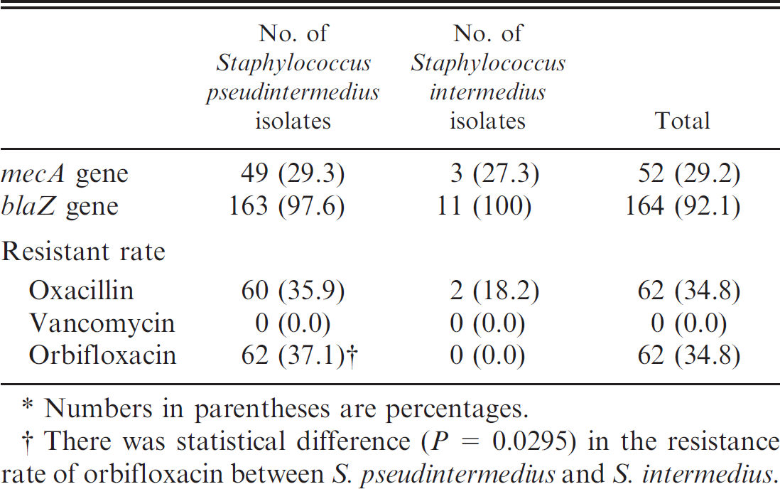
Numbers in parentheses are percentages.
There was statistical difference (P = 0.0295) in the resistance rate of orbifloxacin between S. pseudintermedius and S. intermedius.
As seen in Table 7, among SIG isolates, S. pseudintermedius was the predominant bacteria isolated in the present study, and it displayed minimal antimicrobial differences from S. intermedius (except for Orb by the MIC test). Therefore, transmission of gene elements mediating resistance between these species cannot be ruled out. It is expected that results from the present study will enhance therapeutic guidelines for the treatment of SIG isolate infection, particularly S. pseudintermedius, in companion animals from Korean veterinary hospitals. In addition, such guidelines may also aid in the development of an effective treatment for companion animal patients, prevention of transmission, and avoidance of occurrence of disease caused by SIG. Continuous monitoring and molecular epidemiologic studies should be regularly performed.
Acknowledgements
The current study was supported by the National Veterinary Research and Quarantine Service (B-AD 3–2006–14–04) and the Korea Research Foundation Grant (KRF-2008–531-E00076). Additional support was provided by the Research Institute of Veterinary Science, Department of Veterinary Microbiology, College of Veterinary Medicine, and the BK21 Program for Veterinary Science, Seoul National University, Seoul, Korea. Dr. Jang Won Yoon was supported by RP-Grant 2010 of Ewha Womans University. Sook Shin, Sa-Im, Sa-Eun, Sun Young, Kuk Ju, Min Gi, Bo Youn, Ka Hee, Young Kyung, and Ki Yeon are appreciated for their excellent technical support.
Footnotes
a.
BBL™ Culture Swabs™, BD, Sparks, MD.
b.
Bacto™ Tryptic Soy Broth, BD, Sparks, MD.
c.
BBL™ Baird-Parker Agar, BD, Sparks, MD.
d.
Blood Agar Plate, Komed, Sungnam, Gyeongi-Do, Korea.
e.
DNeasy® Blood & Tissue Kit (250), Qiagen GmBH, Hilden, Germany.
f.
Lysostaphin from Staphylococcus staphylolyticus, Sigma-Aldrich, St. Louis, MO.
g.
BBL™ Sensi-Disc™, BD, Sparks, MD.
h.
Oxacillin, Sigma-Aldrich, St. Louis, MO.
i.
Vancomycin, Sigma-Aldrich, St. Louis, MO.
j.
Orbifloxacin Vetranal®, Riedel-de Haen, Seelze, Niedersachsen, Germany.
k.
Analyse-it Software Ltd., Leeds, United Kingdom.