
Abstract
Geotrichum candidum was cultured from the tonsils of a free-ranging weaner pig that was presented with lethargy and inappetence. Histopathology indicated pyogranulomatous tonsillitis with intralesional fungal hyphae and arthrospores. Geotrichosis is a rare, opportunistic mycosis of immunocompromised hosts, usually human beings.
Keywords
A free-ranging weaner pig from northern coastal New South Wales, Australia, presented with a short history of lethargy and inappetence. After euthanasia, the referring veterinarian carried out a field necropsy. On gross examination, there was ventral cervical swelling, focal ulceration on the snout, and a narrow band of ulceration adjacent to the coronary band of the front hooves. An extensive range of fixed (liver, tonsil, skin [foot and snout], kidney, heart, spleen, stomach, duodenum, jejunum, brain) and fresh (blood, feces, urine, tonsil, jejunum, skin [foot and snout]) tissue samples were submitted for analyses at the State Veterinary Diagnostic Laboratory (Menangle, New South Wales, Australia). The herd was fed a range of rainforest flora supplemented with macadamia nut waste and occasional commercial pig mix. Because of the nature of the cutaneous lesions, testing to exclude vesicular viral diseases was done at the Australian Animal Health Laboratory (Geelong, Victoria) before further diagnostic efforts. Serology, viral isolation, and polymerase chain reaction testing were negative for foot and mouth disease, swine vesicular disease, and vesicular stomatitis.
Multiple organ tissues (liver, tonsil, skin [foot and snout], kidney, heart, spleen, stomach, duodenum, jejunum, brain) were processed routinely for histologic examination. Significant histopathologic findings were noted in the tonsils and skin. There was a multifocal to coalescing granulomatous to pyogranulomatous tonsillitis (Fig. 1), with myriad intralesional and frequently intrahistiocytic fungal elements, which were poorly defined by hematoxylin and eosin (HE). The following tonsillar changes were observed. The parenchyma was altered and expanded by marked, multifocal to coalescing accumulations of neutrophils, macrophages, and lesser number of lymphocytes and plasma cells. The macrophages were distended with intracellular fungal elements, which were elongated to spherical, 5–7 μm in diameter, with a narrow refractile wall, and minimal hyphal components. There were multifocal areas of hypereosinophilia, karyohexis, and karyolysis (interpreted as necrosis). Fungal elements were elongated to spherical, 5–7 μm in diameter, with a narrow refractile wall, and minimal hyphal components (Fig. 2). With Grocott–Gomori methenamine silver staining, the fungal hyphae were irregularly branching and highly septated, which often appeared yeast like (Fig. 3). Cutaneous lesions adjacent to and including the coronary bands consisted of a severe multifocal suppurative dermatitis, with prominent subcorneal pustules. The dermis and subcutis was altered and expanded by severe, multifocal to coalescing accumulations of neutrophils. The interstitial tissue was loosely arranged with variable dilatation of lymphatic vessels (interpreted as edema). The epidermis was distended by moderate, multifocal, subcorneal accumulations of neutrophils (subcorneal pustules).
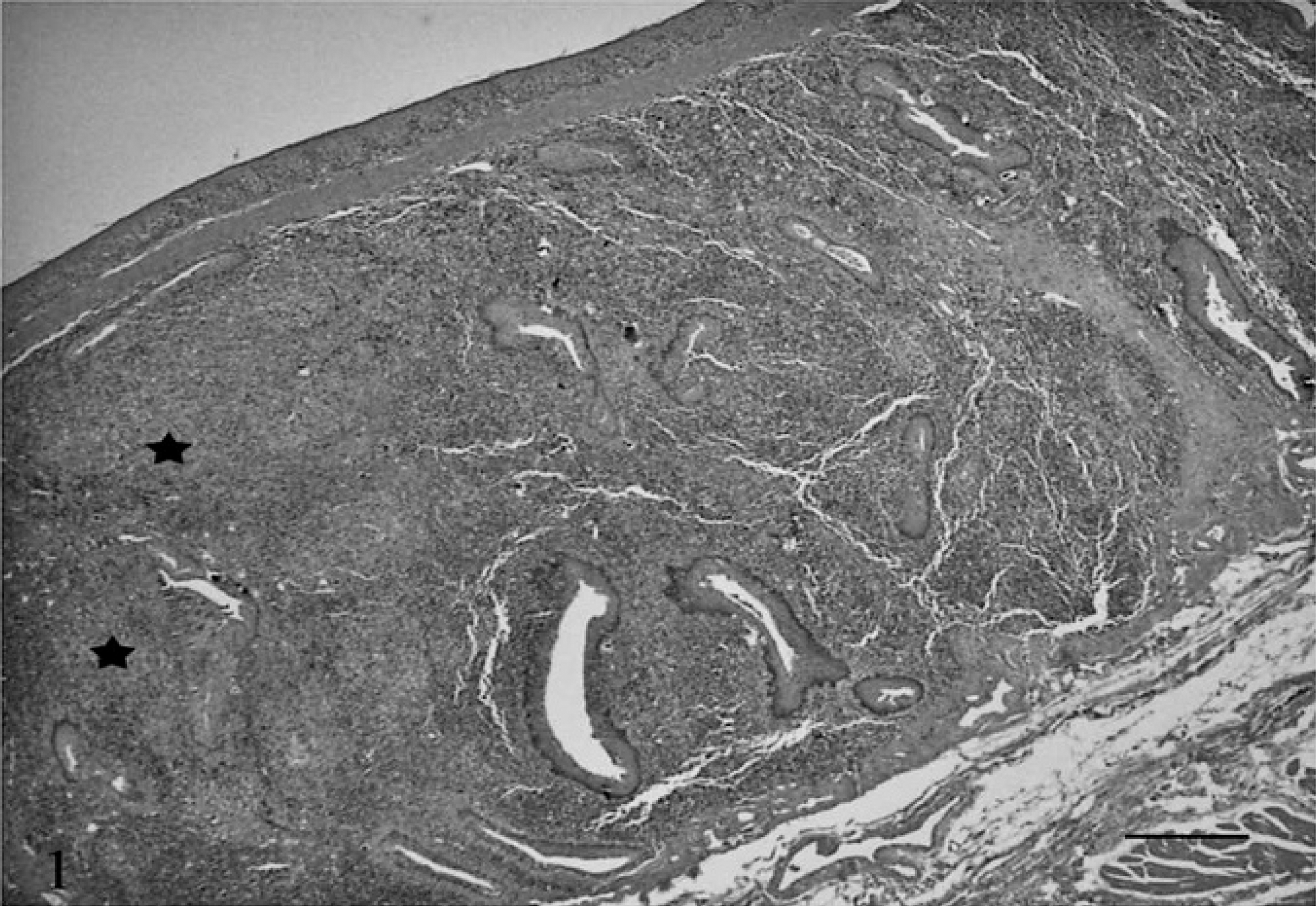
Tonsil; pig. Tonsillar submucosa contains expansile, multifocal to coalescing granulomatous to pyogranulomatous (stars) infiltrates (higher magnification is shown in Fig. 2). Hematoxylin and eosin. Bar = 300 μm.
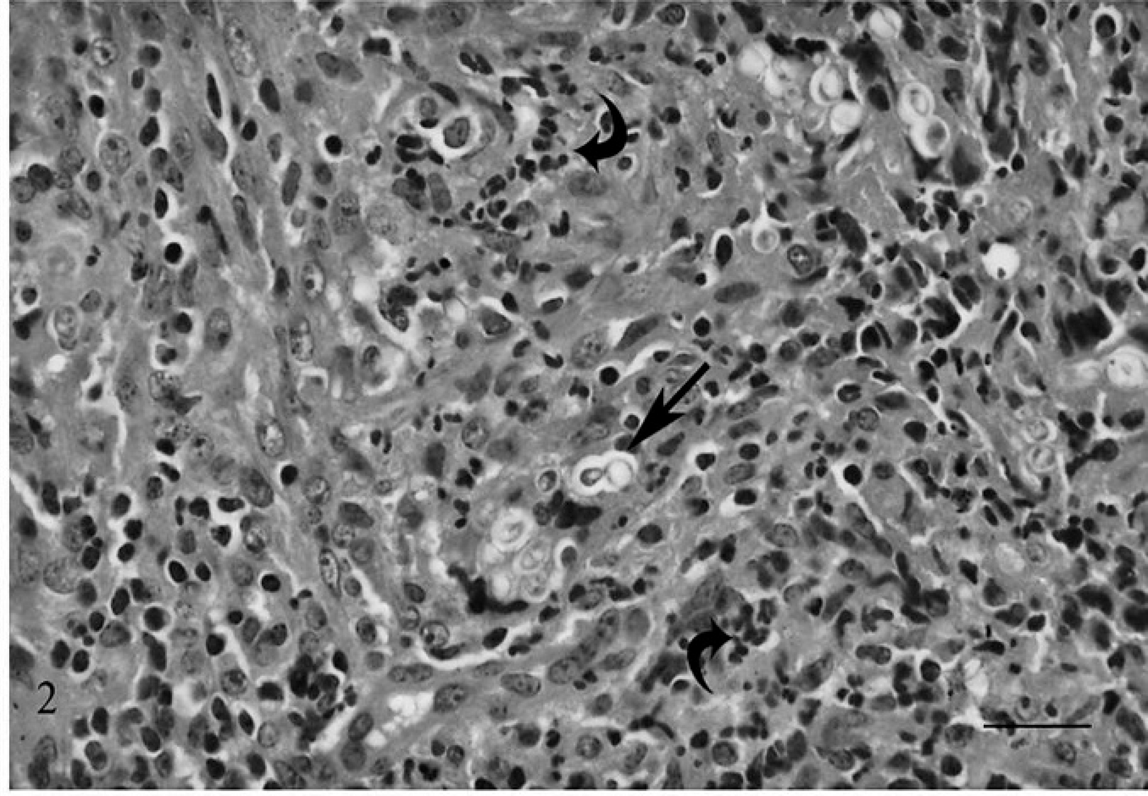
Tonsil; pig. Poorly defined Geotrichum candidum hyphae are noted, frequently budding (straight arrow) within an infiltrate of histiocytes and scattered clusters of neutrophils (curved arrows). Hematoxylin and eosin. Bar = 20 μm.
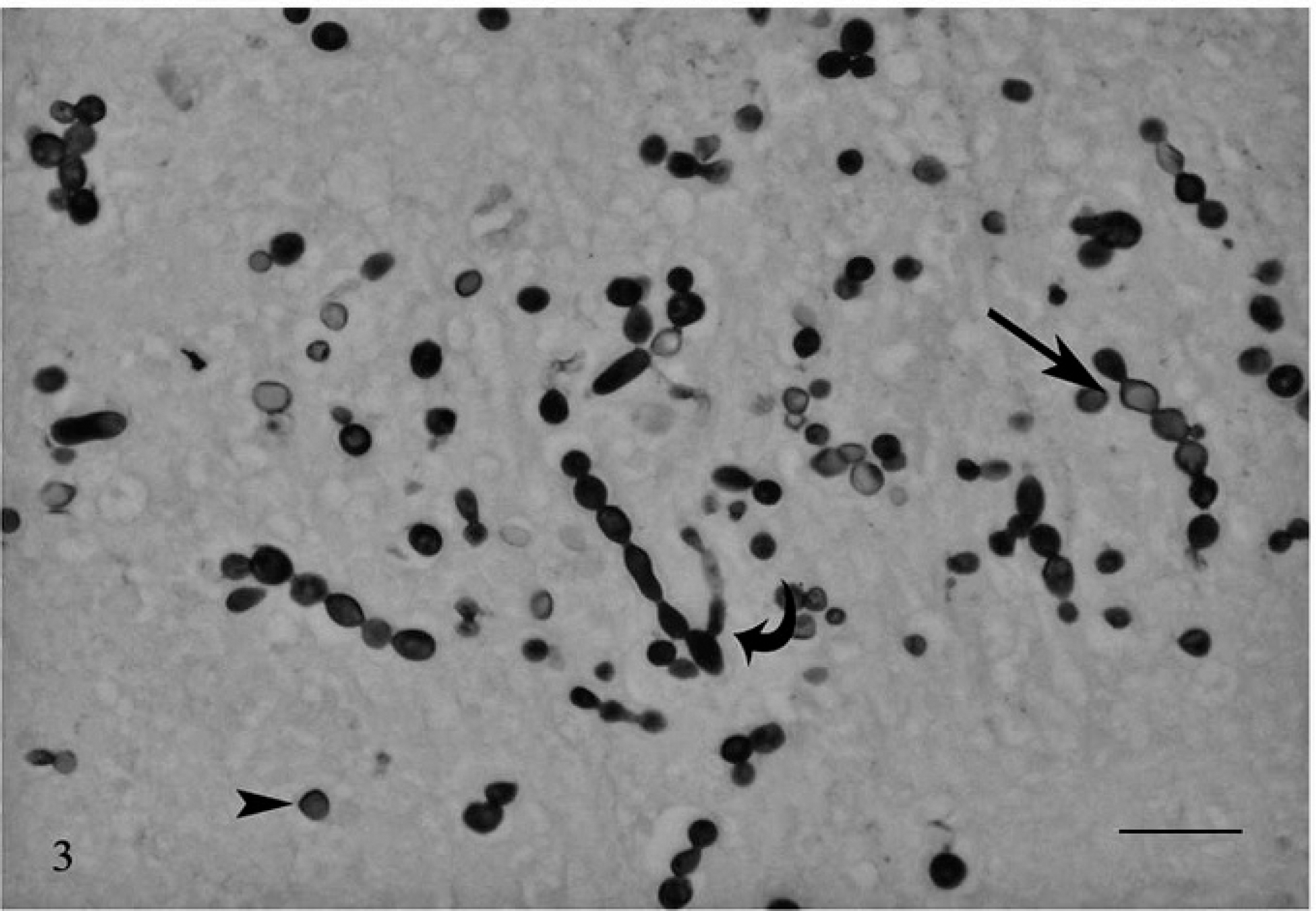
Tonsil; pig. Geotrichum candidum hyphae (straight arrow) with distinctly budding morphology (curved arrow). Individualized arthrospores appear yeast like (arrowhead). Grocott–Gomori methenamine silver. Bar = 20 μm.
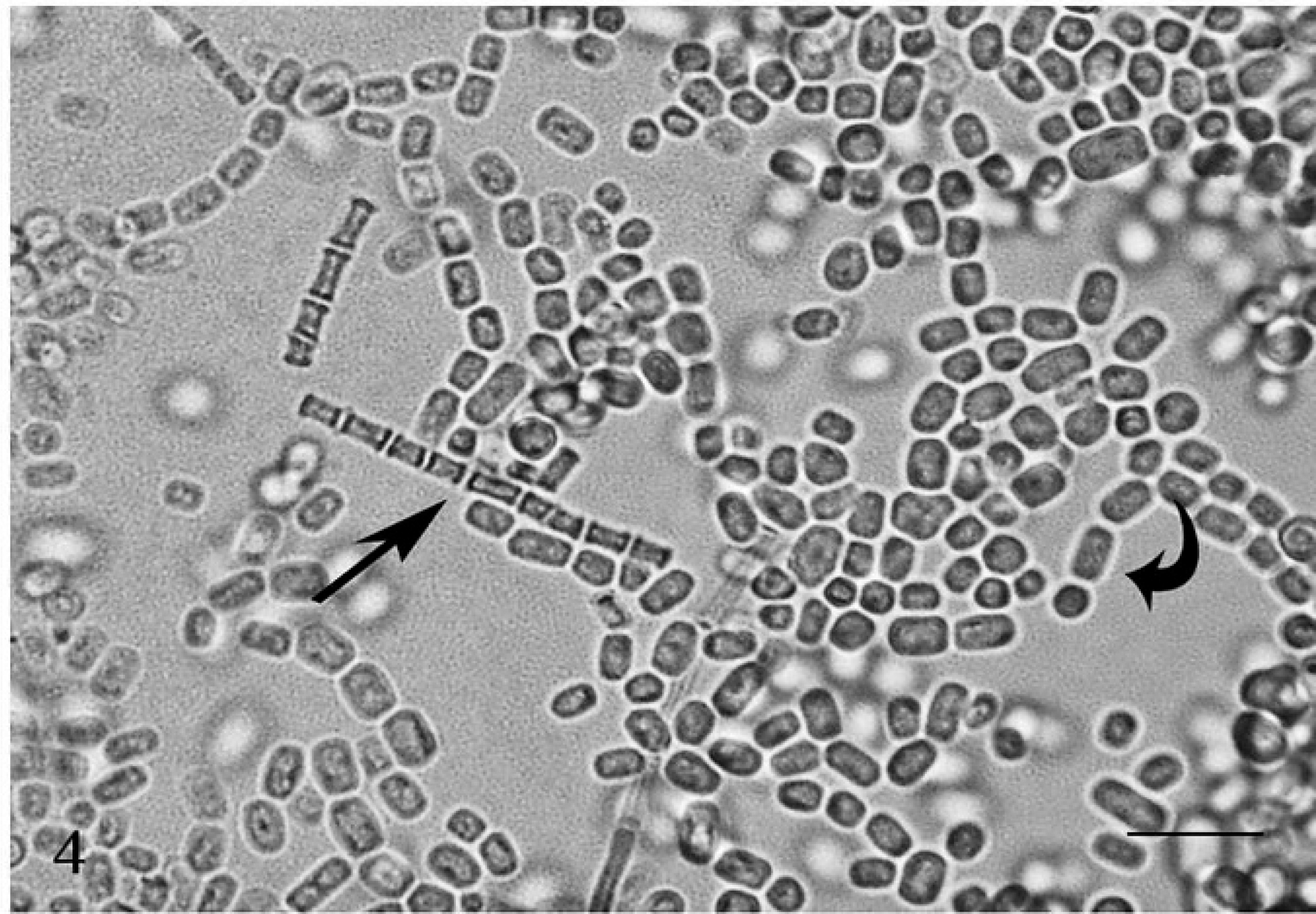
Wet mount. Geotrichum candidum with long septate hyphae (straight arrow) with rectangular arthrospores of variable size and occasional rounded ends (curved arrow). Cotton phenol blue. Bar = 20 μm.
Tonsillar tissue was cultured on Sabouraud media with cycloheximide and incubated at 30°C. 23 By 4 days, a profuse predominant fungal growth of white, flat colonies with a velvety granular surface was noted. Cotton phenol blue preparations from these colonies indicated long septate hyphae with numerous rectangular arthrospores of variable size and occasional rounded ends (Fig. 4). Colonial growth and microscopic characteristics of the fungal colonies were consistent with Geotrichum candidum. 6,23 Cultures from the skin yielded a heavily mixed growth of environmental contaminants.
Geotrichum candidum is a saprophytic fungus found on fruits, vegetables, and dairy products. 14 Infections in animals have been rarely reported in the veterinary literature. Before 1955, the fungus had been isolated from sporadic cases of bovine abortion and mastitis, 1 caseous nodules in the lymph node of a pig, 21 and from the feces of a piglet with diarrhea. 1 A case of disseminated canine geotrichosis was reported in a mixed Labrador Retriever, 17 and the fungus was recovered from another dog with catarrhal enteritis. 1 Geotrichum candidum was cultured from the feces of adult gorillas with diarrhea 19 ; however, a firm association with pathologic lesions was not demonstrated.
Geotrichum remains unusual in human pathology; it is, however, of increasing significance, because it is almost without exception, associated with immunocompromised individuals. 11 Cases of disseminated mycosis, 5,11,16,22 septicemia, 2,4,25 brain abscesses, 15 traumatic joint infection, 12 conjunctivitis, 20 pulmonary infections, 3,5,7,16,20,27 oral infections, 10,20 intestinal infections, 13,20 and cutaneous infections 24 have been described in human beings. Histologic confirmation of G. candidum is difficult because of its morphologic characteristics, which are similar to many potentially pathogenic fungi, including Aspergillus spp., Candida spp., 13,16 Histoplasma capsulatum, 17 coccidioides, and Lobo loboi. 14 Nevertheless, 2 microscopic features of G. candidum aid in its differentiation: 1) poor staining with HE, and 2) irregular hyphae with arthrospores of varying diameters at the poles. 13 Diagnostic confirmation, however, requires a positive culture of G. candidum from the infected tissue site. 13,16 The initial concern in the current case was the feeding of unusually high amounts of “macadamia trash” in the diet. This practice, which is common in macadamia nut–raising regions, is increasingly viewed with alarm by the public, because of the regular use of an insecticide (endosulfan) in the treatment of macadamia nuts (Cubit S: 2009, Endosulfan ruled out as a potential cause of Noosa fish health issues. Available at http://www.apvma.gov.au/news_media/our_view/2009/2009-11-25_endosulfan_ruled_out.php. Accessed September 29, 2010). This raises the issue of residue formation and entry of endosulfan into the food chain. At high doses, endosulfan is neurotoxic and has been linked to nervous signs and fatal poisonings in human beings (Scholl-Buckwald S: 2006, Speaking truth saves lives in the Philippines and India. PAN North America Magazine, pp. 24–27). Endosulfan is a xenoestrogen, which imitates or synergizes the effect of estrogen and has implicated effect on gene expression in female rats. 26 A nonfatal idiopathic syndrome has been associated with the ingestion of macadamia nuts by dogs, characterized by vomiting, ataxia, weakness, hyperthermia, and depression. 8,9,18 In the current case, there was no evidence of toxicity consistent with endosulfan exposure; however, based on the clinical signs, toxicity from ingestion of macadamia nuts remains a possibility. This has not yet been described in pigs and requires further investigation. To the authors' knowledge, the present case is the first to report tonsillitis in a pig induced by G. candidum, an extremely rare, potentially pathogenic, and zoonotic fungal organism.