
Abstract
A female, reticulated python (Python reticularis) of unknown age was presented with a history of lethargy, weakness, and distended coelom. Physical examination revealed severe dystocia and stomatitis. The reticulated python was euthanized due to a poor clinical prognosis. Postmortem examination revealed marked distention of the reproductive tract with 26 eggs (10–12 cm in diameter), pericardial effusion, and a slightly firm, pale tan mass (3–4 cm in diameter) adhered to the endocardium at the base of aorta. Based on histopathologic and transmission electron microscopic findings, the diagnosis of endocardial fibrosarcoma was made.
Neoplasia is frequently encountered in the practice of reptile medicine, 7,13 although neoplastic diseases were once thought to be rare in reptiles. 12 However, there is a paucity of literature on neoplastic conditions in reticulated pythons. The current report describes the microscopic and transmission electron microscopic features of an endocardial fibrosarcoma in a reticulated python.
A female, reticulated python (Python reticularis) of unknown age was presented to the zoological medicine service of Louisiana State University Veterinary Teaching Hospital and Clinics (Baton Rouge, Louisiana) as part of a group of confiscated snakes. On physical examination, the python was dehydrated, weak, and lethargic and had marked distension of the caudal third of the coelom. Examination of the oral cavity revealed moderate stomatitis. Palpation of the coelom revealed soft fluid-like distension. An enema was performed in order to determine if the material associated with the distention was within the gastrointestinal tract. The enema did not yield any expulsion of material. Based on the nonproductive enema, palpation, and the snake's condition, a clinical diagnosis of dystocia or egg binding was made. The reticulated python was euthanized due to a poor clinical prognosis and submitted for necropsy examination.
Postmortem examination revealed a severely distended pericardial sac containing approximately 500 ml of clear yellow fluid. The epicardium and pericardium had multifocal fibrous adhesions. On dissection of heart chambers, there was a slightly firm, pale tan, sessile mass (3–4 cm in diameter) adhered to the endocardium at the base of aorta. The mass was not visible from the epicardial surface. The reproductive tract contained 26 eggs (10–12 cm in diameter). No other major gross findings were noticed. Various tissue samples were fixed in 10% neutral buffered formalin, routinely processed, paraffin embedded, sectioned at 5 μm, and stained with hematoxylin and eosin. Histologically, the endocardial mass was a densely cellular, unencapsulated collection of spindle cells. The spindle cells were arranged in interlacing streams and bundles, with rare storiform patterns supported by fine fibrovascular stroma (Fig. 1). Neoplastic cells were plump and fusiform with small amounts of eosinophilic fibrillar cytoplasm and indistinct cell borders. Nuclei were elongated to oval with finely stippled to vesicular chromatin and a single nucleolus. Mitoses were 3 per 10 high-power fields of view. Anisokaryosis and anisocytosis were moderate. The neoplasm contained multifocal areas of mild necrosis. There were multifocal heterophilic granulomas and moderate scattered heterophilic infiltrates in the underlying endocardium and rarely myocardium. The granulomas were composed of moderate numbers of degenerate heterophils surrounded by small numbers of macrophages with a central area containing moderate amounts of karyorrhectic necrotic cellular debris and occasional mineralization. The endocardial mass was diagnosed as fibrosarcoma. However, peripheral nerve sheath neoplasia was included as a possible differential diagnosis due to the presence of rare storiform patterns. The other histologic changes in the heart included diffuse thickening of pericardium with moderate amounts of fibrous connective tissue. There were multifocal areas of moderate necrosis; hemorrhages; and moderate infiltrates of heterophils, lymphocytes, and plasma cells in the myocardium. In buccal mucosa sections, severe heterophilic bacterial stomatitis was present. No other significant microscopic findings were seen in this animal.
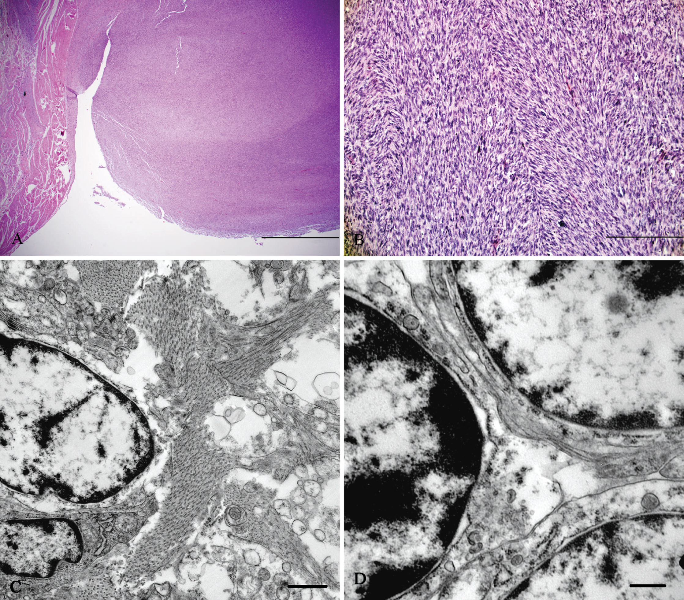
Heart (endocardial mass); reticulated python.
Immunohistochemical staining of formalin-fixed, paraffin-embedded replicate tissue sections was performed for S-100, vimentin, smooth muscle actin, desmin, and neuron-specific enolase, as described by the manufacturer. a The performance of positive and negative controls was within normal limits. However, immunohistochemical staining failed to identify specific lineage of neoplastic cells in the endocardial mass. Similar findings have been reported previously for the immunohistochemical detection of undifferentiated sarcomas in reptiles. 1,16
For transmission electron microscopic examination, 1-mm3 fragments of the endocardial mass were fixed in 3% glutaraldehyde, postfixed in 1% osmium tetroxide, dehydrated in ethanol, and embedded in liquid epoxy resin. b The ultrathin sections (70–90 nm) were stained with lead citrate and uranyl acetate and examined with an electron microscope. c Ultrastructurally, collagen fibers were identified throughout the tissue sections separating the neoplastic cells (Fig. 1). Neoplastic cells contained moderate numbers of mitochondria and profiles of rough endoplasmic reticulum. Nuclei were large, and the prominent heterochromatin was coarsely stippled or in some cases marginated at the inner nuclear membrane (Fig. 1). Neoplastic cells lack intercellular junctional complexes, basal lamia, and lamellated interdigitating cell processes characteristic of a peripheral nerve sheath tumor. Peripheral nerve sheath tumors have been reported rarely in snakes. 7,15 The histologic and electron microscopic morphology of the neoplastic cells in the current case were indicative of fibrosarcoma.
Neoplastic disease in captive reptiles is growing in prevalence as a result of increased life expectancy caused by improved husbandry and management. 16 The most commonly reported ophidian neoplasms are mesenchymal, 7,8 epithelial, 7,16 and lymphoid/hematopoietic. 2,7 Most reported neoplasms among ophidian species are in aged colubrids, followed by crotalids, vipers, and boids. 7 Fibrosarcomas have been reported in snakes affecting the oral cavity, skin, subcutaneous tissue, body wall, and musculoskeletal system. 6,7,10,13 Recently, metastatic fibrosarcoma was reported in a Saharan horned viper (Cerastes cerastes). 14 There are few comprehensive retrospective studies on reptile neoplasia in the veterinary literature. 2,7,16,17 In one retrospective study, the overall prevalence of neoplasia submissions during a 9-year period was highest in snakes (15%), followed by lizards (8.5%), chelonians (2.7%), and crocodilians (2.2%). 7 Fibrosarcoma was considered to be the most common neoplasm reported in snakes. 9 However, a 2004 study showed that soft-tissue sarcomas (11.4%), renal adenocarcinoma (8.2%), and lymphomas (10.6%) were more prevalent than fibrosarcoma (5.8%). 7 Another retrospective study on reptile neoplasia at the Philadelphia zoo revealed that liver was most commonly affected, followed by the integumentary and digestive systems. 17 There is no report of endocardial fibrosarcoma in reticulated pythons. Moreover, there are only rare reports of primary tumors affecting the cardiovascular system in snakes. 2
The possible role played by infectious agents, particularly viruses, has been suggested in the development of reptilian neoplasia. 3,11 “C-type” oncogenic retroviruses were considered a possible cause for the high prevalence of spindle-cell sarcomas in Sacramento Zoo snakes. 16 However, no known casual agent has been routinely detected for most spontaneous tumors diagnosed in reptiles. 13 Ultrastructural examination of the neoplasm in the present case did not reveal the presence of any retroviral particles. Additional testing, such as retroviral culture, was not pursued. Therefore, a specific cause of the neoplasm could not be ascertained. The oral lesions in the python in the current report were compatible with bacterial stomatitis, which is a well-documented disease in snakes. 5 The Gram-negative bacteria Pseudomonas spp., Aeromonas spp., Proteus spp., and Morganella spp. are the most frequently isolated pathogens. The buccal mucosa samples were not submitted for bacterial culture in the present case. Dystocia has been reported frequently in reptiles. 4 The lack of antemortem diagnostics precludes the identification of specific factors that may have contributed to the dystocia. No obstructive lesions were found at necropsy. It can be speculated that stomatitis, pericardial effusion, and endocardial fibrosarcoma are evidence of systemic disease that may have contributed to the dystocia. The clinical signs in the python in the present report were likely due to a combination of the reproductive tract impaction, pericardial effusion, and/or endocardial fibrosarcoma.
Footnotes
a.
EnVision+ System-HRP Labeled Polymer (DAB), Dako North America Inc., Carpinteria, CA.
b.
Epon™, Hexion Specialty Chemicals, Columbus, OH.
c.
JEM-1011, JEOL Ltd., Tokyo, Japan.