
Abstract
Adenovirus-associated enteritis was diagnosed by histopathology of small intestine in a 2-year-old alpaca (Vicugna pacos). Electron microscopy confirmed intracytoplasmic and intranuclear adenoviral particles within enterocytes. Nucleic acid was extracted from paraffin-embedded tissue sections, and a pan-adenovirus nested polymerase chain reaction (PCR) assay was employed to target a partial sequence of the polymerase gene. The PCR product (321 bp) was cloned and sequenced. Comparison of the nucleotide sequence against the National Center for Biotechnology Information (NCBI) nucleotide database demonstrated 68% identity with the isolates Canine adenovirus 1 and Bovine adenovirus 3. Comparison of the predicted amino acid sequence against the NCBI database demonstrated 75% identity with Bovine adenovirus 3. Phylogenetic analysis supported the relatively close relationship of this isolate to Bovine adenovirus 3, but the alpaca isolate was sufficiently distant to be considered a potentially novel adenovirus for this species.
The family Adenoviridae contains 5 genera (Mastadenovirus, Aviadenovirus, Atadenovirus, Siadenovirus, and Ichtadenovirus), which compose adenoviruses isolated from animals and human beings. 1 Mastadenovirus infection is common in mammalian animals but in most cases clinical disease appears only if predisposing factors, such as adverse management problems, overcrowding, transportation, and intercurrent bacterial infections, are present. There are only a small number of reports of adenovirus infections in New World camelids (NWCs),3,5-6,10 but no isolates have yet been formally recognized. 1 The current report describes a potentially novel mastadenovirus infection in an alpaca (Vicugna pacos).
A 2-year-old female lactating alpaca was presented for necropsy. The clinical history indicated that it had developed diarrhea and pyrexia 2 weeks previously and had been treated with antibiotics and nonsteroidal anti-inflammatory drugs; specific details of these treatments and clinical progression over the 2-week period were not provided. It had been the only animal affected in a herd of 50 alpacas of mixed ages. At necropsy, it was in poor body condition and weighed 45 kg (normal reference weight: 55–90 kg). 2 Significant gross findings included submandibular edema and a circular 12 mm in diameter ulcer in the third compartment (C3) of the stomach. Despite the clinical history of diarrhea, fecal pellets of normal consistency were present in the rectum.
Samples of stomach, small and large intestines, kidney, and liver were prepared for histopathology. The C3 gastric ulcer showed extensive necrosis, hemorrhage, and thrombosis extending through the mucosa into the underlying deeper tunics, associated with active septate fungal invasion and more superficial bacterial colonization. The small intestine featured prominent lymphocytic–plasmacytic infiltrates throughout the mucosa, including villus cores. Large numbers of variably sized amphophilic intranuclear inclusions were present within villus enterocytes (Fig. 1) associated with active epithelial exfoliation and necrosis. Crypt epithelium was variably hyperplastic and attenuated with accumulation of cell debris within gland lumina. Hyperplastic mucoid colitis, with mixed inflammatory cell infiltrates of the mucosa and submucosa, was present but without intranuclear epithelial inclusions. Mild protein-losing nephropathy and mild multifocal hepatic necrosis with thrombosis were also observed.
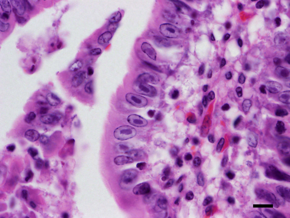
Histopathological examination of small intestine shows intranuclear amphophilic inclusion bodies in enterocytes. Hematoxylin and eosin. Bar = 10 µm.
Electron microscopic examination of small intestine identified small groups of intracytoplasmic and larger intranuclear paracrystalline arrays of electron-dense icosahedral viral particles of 70–90 nm in diameter. The particles were located in enterocytes and were morphologically typical of adenoviruses (Fig. 2). Similar viral particles were also identified by electron microscopic examination of a sample of small intestinal contents.
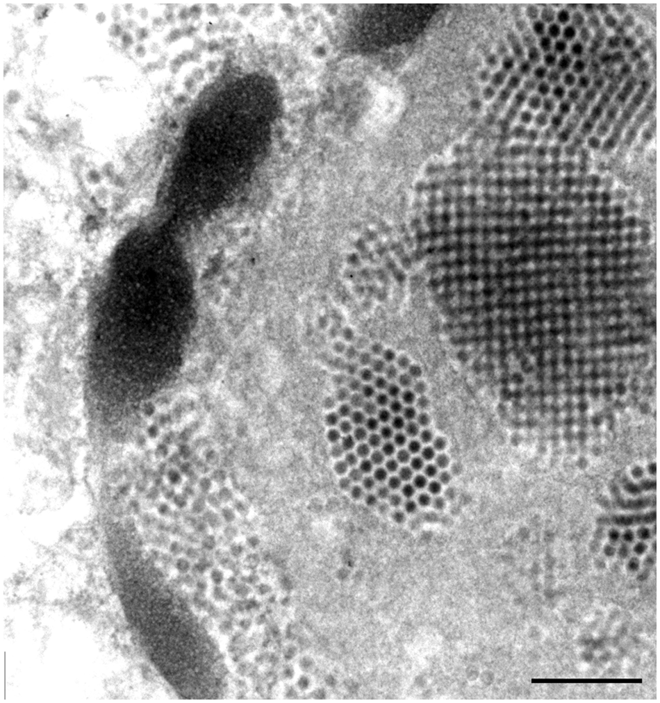
Electron microscopic examination of small intestine demonstrates paracrystalline arrays of adenoviral particles in the nucleus and groups of viral particles in the cytoplasm of enterocytes. Bar = 500 nm.
Nucleic acid was extracted from paraffin-embedded tissue sections of the small intestine. 4 A pan-adenovirus nested polymerase chain reaction (PCR) assay was employed to target a partial sequence of the polymerase gene. 9 The PCR product (321 bp) was visualized by ethidium bromide gel electrophoresis and recovered from the agarose gel. The PCR product was purified using a commercial kit a and cloned using the topoisomerase I, Taq-amplified cloning kit, b performed according to manufacturers’ instructions. Plasmid DNA was extracted using a commercial kit, a and EcoR1 c restriction enzyme digestion was performed to demonstrate the presence of PCR product within the plasmid vector, with visualization by ethidium bromide gel electrophoresis. Plasmid DNA from 3 clones containing the PCR product insert were recovered from agarose gels, purified, and used as templates in direct dye-termination sequence reactions. d The nucleotide sequence (272 nt) of the 3 clones shared 100% sequence identity and was deposited in GenBank (http://www.ncbi.nlm.nih.gov/genbank/) under accession no. FJ711592. Comparison of this sequence against the National Center for Biotechnology Information (NCBI) nucleotide database demonstrated 68% identity with the isolates Canine adenovirus 1 (CAdV-1) and Bovine adenovirus 3 (BAdV-3). Comparison of the predicted amino acid sequence against the NCBI database demonstrated 75% identity with BAdV-3 (AF030154). Phylogenetic analysis of the predicted amino acid sequence supported the relatively close relationship of this isolate to BAdV-3 (Fig. 3), but the alpaca isolate was sufficiently distant to be considered a potentially novel adenovirus for this species.
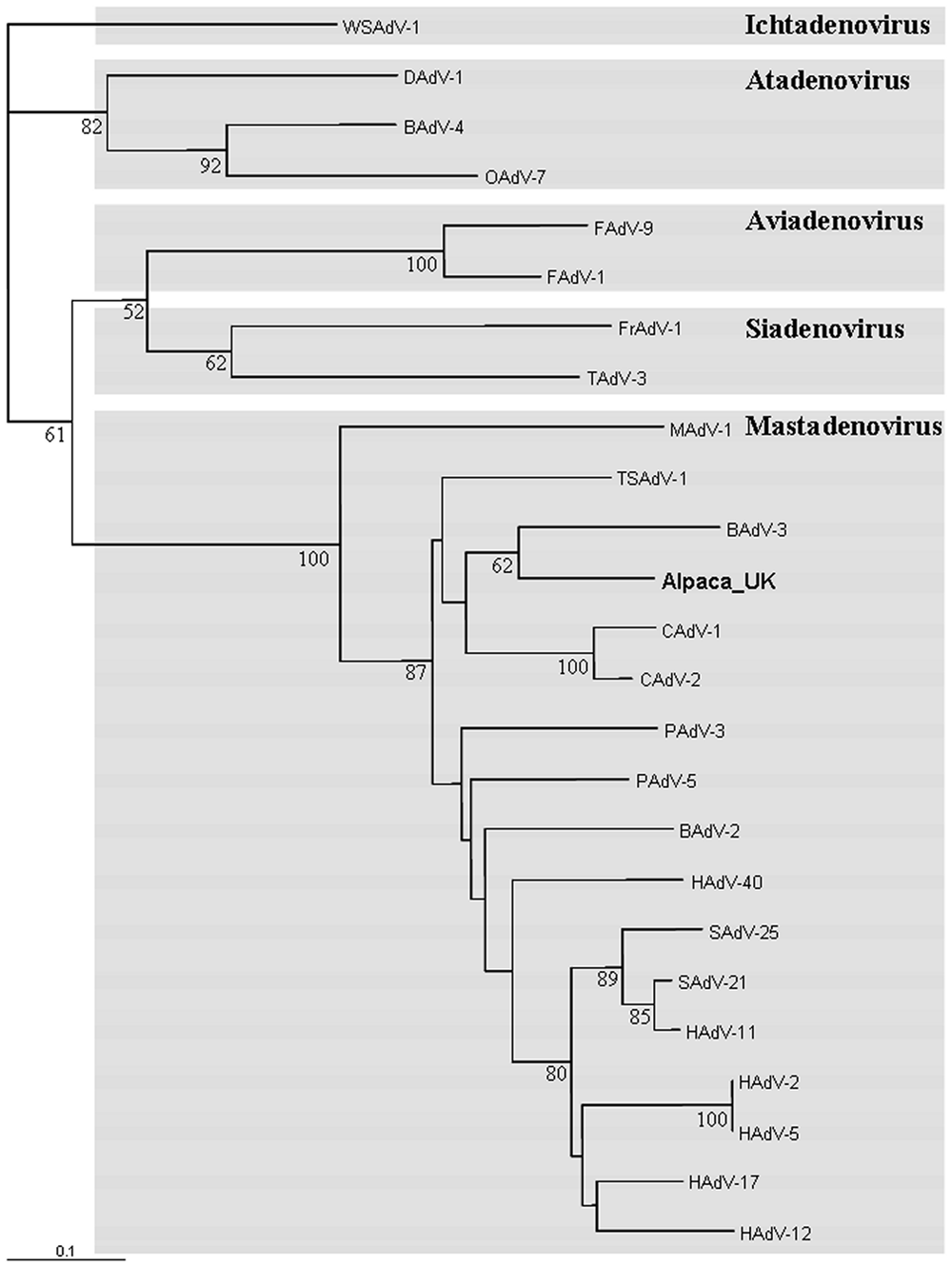
Consensus phylogram of 2,000 Fitch–Margoliash trees generated for a partial amino acid sequence of the polymerase gene (90–91 amino acids). ClustalX was used to align the predicted amino acid sequence with the homologous region from isolates representative of the 5 genera within the family Adenoviridae. The isolate White sturgeon adenovirus was chosen as an outgroup for these analyses. Phylogenetic analysis was performed using ProtDist (Jones–Taylor–Thornton matrix) and Fitch. Bootstrap confidence values were calculated by using Seqboot and Consense (number of replicates = 2,000); values over 50% are indicated. Abbreviations of virus names are indicated at the ends of the branches: B, bovine; C, canine; D, duck; F, fowl; Fr, frog; H, human; M, murine; O, ovine; P, porcine; S, simian, T, turkey; TS, tree shrew; and WS, white sturgeon. Accession numbers: BAdV-2 AF252854; BAdV-3 AF030154; BAdV-4 AF036092; CAdV-1 Y07760; CAdV-2 U77082; DAdV-1 Y09598; FAdV-1 U46933; FAdV-9 AF083975; FrAdV-1 AF224336; HAdV-2 J01917; HAdV-5 M73260; HAdV-11 AY163756; HAdV-12 X73487; HAdV-17 AF108105; HAdV-40 L19443; MAdV-1 NC000942; OAdV-7 U40839; PAdV-3 AF083132; PAdV-5 AF289262; SAdV-21 AR101858; SAdV-25 AR101859; TAdV-3 AF074946; TSAdV-1 AF258784; WSAdV-1 AY082701.
The major diagnostic findings consisted of adenovirus-associated enteritis and ulcerative mycotic gastritis, both of which appeared to have a similar time scale. The relative contribution of these 2 conditions to the alpaca’s demise is unclear but it is likely that they acted synergistically. The mild kidney and liver lesions were most likely terminal events associated with toxemia and disseminated intravascular coagulation. Adenovirus infections are rarely reported in NWCs, and might therefore be assumed to have little clinical significance.3,5-6,10 By contrast, gastric ulceration is a common finding. 7 However, this higher recorded prevalence of gastric ulceration might simply reflect the fact that gastric lesions are easily observed grossly at necropsy without recourse to specialist laboratory testing.
Adenovirus infection, as determined by the presence of viral inclusions, was limited to the small intestine of the alpaca described herein. Electron microscopy and PCR assay were used as confirmatory tests. Immunohistochemistry was not used on this occasion but has been described for other adenoviruses and could potentially associate infection with pathological lesions in NWCs. 8 Enteric involvement has previously been reported in NWCs; adenovirus was isolated from 5 llamas and 1 alpaca with diarrhea. 5 In one case, severe necrotizing enterocolitis was associated with numerous intranuclear inclusion bodies in the intestinal epithelial cells. Adenovirus has also been associated with necrotizing bronchointerstitial pneumonia and hepatocellular necrosis in llamas, 3 and tubulointerstitial nephritis in an alpaca (Melidone R, Jakowski R, Keating J, et al.: 2008, Renal adenoviral infection in an alpaca. Vet Pathol 45:746. Abstract). The case of necrotizing enterocolitis was believed to have been secondary to immunodeficiency. 5 In the alpaca described in this current report, immunosuppression potentially occurred as a feature of chronic debilitation, manifested by weight loss and subcutaneous edema, and possibly explains the concurrent pathological findings of gastric ulceration with adenovirus-associated enteritis. Gastric ulceration often occurs with another disease condition, suggesting that the stress of concurrent disease is a predisposing factor in NWCs. 7 Opportunistic mycotic infection may well have been triggered by recent antibiotic therapy.
There is serological evidence of adenovirus infection in NWCs kept in North America, South America, and Europe. A seroprevalence of 93% was demonstrated in 270 llamas on 21 premises in Oregon. 5 By contrast, only 20 out of 390 llamas (5%) were seropositive from 9 Argentinean farms. 6 A low seroprevalence was also demonstrated in a zoological collection in Turkey: only 1 out of 7 llamas (14%) were seropositive. 10 The difference in seroprevalence between these studies might be explained by the different viral isolates on which the serological tests were based: an isolate that originated from NWCs 5 possibly produced a test of higher sensitivity than those based on bovine adenoviruses.6,10
Two antigenic species of adenovirus have previously been isolated from NWCs, which differed from bovine, ovine, or equine adenoviruses. 5 The present case provides further evidence that NWCs can be infected with their own species of adenovirus, as the isolate was phylogenetically distinct from other adenoviruses within the genus Mastadenovirus on the basis of the predicted amino acid sequence of the partial fragment of the polymerase gene that was examined. These observations are not entirely surprising, given that adenoviruses are mostly specific to, and possibly coevolved with, their hosts. 1 Further characterization of isolates from NWCs is required to confirm a new host-specific adenovirus species. Further study is also required to determine the epidemiological and pathological significance of these viruses.
Footnotes
Acknowledgements
The authors would like to thank Dr. A. Schock for assistance with photography.
a.
QIAquick Gel Extraction kit, Plasmid Mini kit; Qiagen Ltd., Crawley, West Sussex, UK.
b.
TOPO TA cloning kit, Invitrogen, Life Technologies Ltd., Paisley, Renfrewshire, UK.
c.
Promega, Southampton, Hampshire, UK.
d.
Big Dye Terminator Cycle Sequencing Ready Reaction, Applied Biosystems, Life Technologies Ltd., Paisley, Renfrewshire, UK.
Declaration of conflicting interests
The author(s) declared no potential conflicts of interest with respect to the research, authorship, and/or publication of this article.
Funding
The author(s) disclosed receipt of the following financial support for the research, authorship, and/or publication of this article: This work was funded under the Defra scanning surveillance program.